DOI:10.1002/JLB.2A0118-031RR
Activated
NK
cells
kill
hepatic
stellate
cells
via
p38/PI3K
signaling
in
a
TRAIL-involved
degranulation
manner
Tianyang Li
1,2Yang Yang
1Hongxiao Song
1Haijun Li
1An Cui
1Yanhou Liu
1Lishan Su
1,3Ian Nicholas Crispe
1,4Zhengkun Tu
11Institute of Translational Medicine, First Hospital, Jilin University, Changchun, Jilin, China
2Infectious Disease, The Second Affiliated Hospital of Guangzhou Medical University, Guangzhou, Guangdong, China
3Lineberger Comprehensive Cancer Center, School of Medicine, University of North Carolina at Chapel Hill, Chapel Hill, North Carolina, USA 4Department of Pathology, University of Washington, Seattle, Washington, USA
Correspondence
Zhengkun Tu, Institute of Translational Medicine, The First Hospital, Jilin University, 519 E. Minzhu Ave, Changchun 130061, China.
Email: [email protected]
Abstract
NK cells are important in regulating hepatic fibrosis via their cytotoxic killing of hepatic stellate
cells (HSCs). NK cells are activated by both cytokines such as IL-12 and IL-18, and innate immune
stimuli such as ligation of TLRs. The secretion of IL-18 depends upon activation of the
inflamma-some, whereas TLRs are stimulated by microbial products. In the case of NK cells, IL-18 acts
syn-ergistically with stimulation of TLR3 to cause cell activation and cytotoxic function. In the present
study, we activated NK cells to kill HSCs via IL-18 and TLR3 ligand stimulation, and dissected the
signaling pathways or molecules critical for such activation or killing. We find that such
activa-tion depends on signaling via the p38/PI3K/AKT pathway, and that the activated NK cells mediate
HSC death in a TRAIL-involved mechanism. As liver fibrosis is a major global health problem with
no good solution, these results emphasize that the p38/PI3K/AKT pathway in NK cells may be a
novel drug target to promote fibrosis regression.
K E Y W O R D S
IL 18, p38 MAPK, TRAIL, TLR3
1
I N T RO D U C T I O N
Liver fibrosis, which result from chronic liver injury of any etiology such
as viral infection, alcoholic liver disease, and NASH, is a major global
health problem for which there is no effective treatment.1As liver
fibrosis progresses, liver cirrhosis and liver cancer regularly demand
liver transplant.2Hepatic stellate cells (HSCs) are essential in the
pro-gression of liver fibrosis. HSCs are activated by inflammatory cytokines
and mediators to trans-differentiate into myofibroblasts, and become
the major source of extracellular matrix including collagens in the liver
during the development of fibrosis.3,4
As an important component of the innate immune system, NK cells
respond rapidly to transformed or virus-infected cells, and kill such
tar-get cells without restriction by either major histocompatibility
anti-gen or a need for presensitization.5NK cells have been implicated in
suppressing the pathogenesis of liver fibrosis,6but the exact
mecha-nism remains elusive. It has been reported that NK cells selectively kill
early activated (transitional) or senescent-activated HSCs rather than
quiescent or fully activated HSCs (myofibroblasts).7Activated HSCs
express less NK cell inhibitory ligand MHC-1,8but strongly express
Abbreviations: HSC, hepatic stellate cell; poly I:C, polyinosinic–polycytidylic acid
ULBP-2 (UL16 binding protein 2), MICA/B (MHC class I
polypeptide-related sequence A/B),9and RAE-1 (retinoic acid early inducible 1),7,10
which activate NK cells through engagement with NKG2D. This leads
to killing of activated HSCs by NKG2D-dependent degranulation of
NK cells.7,9Additionally, NK cells induce HSCs apoptosis through FasL
with activated HSCs’ intense expression of Fas, as well as
TRAIL-mediated killing.7,9NK cells also restrain HSCs activation through IFN-𝛾secretion.11However, the effects of NK cells on HSCs through the established mechanisms are attenuated as NK cells are deactivated
as liver fibrosis inexorably progresses, and impaired antifibrotic
func-tions of NK cells are associated with accelerated progression of liver
fibrosis.12Therefore, the restoration and promotion of NK cell activity
might promote the regression of liver fibrosis.
IL-18 belongs to the IL-1 family. Once its 24 kDa precursor is
cleaved to the 18 kDa form by IL-1𝛽 converting enzyme (ICE, also
known as caspase-1), IL-18 acquires biologic function to promote
proliferation and cytolytic activity of NK and T cells combined with
IL-12 or IL-2.13,14The immunostimulatory properties of polyinosinic–
polycytidylic acid (poly I:C), a surrogate TLR3 ligand, have been verified
in various pathologic circumstances in vivo.15Furthermore, NK cell
activation in response to IL-18/IL-12 or IL-15/IL-12 stimulation is
by which IL-18 and poly I:C synergistically activate NK cells is not
clearly defined.
In the present study, we investigated the mechanisms by which NK
cells are activated by IL-18 and poly I:C to kill HSCs using human NK
cells from healthy donors and primary HSCs or LX2 cells. Our results
showed that IL-18 and poly I:C synergistically activate NK cells via the
p38/PI3K/Akt signaling pathway, and the activated NK cells kill HSC
in a TRAIL-involved degranulation manner. We thus propose that p38
MAP/PI3 kinase might be a novel therapeutic target for intervention of
human liver fibrosis targeting NK cells.
2
M AT E R I A L S A N D M E T H O D S
2.1
Human subjects
Buffy coats from healthy donors were provided by the Changchun
Blood Center, and informed consent was provided according to the
protocols of the Changchun Blood Center. Liver perfusion from
cadaver donors were collected as we described previously.18All
stud-ies were conducted according to the experimental practices and
stan-dards that were approved by the Medical Ethics Committee of First
Hospital of Jilin University (approval code: 2015–125).
2.2
Cell isolation and purification
PBMCs were freshly isolated from peripheral blood of healthy
indi-viduals by Ficoll-Paque Plus (GE Healthcare, Uppsala, Sweden)
den-sity gradient separation. Liver mononuclear cells were freshly isolated
from liver perfusates as we described previously.18For magnetic cell
sorting, NK cells were purified by NK cell isolation kit (Miltenyi Biotec,
Bergisch Gladbach, Germany). The purity of NK cells was
approxi-mately 95% as determined by flow cytometry (Supplemental Fig. S1A).
2.3
Cell culture
Purified NK cells were cultured in RPMI 1640 medium (Corning Life
Science, Lowell, MA, USA) with 10% FBS and 1% Pen-Strep (GE
Health-care). After stimulated with IL-18 (R&D System, Minneapolis, MN,
USA) or poly I:C (Sigma–Aldrich, Saint Louis, MO, USA) for 24 h in 24
wells plate, cells were collected for FACS analysis, the supernatants
were harvested for cytokine detection. To inhibit PI3K or p38 MAPK,
NK cells were pretreated in the presence of 50𝜇M LY294002 (Sigma–
Aldrich) or 20𝜇M SB202190 (Sigma–Aldrich) for 1 h and then
stimu-lated with IL-18 and/or poly I:C.
LX2 cells were cultured in DMEM medium (Corning Life Science)
supplemented with 10% FBS. Primary human HSCs purchased from
ScienCell (San Diego, CA, USA), and were cultured within 3 passages
in Stellate Cell Medium (ScienCell). The definition of LX2 cells and
pri-mary HSCs were shown as Supplemental Fig. S1B.
2.4
Cell coculture
NK cells stimulated with IL-18 and/or poly I:C were collected and
resuspended in fresh medium, and then cocultured with LX2 or
pri-mary HSCs, which were labeled with 1.25𝜇M CFSE (Lifescience
Tech-nology, Carlsbad, CA, USA), in 24 wells plate atE:T=1:1 for 5 h. For
transwell coculture experiments, NK cells were in the upper
cham-ber, whereas LX2 cells or primary HSCs were in the lower chamber
using the plates with 0.4𝜇m pore diameter (Corning Life Science). For
the blocking experiments, IL-18/poly I:C-stimulated NK cells were
pre-treated with 50𝜇M 3,4-DCI (Sigma–Aldrich) or 1𝜇g/ml anti-TRAIL
(R&D System; Catalog number: AF375) for 1 h, and then cocultured
with LX2 or primary HSCs. For IFN-𝛾-blocking coculture experiments,
IL-18/poly I:C-primed NK cells were cocultured with LX2 or primary
HSCs with 1𝜇g/ml anti-IFN-𝛾supplied in medium. Soluble TRAIL (R&D
System; Catalog number: 375-TL-010) was added to HSCs half hour
earlier than NK cells in coculture.
2.5
Flow cytometry
Cell staining and flow cytometry analysis were performed as
described.19 Briefly, the phenotypic and functional markers of
NK cell were characterized by staining with the following antibodies:
mouse anti-human NKp30-PE (BD Pharmingen; Catalog number:
558407; BD Biosciences, San Jose, CA, USA), mouse anti-human
NKp46-APC (BD Pharmingen; Catalog number: 558051), mouse
anti-human NKG2D-APC (BD Pharmingen; Catalog number: 558071),
mouse anti-human CD94-FITC (BD Pharmingen; Catalog number:
555888), mouse anti-human CD69-V450 (BD Horizon; Catalog
num-ber: 560740), mouse anti-human CD253-PE (BD Pharmingen; Catalog
number: 550516), mouse anti-human CD16-PE-Cy7 (BD
Pharmin-gen; Catalog number: 557744). Intracellular IFN-𝛾 was stained by
mouse anti-human IFN-𝛾-APC (BD Pharmingen; Catalog number:
551385). Phosphorylation of p38 MAPK or Akt were characterized
by staining with mouse anti-Akt-PE-CF594 (pS473; BD Phosflow;
Catalog number: 562465) or mouse anti-p38 MAPK-Pacific Blue
(pT180/pY182; BD Phosflow; Catalog number: 560313). For CD107a
detection, mouse anti-human CD107a-FITC (BD Pharmingen; Catalog
number: 555800) was added into medium for 5 h, and Golgi-Plug was
involved in the last 4 h, and cells were collected for flow cytometry.
For cell apoptosis analysis, LX2 and primary HSCs were stained with
Annexin V and PI using Apoptosis Detection Kit (BD Pharmingen;
Catalog number: 556547). Flow cytometry was performed using a BD
LSRFortessa flow cytometer (BD Biosciences). The data acquired were
analyzed with FlowJo (Treestar software, Ashland, OR, USA).
2.6
ELISA
The cell culture supernatants were collected, and IFN-𝛾
concentra-tions were measured by Human IFN-gamma ELISA Ready-Set-GoR
(eBioscience, San Diego, CA, USA) according to the manufacturer’s
instructions.
2.7
NK cell siRNA transfection
Transfection were performed as described.20 Briefly, NK cells were
seeded in 24-well plates, and transfected with SignalSilenceR p38
MAPK siRNA I (Cell Signalling Technology, Danvers, MA, USA) or
AllStars Negative Control siRNA (QIAGEN, Germantown, MD, USA)
using HiPerFect Transfection Reagent (QIAGEN) before IL-18/poly
F I G U R E 1 IL-18 and poly I:C synergistically activate NK cells.Purified NK cells were treated with IL-18 or/and poly I:C at indicated concentra-tion for 24 h. IFN-𝛾expression was measured by intracellular stain (A). IFN-𝛾secretion in the supernatant was detected by ELISA (B). CD69/TRAIL NKp46/NKp30/NKG2D/CD94 expression on NK cells was analyzed by flow cytometry (C). Results were shown as mean±SEM of 6 independent experiments performed with 6 different donor samples. **P<0.01 and ***P<0.001, pairedt-test
2.8
Western blot analysis
Purified NK cells stimulated with IL-18 and/or poly I:C were collected
and lysed for Western blotting analysis as described previously.21
Briefly, cells were collected by centrifugation and resuspended in
RIPA buffer (Cell Signalling Technology) to extract protein. Protein
extracts were resolved on a 10% SDS-PAGE under constant
Volt-age condition. After electrophoresis, protein was transferred to a
transfer membrane (Merck Millipore, Darmstadt, Germany). Blots
were incubated at room temperature for 1 h and at 4◦C overnight
with anti-p38 (Cell Signalling Technology; Catalog number: 9212),
Akt (Cell Signalling Technology; Catalog number: 9272),
anti-MAPKAPK-2 (Cell Signalling Technology; Catalog number: 3042),
anti-phosphorylated p38 (Cell Signalling Technology; Catalog
num-ber: 4511), anti-phosphorylated Akt (Cell Signalling Technology;
Cata-log number: 4060), anti-phosphorylated MAPKAPK-2 (Cell Signalling
Technology; Catalog number: 3041), and anti-beta-actin (Cell
Sig-nalling Technology; Catalog number: 4970). After washed 3 times by
0.1% tween 20 (Amresco, Solon, OH, USA) PBS, peroxidase-conjugated
anti-rabbit IgG (Proteintech, Rosemont, IL, USA; Catalog number:
10545-2-AP) was involved for another 1 h at room temperature.
Immunoreactivity was determined by using ECL kit (PerkinElmer,
Waltham, MA, USA) by XRS+(BIO-RAD, Hercules, USA). Results were analyzed by Image Lab (BIO-RAD).
2.9
Live cell imaging
Live cell imaging was performed as described.22LX2 cells were seeded
in glass bottom dish (In Vitro Scientific, Mountain View, CA, USA)
and cultured for 30 min. Then prestimulated NK cells as described
above were added at NK: HSC ratio of 2:1 and immediately
coincu-bated by using DeltaVision Elite (GE Healthcare, Buckinghamshire,
UK). Cells were maintained in a 37◦C, 5% CO2chamber while videos
were recorded.
2.10
Statistical analysis
All data were analyzed using the D’Agostino and Pearson omnibus
nor-mality test.Pvalues of<0.05 were considered statistically significant.
Mean values were compared using either a pairedt-test (2 groups) or
ANOVA (more than 2 groups), followed by a Bonferroni correction for
multiple comparisons test. All statistical tests were performed using
GraphPad Prism software (San Diego, CA, USA).
2.11
Online supplemental material
Supplemental Figs. S1–S3 and Supplemental Videos S1–S3 were
included as Online Supplemental Material.
3
R E S U LT S
3.1
IL-18 and poly I:C synergistically activate NK
cells to express IFN-
𝜸
, TRAIL, and CD69
To investigate whether IL-18 and poly I:C activate NK cells, we first
purified NK cells from peripheral blood of healthy donors, and the
distribution of CD56dimCD16+/ CD56brightCD16–subsets was shown
F I G U R E 2 IL-18 and poly I:C synergistically activate p38 MAPK/PI3K/Akt to induce NK cell activation.Purified NK cells (n=3) were stimulated with IL-18 or/and poly I:C for 30 min, the expression of p38 MAPK, Akt, STAT3, mTOR, and the phosphorylation of p38 MAPK, Akt, STAT3, mTOR was determined by Western blot.𝛽-Actin was used as loading control (A). NK cells were pretreated with PI3Ki (LY294002) or p38i (SB202190) for 1 h, and then stimulated with IL-18 and/or poly I:C, the expression and phosphorylation of Akt, MAPKAPK-2, p38 MAPK was measured by Western blot (n=3, B) or flow cytometry (n=3, C). NK cells were transfected with control siRNA or p38 siRNA before stimulated by IL-18 and poly I:C, the expression of p38 MAPK, Akt,𝛽-actin, and the phosphorylation of Akt were detected by Western blot (B). The expression of IFN-𝛾in NK cells (n=6) was measured by intracellular cytokine staining (D) and ELISA (E). CD69 and TRAIL expression on NK cells (n=6) were detected by using flow cytometer analysis (F). Results are shown as mean±SEM of the indicated numbers of independent experiments performed with different donor samples. *P<0.05, **P<0.01, and ***P<0.001, pairedt-test; ns, not significant
with the optimal concentration of IL-18 (100 ng/ml) and/or poly I:C
(50𝜇g/ml) as determined in Supplemental Fig. S2C. IFN-𝛾production
was analyzed by intracellular staining and ELISA, the activating
recep-tor and inhibirecep-tory receprecep-tor expressed on NK cells were examined by
flow cytometry. Consistent with liver NK cells,23IL-18 and poly I:C
synergistically induce IFN-𝛾expression (P<0.01; Fig. 1A) and
produc-tion (P<0.01; Fig. 1B) as well as TRAIL expression (P<0.01; Fig. 1C) of
peripheral NK cells, compared with IL-18 or poly I:C alone. Moreover,
we observed that IL-18 and poly I:C synergistically up-regulate CD69
expression (P<0.01), but fail to affect Nkp30/ Nkp46/ NKG2D/ CD94
expression on NK cells (Fig. 1C).
3.2
IL-18 and poly I:C synergistically activate NK
cells via p38 MAPK/PI3K signaling pathway
We further investigated the mechanism by which IL-18 and poly I:C
synergistically activate NK cells. Purified NK cells were stimulated
with IL-18 and/or poly I:C, the protein expression and phosphorylation
levels of Akt, p38, STAT3, and mTOR were determined by Western
blots. The results showed that the phosphorylation of p38 and Akt,
but not STAT3 and mTOR, are activated by the synergy of IL-18 and
poly I:C, whereas neither IL-18 nor poly I:C alone did (P<0.05; Fig 2A).
To define the signaling pathway of IL-18/poly I:C-induced NK cell
acti-vation, the inhibitors of p38 (SB202190) and PI3K (LY294002) were
added to NK cells before stimulation with IL-18 and poly I:C. The
phos-phorylation levels of Akt, p38, and MAPKAPK-2 (directly
phosphory-lated by p-p38) were examined by Western blot and flow cytometry,
IFN-𝛾production by intracellular staining and ELISA, and the
expres-sion of CD69 and TRAIL by flow cytometry. We observed that either
SB202190 or LY294002 inhibit IL-18/poly I:C-induced Akt
phospho-rylation (P<0.05), but LY294002 fail to inhibit IL-18/poly I:C-induced
activation of p38/MAPKAPK-2 (Figs. 2B and C). Phosphorylation
levels of Akt were reduced when NK cells were transfected with p38
siRNA (Fig. 2B), suggesting that PI3K/Akt is downstream signaling of
p38 in IL-18/poly I:C-activated NK cells. Moreover, both LY294002 and
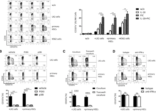
F I G U R E 3 HSCs activate IL-18/poly I:C-primed NK cell degranulation.NK cells stimulated with IL-18 and/or poly I:C were collected and resus-pended in fresh medium, and then cocultured with LX2, primary hepatic stellate or K562 cells at E:T=1:1, the expression of CD16 and CD107a was analyzed by flow cytometry (A). NK cells were pretreated with SB202190 or LY294002 before IL-18 and poly I:C stimulation, and then cocul-tured with LX2 or primary HSCs, CD107a expression was shown (B). CD107a expression was shown when transwell-coculture was used to sep-arate NK cells from LX2 or primary HSCs (C). Anti-IFN-𝛾or isotype antibody were added in coculture, and CD107a expression of NK cells were detected (D). Results were shown as mean±SEM of 6 independent experiments performed with 6 different donor NK cells. **P<0.01, ***P<0.001, pairedt-test
Fig. 2D) and production (P<0.01; Fig. 2E) as well as TRAIL expression
(P<0.001; Fig. 2F). The CD69 expression induced by IL-18/poly I:C,
however, is only inhibited by SB202190 (P<0.001) but not LY294002
(Fig. 2F). It indicates that IL-18/poly I:C-activated p38/PI3K/AKT
signaling pathway is involved in IFN-𝛾and TRAIL expression, and an
alternative p38 signaling pathway in CD69 expression on NK cells.
3.3
HSCs activate IL-18/poly I:C-primed
NK cell degranulation
During liver injury, the activation of HSCs in response to hepatocyte
damage results in the increased NK cell stimulation and decreased
NK cell inhibition.4 We further determined whether HSCs activate
IL-18/poly primed NK cell degranulation. IL-18 and/or poly
I:C-pretreated NK cells were cocultured with LX2 cells, primary HSCs, or
K562 cells. CD107a expression level of NK cells was used to evaluate
the primed NK cell degranulation using flow cytometer analysis. As
the results shown in Fig. 3A, IL-18/poly I:C-pretreated NK cells, which
were cocultured with HSCs or K562 cells, show significantly higher
CD107a expression than that of IL-18 or poly I:C alone pretreated
NK cells (P<0.01), whereas both LX2 cells and primary HSCs induce
slightly CD107a expression of IL-18 or poly I:C pretreated NK cells,
but not rest NK cells. As control, IL-18 and/or poly I:C could not
directly induce the CD107a expression of NK cells without target cell
stimulus, K562 cells induce the CD107a expression of rest NK cell. We
also observed that CD107a expression is also suppressed when NK
cells were treated with inhibitors of PI3K or p38 before stimulated by
IL-18 and poly I:C (P<0.01; Fig. 3B). CD107a expression is completely
blocked using transwell to separate IL-18/poly I:C-pretreated NK cells
from LX2 cells or primary HSCs (P<0.001; Fig. 3C). But neutralization
of IFN-𝛾in coculture did not affect CD107a expression (Fig. 3D). This
suggests that HSCs induce the degranulation of IL-18/poly I:C-primed
NK cells in a cell–cell contact-dependent manner, and that IL-18/poly
I:C-activated p38/PI3K/AKT signaling is required for HSC-induced NK
cell degranulation.
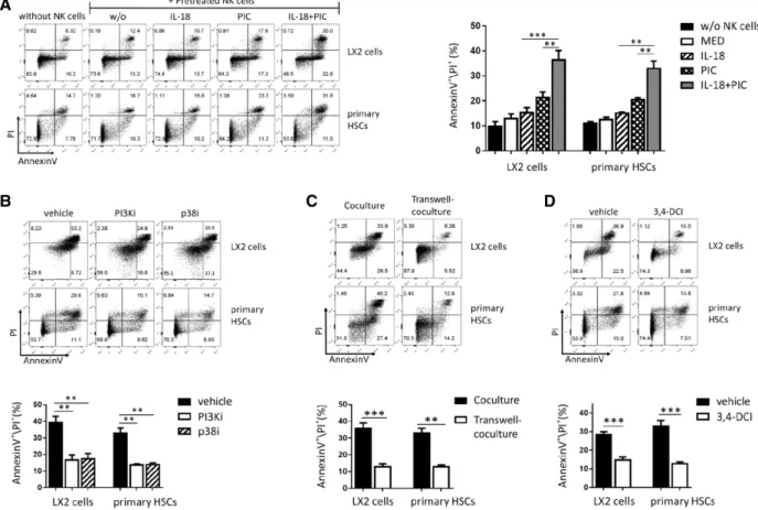
3.4
IL-18/poly I:C-primed NK cells reciprocally
induce HSCs death through degranulation
Next, to investigate IL-18/poly I:C-primed NK cell cytotoxicity to
HSCs, CFSE-labeled LX2 cells or primary HSCs were cocultured with
F I G U R E 4 IL-18/poly I:C-primed NK cells reciprocally induce HSC death through degranulation.Primary HSCs or LX2 cells were cocultured with IL-18- and/or poly I:C-pretreated NK cells, the cell death was evaluated by AnnexinV and PI staining. The cell death of primary HSCs and LX2 cells was shown (A). NK cells were pretreated with PI3Ki (LY294002) or p38i (SB202190) before IL-18 and poly I:C stimulation, and then cocultured with LX2 or primary HSCs, the cell death of HSCs was shown (B). Primary HSC and LX2 cell deaths were shown when transwell-coculture was used to separate LX2 or primary HSCs from NK cells (C). IL-18 and poly I:C-stimulated NK cells were pretreated with 3,4-DCI before cocultured with LX2 or primary HSCs, and the cell death of LX2 and primary HSCs was shown (D). Results were shown as mean±SEM of 6 independent experiments performed with 6 different donor NK cells. **P<0.01, ***P<0.001, pairedt-test; ns, not significant
in Supplemental Fig. S2D, the cell death of LX2 and primary HSCs
were evaluated with PI+/AnnexinV+. We observed that the activated
NK cell-induced HSC death is dependent on the NK cells/HSCs ratio
(Supplemental Fig. S2E), and E/T ratio=1:1 was used in the follow-ing NK cells/HSCs coculture experiments. IL-18/poly I:C-pretreated
NK cells significantly induce the cell death of primary HSCs and LX2,
compared with IL-18 or poly I:C alone pretreated NK cells (P<0.01;
Fig. 4A), whereas pretreated NK cells with medium did not do that.
The cell death of primary HSCs and LX2 was suppressed by PI3K
or p38 inhibitors, which were added to NK cells before stimulated
with IL-18 and poly I:C (P< 0.01; Fig. 4B). Transwell, which was
used to separate primary HSCs or LX2 cells from IL-18/poly
I:C-activated NK cells in coculture, completely inhibits primary HSCs and
LX2 cell death (P<0.01; Fig. 4C). Furthermore, when IL-18/poly
I:C-activated NK cells were pretreated with GranzymeB inhibitor
(3,4-dichloroisocoumarin) before cocultured with LX2 cells or primary
HSCs, the cell death of primary HSCs and LX2 is entirely inhibited as
well (P<0.001; Fig. 4D). However, neutralization of IFN-𝛾by anti-IFN-𝛾 antibody in cell coculture did not affect the cell death of LX2 and primary HSCs (Supplemental Fig. S3A). These data indicate that NK
cell cytotoxicity to HSCs occurs through degranulation in a cell–cell
contact-dependent manner, but not directly through IFN-𝛾production.
3.5
IL-18/poly I:C-primed NK cells kill HSCs in a
TRAIL-involved degranulation manner
Previous studies reported that NK cells induce a rapid apoptosis of
HSCs through TRAIL7,9or degranulation.8,20We thus investigated the
role of TRAIL in NK cells killing HSCs. Primary HSCs and LX2 cells
expressed TRAIL-R2 and TRAIL-R4 (Supplemental Fig. S3B). Using
anti-TRAIL antibody or soluble TRAIL (sTRAIL) in HSC/NK cell
cocul-ture for the blockades of TRAIL or TRAIL receptor, which are expressed
on NK cells or HSCs, respectively, the expression levels of CD107a or
PI/AnnexinV were used to evaluate NK cell degranulation or HSC cell
death. We observed that the blockades of TRAIL and TRAIL
recep-tor with anti-TRAIL and sTRAIL inhibit the cell death of LX2 and
pri-mary HSCs (P<0.01; Fig. 5A and B), as well as NK cell degranulation
(P<0.05; Fig. 5C and D).
To further analyze the immune synapse of HSC/NK cell interaction,
we visualized LX2 cells and PKH26-labeled NK cells coculture by live
cell imaging for 150 min. As shown in Fig. 5E and Supplemental Video,
IL-18/poly I:C-activated NK cells rapidly induce LX2 cell budding after
adhesion (Supplemental Video S2) contrast to resting NK cells
(Sup-plemental Video S1). Consistently, the blockade of TRAIL inhibited
F I G U R E 5 IL-18/poly I:C-primed NK cells kill hepatic stellate cells in a TRAIL- involved degranulation manner.IL-18 and poly I:C-primed NK cells were treated by anti-TRAIL or isotype antibody before coculture. HSC death was measured (A), and NK cell degranulation was evaluated by CD107a expression (C). Soluble TRAIL was added to cell coculture, the cell death of HSCs (B) and CD107a expression of NK cells (D) was shown. Results were shown as mean±SEM of 6 independent experiments performed with 6 different donor NK cells. *P<0.05, **P<0.01, ***P<0.001, pairedt-test. The kinetics of the activated NK cell-mediated LX2 cell apoptosis were observed by using live-cell imaging (E)
NK cells were cocultured transiently with CFSE-labeled LX2 cells and
washed gently to remove unattached NK cells. The results showed that
anti-TRAIL decrease the number of adhesive NK cells on LX2 cells
(Sup-plemental Fig. S3C).
Taken together, these data suggest that the engagement of TRAIL
expressed on activated NK cells with TRAIL receptors expressed on
HSCs play a role in the immune synapse formation, which initiates NK
cell degranulation to kill HSCs.
3.6
IL-18/poly I:C-primed hepatic NK cells kill HSCs
in a TRAIL-involved degranulation manner
Finally, to testify whether IL-18 and poly I:C increase hepatic NK cell
cytotoxicity to HSCs as peripheral NK cells did, we isolated hepatic
NK cells from liver perfusate of cadaver donors to establish hepatic
NK cells and primary HSCs coculture as we did in peripheral NK cells.
As expected, primary HSCs activate IL-18/poly I:C-primed hepatic NK
cell degranulation (P<0.05; Fig. 6A). IL-18/poly I:C-primed hepatic NK
cells induce the cell death of primary HSCs (P<0.01; Fig. 6B).
More-over, blockade of TRAIL with anti-TRAIL antibody inhibit hepatic NK
cell degranulation (P<0.05; Fig. 6C) and the cell death of primary HSCs
(P<0.01; Fig. 6D).
4
D I S C U S S I O N
We have previously reported that poly I:C induces IL-18 expression
in Kupffer cells to activate liver NK cells.19Liver NK cells are also
synergistically activated by IL-18 and poly I:C to produce IFN-𝛾
and express TRAIL on their surface.23But the mechanism by which
IL-18 and poly I:C synergistically activate NK cells remains to be
defined. In the present study, our results showed that consistent
with hepatic NK cells, the synergy of IL-18 and poly I:C activate NK
cells (Fig. 1). We further found that IL-18 and poly I:C synergistically
induce NK cell activation through P38/PI3K/AKT signaling pathway
(Fig. 2). IL-18 signaling is MyD88 (Myeloid differentiation primary
response gene 88) dependent,13 whereas poly I:C/TLR3
signal-ing is TIR-domain-containsignal-ing adapter-inducsignal-ing interferon-𝛽 (TRIF)
dependent and MyD88 independent.16 Downstream of TRIF and
F I G U R E 6 IL-18/poly I:C-primed hepatic NK cells kill HSCs in a TRAIL-involved degranulation manner.Liver NK cells were purified from speci-men, then primed by IL-18 and/or poly I:C before cocultured with primary HSCs as above. CD107a expression of liver NK cells (A) and PI/AnnexinV level of primary HSCs (B) were detected. Anti-TRAIL or isotype antibody were used to treat IL-18/poly I:C-primed liver NK cells before cocultured with primary HSCs, then CD107a expression of liver NK cells (C) and the cell death of primary HSCs (D) was measured. Results were shown as mean±SEM of 5 independent experiments performed with 5 different donor NK cells. *P<0.05, **P<0.01, pairedt-test
and p3827could be the coordinates of these 2 pathways. IL-12 plus
IL-18 stimulate greater IFN-𝛾secretion by resting NK cells through
stabilization of IFN-𝛾 mRNA via p38 MAPK,28 and highly augment
human NK cell cytotoxicity and degranulation in vitro.29p38 MAPK
activation controls poly I:C-enhanced cytotoxicity and poly I:C/
IL-12-costimulated IFN-𝛾secretion in human NK cells.30There is emerging
evidence about cross-talk between p38 MAPK and PI3K/Akt signaling.
TLR2-mediated interplay between MAPK and PI3K signaling axis
controls ESAT-6 (early secreted antigenic target protein 6) induced
expression of cyclooxygenase-2 in macrophages.31 PI3K/Akt/eNOS
inhibit p38 MAPK and maintain the integrity of vasculature in mouse
lung.32 P38/PI3K/Akt signaling activates heat shock protein 27 to
antagonize melatonin-induced apoptosis of gastric cancer cells.33
The synergy between Poly I:C/TLR3/TRIF and IL-18/MyD88
sig-naling pathways may be mediated via IRF5 to activate the NF-𝜅B
transcription factor through the canonical pathway,26 which we
identified it as p38/PI3K/Akt signaling pathway. To our knowledge, this
is the first description of NK cell activation via the p38 MAPK/PI3K
signaling pathway.
NK cells play an important role in inhibition of liver fibrosis by killing
HSCs.4,11The present study provided several lines of evidence to
sup-port a novel mechanism by which IL-18/poly I:C activated NK cells via
p38/PI3K/AKT signaling kill HSCs. First, HSCs activate IL-18/poly
I:C-pretreated NK cell degranulation (Fig. 3), and IL-18/poly I:C-activated
NK cells reciprocally induce HSCs death through degranulation
(Fig. 4). The interaction between NK cell and HSCs is dependent on
cell-cell contact and IL-18/poly I:C-activated p38/PI3K/AKT signaling
in NK cells, but independent on IFN-𝛾production. The direct cytotoxic
degranulation of NK cells killing HSCs depends on the engagement
between NKG2D of NK cells with ULBP-2, MICA/B, and RAE-1 of
HSCs and NK activation via p38/PI3K/AKT signaling pathway to
release perforin and Granzyme.4,8,11,20Although IFN-𝛾enhances NK
cells TRAIL expression23and cytotoxicity to HSCs,7,34it has no direct
cytotoxic effect against HSCs in 5 h coculture (Supplemental Fig. S3A).
It has been demonstrated that activated NK cells killing HSC
occurred 2 separate pathways: direct cytotoxic degranulation of NK
cells4,8,11,20and TRAIL-induced apoptosis.7,9 However, the present
study showed that blockades of engagement between TRAIL with
TRAIL receptor inhibit NK cell-induced HSC apoptosis as well as
NK cell degranulation (Figs. 5A–D). Using the live-cell image we
found that IL-18/poly I:C-activated NK cells rapidly induce LX2 cells
death, and the blockade of TRAIL obviously inhibit the interaction
between NK cells and LX2 cells (Fig. 5E). NK cells’ natural cytotoxicity
is activated by interactions between different ligand–receptor pairs,
and requires composed signals of adhesion, granule polarization and
degranulation.35As a prerequisite for NK cell effector functions, the
engagement of TRAIL and TRAIL receptor is suggested supplying
firm connection with HSCs (Supplemental Fig. S3C). Previous study
indicated that ligation of membrane TRAIL and its receptors
trans-duced a costimulation signal and acted as a coreceptor of TCR in
T cells.36Ligation of the TRAIL by its soluble receptor, DR4-Fc, alone
induced phosphorylation of Lck and ZAP70, resulting in activation of
the downstream NF-kB pathway. Integration of the TRAIL with TCR
signaling is via enhanced lipid raft recruitment of Lck, which integrates
mitogenic NF-kB-dependent signals from the TCR and TRAIL in
T lymphocytes.37In activated NK cells, membrane TRAIL supplements
the perforin/granzyme cytotoxic pathway, contributing to
TRAIL-resistant neuroblastoma cell lysis.38 These findings suggest that in
addition to directly induce HSC apoptosis, NK cell membrane TRAIL
binding to its receptors is involved in NK cell cytotoxic degranulation
to HSCs. But the mechanism needs to be further investigated.
On the basis of CD56 and CD16 expression, human NK cells can
be divided into 2 functional subsets of cytokine secretion and
cyto-toxic effector. Overall, approximately 90% of peripheral blood NK cells
are CD56dimCD16+cells, which efficiently kill target cells with low
secretion of cytokines. In contrast, CD56brightCD16–NK cells, which
produce large amounts of cytokines with low cytolytic activity,
com-pose of<10% peripheral blood NK cells, but represent up to 70% of
total NK cells in the liver.39NK cells traffic between liver and
periph-eral blood,39,40also redistribute in liver and spleen during
TLR3-ligand-induced inflammation of liver.41NK cells are widely distributed in both
lymphoid (bone marrow and liver) and non-lymphoid organs
(periph-eral blood, lung, and uterus), and periph(periph-eral NK cells could accumulate
into liver and attenuate hepatic fibrosis.41,42Although the main subset
of human blood NK cells is represented by NK56dimcells that are
phe-notypically and functionally different from liver resident NK cells that
are mainly represented by NK56brightsubset, hepatic NK cells are also
synergistically activated by IL-18 and poly I:C to produce IFN-𝛾and
express TRAIL on their surface.23In this study, we showed the
signal-ing pathway through which this occurs, and reproduced that IL-18 and
poly I:C synergistically induce the degranulation in hepatic NK cells to
kill HSCs in TRAIL-involved manner as peripheral NK cells did (Fig. 6).
It makes more relevant in pathophysiologic context of liver fibrosis.
NK cells play a paradoxical role in the development of liver fibrosis.
On one hand, NK cells can enhance liver injury by killing stressed
hep-atocytes via engagement of NKG2D, NKp30, and/or TRAIL, leading to
the development of fibrosis.43On other hand, the cytotoxic activity of
NK cells plays an important role in inducing HSC apoptosis and thus
curtailing the progression of fibrosis.7However, NK cells are
deacti-vated in chronic liver injury of some etiologies such as viral infection,
and incapable of killing the activated HSCs.12The better
understand-ing of the contributions of NK cells to liver fibrosis will benefit for
developing therapeutics that target the restoration and promotion of
NK cells. Targeting of p38 MAP/PI3 kinase in NK cell activity may be as
an intervention strategy against liver fibrosis in the clinic setting.
AU T H O R S H I P
T.L. and Z.T. conceptualized the study. T.L., Y.Y., H.S., H.L., A.C., and
Y.L. investigated for the study. T.L., Y.Y., and Z.T. did the data
curation-formal analysis. Project administration/oversight was carried out by
T.L. and Z.T. T.L., Y.Y., and Z.T. wrote the manuscript and prepared
its original draft. I.N.C., L.S., and Z.T. wrote, reviewed, and edited the
manuscript. T.L. and Y.Y. contributed equally to this work.
AC K N O W L E D G M E N T S
This work was supported by the National Natural Science Foundation
of China [grant numbers 81571535 and 81373143 to Z.T.]. The
fun-ders had no role in study design, data collection and analysis, decision
to publish, or preparation of the paper.
D I S C LO S U R E S
The authors declare no conflicts of interest.
R E F E R E N C E S
1. Charlton MR, Burns JM, Pedersen RA, Watt KD, Heimbach JK, Dierkhising RA. Frequency and outcomes of liver transplantation for nonalcoholic steatohepatitis in the United States.Gastroenterology. 2011;141:1249–1253.
2. White DL, Thrift AP, Kanwal F, Davila J, El-Serag HB. Incidence of hep-atocellular carcinoma in all 50 United States, from 2000 through 2012. Gastroenterology. 2017;152:812–820.
3. Friedman SL. Mechanisms of hepatic fibrogenesis.Gastroenterology. 2008;134:1655–1669.
4. Fasbender F, Widera A, Hengstler JG, Watzl C. Natural killer cells and liver fibrosis.Front Immunol. 2016;7:19.
5. Vivier E, Tomasello E, Baratin M, Walzer T, Ugolini S. Functions of nat-ural killer cells.Nat Immunol. 2008;9:503–510.
6. Marra F, Aleffi S, Galastri S, Provenzano A. Mononuclear cells in liver fibrosis.Semin Immunopathol. 2009;31:345–358.
7. Radaeva S, Sun R, Jaruga B, Nguyen VT, Tian Z, Gao B. Natural killer cells ameliorate liver fibrosis by killing activated stellate cells in NKG2D-dependent and tumor necrosis factor-related apoptosis-inducing ligand-dependent manners. Gastroenterology. 2006;130: 435–452.
8. Melhem A, Muhanna N, Bishara A, et al. Anti-fibrotic activity of NK cells in experimental liver injury through killing of activated HSC.J Hepatol. 2006;45:60–71.
9. Glassner A, Eisenhardt M, Kramer B, et al. NK cells from HCV-infected patients effectively induce apoptosis of activated primary human hep-atic stellate cells in a TRAIL-, FasL- and NKG2D-dependent manner. Lab Invest. 2012;92:967–977.
10. Mitra A, Satelli A, Yan J, et al. IL-30 (IL27p28) attenuates liver fibro-sis through inducing NKG2D-rae1 interaction between NKT and acti-vated hepatic stellate cells in mice.Hepatology. 2014;60:2027–2039.
11. Gao B, Radaeva S. Natural killer and natural killer T cells in liver fibro-sis.Biochim Biophys Acta. 2013;1832:1061–1069.
12. Jeong WI, Park O, Suh YG, et al. Suppression of innate immunity (natu-ral killer cell/interferon-gamma) in the advanced stages of liver fibrosis in mice.Hepatology. 2011;53:1342–1351.
13. Kalina U, Kauschat D, Koyama N, et al. IL-18 activates STAT3 in the natural killer cell line 92, augments cytotoxic activity, and medi-ates IFN-gamma production by the stress kinase p38 and by the extracellular regulated kinases p44erk-1 and p42erk-21.J Immunol. 2000;165:1307–1313.
14. Gu Y, Kuida K, Tsutsui H, et al. Activation of interferon-gamma induc-ing factor mediated by interleukin-1beta convertinduc-ing enzyme.Science (New York, NY). 1997;275:206–209.
16. Guillerey C, Chow MT, Miles K, et al. Toll-like receptor 3 regulates NK cell responses to cytokines and controls experimental metastasis. Oncoimmunology. 2015;4:e1027468.
17. Girart MV, Fuertes MB, Domaica CI, Rossi LE, Zwirner NW. Engage-ment of TLR3, TLR7, and NKG2D regulate IFN-gamma secre-tion but not NKG2D-mediated cytotoxicity by human NK cells stimulated with suboptimal doses of IL-12.J Immunol. 2007;179: 3472–3479.
18. Shi Y, Zhang P, Wang G, et al. Description of organ-specific pheno-type, and functional characteristics of tissue resident lymphocytes from liver transplantation donor and research on immune tolerance mechanism of liver.Oncotarget. 2018;9:15552–15565.
19. Tu Z, Bozorgzadeh A, Pierce RH, Kurtis J, Crispe IN, Orloff MS. TLR-dependent cross talk between human Kupffer cells and NK cells.J Exp Med. 2008;205:233–244.
20. Muhanna N, Abu Tair L, Doron S, et al. Amelioration of hepatic fibrosis by NK cell activation.Gut. 2011;60:90–98.
21. Zhang Q, Wang Y, Zhai N, et al. HCV core protein inhibits polarization and activity of both M1 and M2 macrophages through the TLR2 signal-ing pathway.Sci Rep. 2016;6:36160.
22. Melki MT, Saidi H, Dufour A, Olivo-Marin JC, Gougeon ML. Escape of HIV-1-infected dendritic cells from TRAIL-mediated NK cell cytotox-icity during NK-DC cross-talk–a pivotal role of HMGB1.PLoS Pathog. 2010;6:e1000862.
23. Tu Z, Hamalainen-Laanaya HK, Crispe IN, Orloff MS. Synergy between TLR3 and IL-18 promotes IFN-gamma dependent TRAIL expression in human liver NK cells.Cell Immunol. 2011;271:286–291.
24. Suet Ting Tan R, Lin B, Liu Q, et al. The synergy in cytokine production through MyD88-TRIF pathways is co-ordinated with ERK phosphorylation in macrophages.Immunol Cell Biol. 2013;91: 377–387.
25. Chen K, Huang J, Liu Y, Gong W, Cui Y, Wang JM. Synergy of TRIF-dependent TLR3 and MyD88-dependent TLR7 in up-regulating expression of mouse FPR2, a promiscuous G-protein-coupled recep-tor, in microglial cells.J Neuroimmunol. 2009;213:69–77.
26. Ouyang X, Negishi H, Takeda R, Fujita Y, Taniguchi T, Honda K. Cooperation between MyD88 and TRIF pathways in TLR syn-ergy via IRF5 activation.Biochem Biophys Res Commun. 2007;354: 1045–1051.
27. Planes R, Ben Haij N, Leghmari K, Serrero M, BenMohamed L, Bahraoui E. HIV-1 Tat protein activates both the MyD88 and TRIF pathways to induce tumor necrosis factor alpha and interleukin-10 in human mono-cytes.J Virol. 2016;90:5886–5898.
28. Mavropoulos A, Sully G, Cope AP, Clark AR. Stabilization of IFN-gamma mRNA by MAPK p38 in IL-12- and IL-18-stimulated human NK cells.Blood. 2005;105:282–288.
29. Mirjacic Martinovic K, Babovic N, Dzodic R, Jurisic V, Matkovic S, Konjevic G. Favorable in vitro effects of combined IL-12 and IL-18 treatment on NK cell cytotoxicity and CD25 receptor expression in metastatic melanoma patients.J Transl Med. 2015;13:120.
30. Pisegna S, Pirozzi G, Piccoli M, Frati L, Santoni A, Palmieri G. (2004) p38 MAPK activation controls the TLR3-mediated up-regulation of cytotoxicity and cytokine production in human NK cells.Blood;104: 4157–4164.
31. A SK, Bansal K, Holla S, Verma-Kumar S, Sharma P, Balaji KN. ESAT-6 induced COX-2 expression involves coordinated interplay between PI3K and MAPK signaling.Mol Immunol. 2012;49:655–663.
32. Peng XQ, Damarla M, Skirball J, et al. Protective role of PI3-kinase/Akt/eNOS signaling in mechanical stress through inhibition of p38 mitogen-activated protein kinase in mouse lung.Acta Pharmacol Sin. 2010;31:175–183.
33. Deng W, Zhang Y, Gu L, et al. Heat shock protein 27 downstream of P38-PI3K/Akt signaling antagonizes melatonin-induced apoptosis of SGC-7901 gastric cancer cells.Cancer Cell Int. 2016;16:5.
34. Jeong WI, Park O, Radaeva S, Gao B. STAT1 inhibits liver fibrosis in mice by inhibiting stellate cell proliferation and stimulating NK cell cytotoxicity.Hepatology. 2006;44:1441–1451.
35. Bryceson YT, March ME, Ljunggren HG, Long EO. Activation, coactiva-tion, and costimulation of resting human natural killer cells.Immunol Rev. 2006;214:73–91.
36. Tsai HF, Lai JJ, Chou AH, Wang TF, Wu CS, Hsu PN. Induction of costimulation of human CD4 T cells by tumor necrosis factor-related apoptosis-inducing ligand: possible role in T cell activation in systemic lupus erythematosus.Arthritis Rheum. 2004;50:629–639.
37. Huang SC, Tsai HF, Tzeng HT, Liao HJ, Hsu PN. Lipid raft assembly and Lck recruitment in TRAIL costimulation mediates NF-kappaB activa-tion and T cell proliferaactiva-tion.J Immunol. 2011;186:931–939.
38. Sheard MA, Asgharzadeh S, Liu Y, et al. Membrane-bound TRAIL sup-plements natural killer cell cytotoxicity against neuroblastoma cells.J Immunother. 2013;36:319–329.
39. Peng H, Wisse E, Tian Z. Liver natural killer cells: subsets and roles in liver immunity.Cell Mol Immunol. 2016;13:328–336.
40. Cichocki F, Sitnicka E, Bryceson YT. NK cell development and function–plasticity and redundancy unleashed. Semin Immunol. 2014;26:114–126.
41. Wang J, Xu J, Zhang W, Wei H, Tian Z. TLR3 ligand-induced accumula-tion of activated splenic natural killer cells into liver.Cell Mol Immunol. 2005;2:449–453.
42. Tosello-Trampont AC, Krueger P, Narayanan S, Landes SG, Leitinger N, Hahn YS. NKp46(+) natural killer cells attenuate metabolism-induced hepatic fibrosis by regulating macrophage activation in mice. Hepatol-ogy. 2016;63:799–812.
43. Fernandez-Alvarez S, Gutierrez-de Juan V, Zubiete-Franco I, et al. TRAIL-producing NK cells contribute to liver injury and related fibro-genesis in the context of GNMT deficiency. Lab Invest. 2015;95: 223–236.
S U P P O RT I N G I N F O R M AT I O N
Additional information may be found online in the Supporting
Informa-tion secInforma-tion at the end of the article.
How to cite this article: Li T, Yang Y, Song H, et al. Activated
NK cells kill hepatic stellate cells via p38/PI3K signaling in a
TRAIL-involved degranulation manner.J Leukoc Biol. 2019;105: