CYTOPROTECTIVE STRATEGIES THAT SELECTIVELY RECOGNIZE AND SUPPRESS PROTEIN AGGREGATION IN THE CELL
Daniel W. Summers
A dissertation submitted to the faculty of the University of North Carolina at Chapel Hill in partial fulfillment of the requirements for the degree of Doctor of Philosophy in the Department of Cell and
Developmental Biology
Chapel Hill 2011
ii ABSTRACT Daniel W Summers
Cytoprotective Strategies that Selectively Recognize and Suppress Protein Aggregation in the Cell (Under the direction of Douglas Cyr PhD)
ii
ii
ACKNOWLEDGEMENTS
I would like to thank my mentor Dr. Douglas Cyr for his guidance and support during my graduate career. I also like to thank members of the Cyr laboratory for their assistance and friendship over the past six years.
iii
TABLE OF CONTENTS
Page
LIST OF FIGURES………..…viii
LIST OF ABBREVIATIONS………....ix
CHAPTER 1. Polypeptide transfer from Hsp40 to Hsp70 molecular chaperones……….1
1.1 Abstract………2
1.2 Hsp70s and protein folding………..3
1.3 Hsp40s: diverse Hsp70 binding partners………..5
1.4 Polypeptide transfer and release from Hsp70………..8
1.5 Concluding remarks………...10
1.6 References………..…11
2. Prion propagation by Hsp40 molecular chaperones………..13
2.1 Abstract………..14
2.2 Molecular chaperones and yeast prions………..14
2.3 Protein quality control by Hsp40 molecular chaperones………17
2.4 Hsp40 activity in propagation of [PSI+] and [URE3] prions……….20
2.5 Selective recognition of the [RNQ+] prion by opposing Hsp40 activities………..21
2.6 Hsp40s protect cells from toxic prion conformers……….…25
2.7 Concluding remarks and future directions……….…26
2.8 References………..…27
3. Use of yeast as a system to study amyloid toxicity………...33
iv
3.2 Introduction………35
3.3 Basic methods for culturing yeast………..……37
3.4 Growth and viability assays………..….40
3.5 Morphological analysis of protein aggregation………..42
3.6 Biochemical analysis of amyloid-like aggregates………..44
3.7 Final conclusions………50
3.8 References………..…………51
4. The type I Hsp40 Ydj1 utilizes a farnesyl moiety and zinc finger-like region to suppress prion toxicity………...……54
4.1 Abstract………..………55
4.2 Introduction………56
4.3 Results………59
4.4. Discussion……….75
4.5 Materials and methods………85
4.6 References………..89
5. A microtubule-dependent quality control pathway protects misfolded, cytosolic proteins from aggregation………..………..93
5.1 Abstract………..…94
5.2 Introduction………...……….98
5.3 Results………...……….92
5.4 Discussion………122
5.5 Materials and methods……….………126
v
6. Final Conclusions………135 6.1 Introduction ……….…136 6.2 The Hsp40 co-chaperone Ydj1 utilizes a tripartite binding mechanism
to interact with non-native polypeptides and suppress aggregation ……….…137
6.3 The cooperative action of molecular chaperones and the microtubule
cytoskeleton protect cells from aggregation of misfolded proteins …………..…...140
vi
LIST OF FIGURES
Figure 1.1 Hsp70 polypeptide binding and release is regulated through cycles of
ATP hydrolysis exchange………..4
Figure 1.2 Domain structures of several human Hsp40s………6
Figure 2.1 Domain structures of the Type I Hsp40 Ydj1 and Type II Hsp40 Sis1………..19
Figure 2.2 Domain structure of Rnq1 from S. cerevisiae……….22
Figure 3.1 Expression of amyloid-forming proteins is toxic to yeast……….…..41
Figure 3.2 Visualization of protein aggregates by fluorescence microscopy……….…..43
Figure 3.3 Biochemical analysis of SDS-insoluble, amyoid-like aggregates……….…..45
Figure 4.1 The Rnq1 PrD assembles into benign [RNQ+] prion……….….60
Figure 4.2 The Rnq1 PrD forms SDS-insoluble, amyloid-like [RNQ+] prion………..61
Figure 4.3 Ydj1 interacts with the PrD of Rnq1………...63
Figure 4.4 Ydj1 specifically binds the PrD of Rnq1……….65
Figure 4.5 Deletion of YDJ1 sensitizes yeast to overexpression of the PrD………66
Figure 4.6 The Ydj1 CAAX box is required to suppress PrD toxicity……….68
Figure 4.7 A functional J-domain is required for Ydj1 to suppress PrD toxicity……….70
Figure 4.8 Deletion of the farnesyltransferase subunit RAM1 disrupts binding to PrD-GFP………72
Figure 4.9 Mutation in the Ydj1 ZFLR disrupts binding to the PrD………....74
Figure 4.10 Binding between Ydj1 and Sup35 requires the Ydj1 ZFLR and farnesylation……….……...76
Figure 4.11 The Ydj1 ZLFR is required to suppress HD53Q aggregation……….………76
vii
Figure 4.13 Chaperone-dependent polyubiquitination requires the Ydj1 ZFLR
and peptide-binding pocket………79
Figure 5.1 Ydj1 holds a misfolded, cytosolic protein in a soluble state………..99
Figure 5.2 Ydj1-151 delays slGFP degradation………..100
Figure 5.3 Ubr1 and San1 E3 ligases participate in degradation of slGFP reporter………...…102
Figure 5.4 Ubiquitination and accumulation of slGFP in ∆ubr1 and ∆san1 backgrounds………...…..….…….103
. Figure 5.5 Benomyl treatment causes slGFP to form insoluble aggregates……….…..…105
Figure 5.6 Benomyl-induced slGFP aggregation is [RNQ+] independent……….106
Figure 5.7 Disrupting microtubule dynamics inhibits slGFP degradation……….108
Figure 5.8 Benomyl selective impacts solubility and degradation of misfolded proteins…...109
Figure 5.9 Expression of dominant-lethal Tub1 does not affect GFP-VHL turnover…………110
Figure 5.10 Degradation and localization of CupGFP is unaffected by benomyl………113
Figure 5.11 Benomyl disrupts interaction between slGFP and the ubiquitin receptors Rad23 and Rpn10……….….115
Figure 5.12 Benomyl treatment selectively disrupts chaperone interactions with slGFP……….117
Figure 5.13 Chaperone interaction with slGFP after benomyl treatment ……….……..119
Figure 5.14 Elevation of Sis1 levels accelerates slGFP turnover….……….…...121
Figure 5.15 Model for PQC pathways that participate in degradation of misfolded cytosolic proteins and consequences of interference with specific components………..…123
viii
LIST OF ABBREVIATIONS
Hsp Heat shock protein
Gln glutamine
Asn asparagine;
G/F glycine/phenylaline; G/M glycine/methionine ZFLR zinc finger-like region; G/M-rich glycine/methionine-rich; PrD prion domain,
UPS ubiquitin-proteasome system, PQC protein quality control, SDS sodium dodecyl sulfate ATP adenosine triphosphate ADP adenosine diphosphate ZFLR zinc finger-like region CTD C-terminal domain DD dimerization domain aa amino acid
kDa kilodalton
mm millimolar
μM micromolar
GPD glyceraldehyde 3-phosphate dehydrogenase ADH alcohol dehydrogenase
ix 2μ episomal
OD ocular density
M molar
GFP green fluorescent protein
mRFP monomeric red fluorescent protein YFP yellow fluorescent protein
ThT thioflavin T DTT dithiothreitol
PMSF phenylmethanesulfonyl fluoride NaCl sodium chloride
EDTA ethylenediaminetetraacetic acid CuSO4 copper sulfate
SDS-PAGE SDS-polyacrylamide gel electrophoresis
hr hour
min minute
PVDF polyvinylidene fluoride
SDD-AGE semi-denaturing agarose gel electrophoresis HCl hydrogen chloride
Chapter One
Polypeptide transfer from Hsp40 to Hsp70 molecular chaperones
2 1.1 Abstract
3 1.2 Hsp70s and protein folding
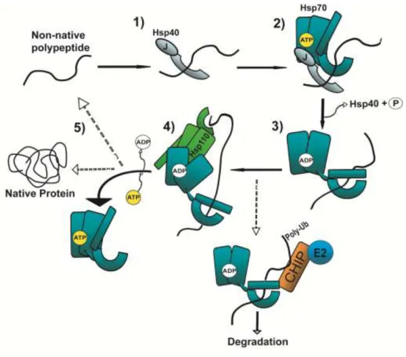
Heat shock protein 70 family members generally referred to as Hsp70 interact with different combinations of Hsp40s and other folding and degradatory co-chaperones to facilitate multiple processes required for protein homeostasis (1,2). Defects in protein homeostasis can trigger aberrant protein aggregation, a hallmark of a broad class of diseases known collectively as “Conformational Disorders”. Hsp70 molecular chaperones protect cells against the accumulation of proteotoxic species
by maintaining a delicate balance between protein synthesis, folding, and degradation (3). Yet the mechanism(s) by which functionally distinct Hsp40s (also known as J-proteins) bind and deliver a diverse array of proteins to the polypeptide-binding site of Hsp70 remains a major unanswered question.
Polypeptide binding and release by Hsp70 is coordinated via an array of co-chaperones that tightly regulate the Hsp70 ATP hydrolytic cycle (Fig. 1.1) (4-6). This process is initiated when a non-native polypeptide is bound by an Hsp40 co-chaperone (1). This large and structurally diverse family is defined by a highly conserved region called a J-domain that interacts with the Hsp70 nucleotide-binding domain (NBD) and stimulates intrinsic Hsp70 ATPase activity (1,7,8). Hydrolysis of ATP to ADP in the Hsp70 NBD induces a conformational change in the Hsp70 substrate-binding domain (SBD) thus increasing Hsp70’s affinity for the substrate (9,10). In most cases, polypeptides are
4
5 1.3 Hsp40s: diverse Hsp70 binding partners
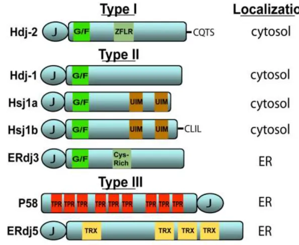
Through direct binding to native substrates, Hsp40s serve as conduits that funnel non-native clients to Hsp70. Despite sharing a conserved J-domain, the Hsp40 family is extensive and members possess unique domains that bind diverse clients and target Hsp70 to distinct cellular machineries. Hsp40s are categorized based on homology to the founding member in Escherichia coli, DnaJ (7,8). Type I Hsp40s possess a J-domain, a glycine- and phenylalanine-rich (G/F-rich) region and cysteine-rich, zinc finger-like region (ZFLR). Type II Hsp40s contain the J-domain and G/F-rich region whereas Type III Hsp40s retain only the J-domain. Action of the Hsp40 J-domain alone is sufficient to enable Hsp70 perform its essential cellular functions (15). However, the specialized Hsp40 polypeptide binding domain discussed above impart significant influence over Hsp40 quaternary structure and play critical roles in directing Hsp70 to carry out a variety of specialized functions (16),17.
6
7
8 1.4 Polypeptide transfer and release from Hsp70
Once Hsp40s bind non-native polypeptides, bound clients must be transferred to Hsp70 for subsequent processing. To begin to address how this occurs, given the extensive structural diversity within the Hsp40s, Petrova et al (18) and Jin et al (19) recently examined how two different ER-localized Hsp40s release substrates upon interaction with the Hsp70 BiP. In these studies, the Hsp40s P58 and ERdj3 specifically bound misfolded or denatured substrates. Efficient substrate release from either Hsp40 relied upon interaction between BiP and the J-domain, specifically when BiP was ATP-bound. Mutational analysis of BiP revealed that substrate release was functionally coupled to BiP’s
ATPase activity suggesting that a J-domain–BiP complex was not sufficient to dissociate the substrate. How does Hsp70 binding and ATP hydrolysis provoke Hsp40 to release its substrate? One hypothesis is that binding to Hsp70 induces an allosteric shift in the Hsp40 that reduces substrate affinity in the polypeptide-binding domain. However, Petrova et al (16) found that the location of the J-domain within the Hsp40 was inconsequential to BiP-induced substrate release. Furthermore, the extensive variation present in the Hsp40 family argues against a unified allosteric mechanism. However, J-domain–Hsp70 binding might bring the substrate in close proximity to the Hsp70 SBD. Following ATP hydrolysis, the increase in substrate affinity by Hsp70 could then out-compete the Hsp40 for binding.
9
10 1.5 Concluding Remarks
11 1.6 References
1. Cyr, D. M., Langer, T., and Douglas, M. G. (1994) Trends Biochem Sci 19, 176-181
2. Balch, W. E., Morimoto, R. I., Dillin, A., and Kelly, J. W. (2008) Science 319, 916-919
3. Cyr, D. M., Hohfeld, J., and Patterson, C. (2002) Trends Biochem Sci 27, 368-375
4. Langer, T., Lu, C., Echols, H., Flanagan, J., Hayer, M. K., and Hartl, F. U. (1992) Nature 356, 683-689
5. Cyr, D. M., Lu, X., and Douglas, M. G. (1992) J Biol Chem 267, 20927-20931
6. Liberek, K., Galitski, T. P., Zylicz, M., and Georgopoulos, C. (1992) Proc Natl Acad Sci U S A 89, 3516-3520
7. Walsh, P., Bursac, D., Law, Y. C., Cyr, D., and Lithgow, T. (2004) EMBO Rep 5, 567-571
8. Qiu, X. B., Shao, Y. M., Miao, S., and Wang, L. (2006) Cell Mol Life Sci 63, 2560-2570
9. Vogel, M., Bukau, B., and Mayer, M. P. (2006) Mol Cell 21, 359-367
10. Liu, Q., and Hendrickson, W. A. (2007) Cell 131, 106-120
11. Szabo, A., Langer, T., Schroder, H., Flanagan, J., Bukau, B., and Hartl, F. U. (1994) Proc Natl Acad Sci U S A 91, 10345-10349
12. Cyr, D. M. (2008) Cell 133, 945-947
13. Rosser, M. F., Washburn, E., Muchowski, P. J., Patterson, C., and Cyr, D. M. (2007) J Biol Chem 282, 22267-22277
14. Han, S., Liu, Y., and Chang, A. (2007) J Biol Chem 282, 26140-26149
12
16. Ramos, C. H., Oliveira, C. L., Fan, C. Y., Torriani, I. L., and Cyr, D. M. (2008) J Mol Biol 383, 155-166
17. Ushioda, R., Hoseki, J., Araki, K., Jansen, G., Thomas, D. Y., and Nagata, K. (2008) Science 321, 569-572
18. Petrova, K., Oyadomari, S., Hendershot, L. M., and Ron, D. (2008) Embo J 27, 2862-2872
19. Jin, Y., Awad, W., Petrova, K., and Hendershot, L. M. (2008) Embo J 27, 2873-2882
20. Caplan, A. J., Tsai, J., Casey, P. J., and Douglas, M. G. (1992) J Biol Chem 267, 18890-18895
21. Flom, G. A., Lemieszek, M., Fortunato, E. A., and Johnson, J. L. (2008) Mol Biol Cell 19, 5249-5258
22. Chapple, J. P., and Cheetham, M. E. (2003) J Biol Chem 278, 19087-19094
23. Freeman, B. C., Myers, M. P., Schumacher, R., and Morimoto, R. I. (1995) Embo J 14, 2281-2292
24. Fan, C. Y., Ren, H. Y., Lee, P., Caplan, A. J., and Cyr, D. M. (2005) J Biol Chem 280, 695-702
Chapter Two
Prion Propagation by Hsp40 Molecular Chaperones
14 2.1 Abstract
15 2.2 Molecular chaperones and yeast prions
Proteins adopt a diverse and dynamic array of structural conformations. Prions are unique in that these proteins induce conversion of the soluble, native structure into the prion conformer with a high propensity to self-assemble into beta-sheet-rich, amyloid-like fibrils.(1) Extensive investigation of prion biogenesis in the budding yeast Saccharomyces cerevisiae has uncovered some of the basic mechanisms underlying prion assembly into amyloid-like fibrils and inheritance of the prion state. One intriguing development in this story was the intimate role for heat shock protein (HSP) molecular chaperones in these pathways.(2,3) Indeed, numerous yeast prions are dependent upon molecular chaperones for efficient maintenance and propagation of prion structures.(4,5) On the other hand, overexpression of some molecular chaperones “cure” yeast of the heritable prion suggesting
molecular chaperones antagonize prion assembly.(4,6,7) How such opposing activities efficiently coordinate prion assembly into amyloid-like fibrils and propagation of the prion state inside the cell is an outstanding question in the field. Study of this process is significant because amyloid-like fibrils accumulate in numerous conformational disorders.(8,9) However, the connection between amyloid assembly and neuronal cell death is still controversial as several recent studies implicate the assembly of amyloid-like fibrils as benign or even protective.(10-12) In addition, prions found in S. cerevisiae possess domains enriched in glutamines (Gln) and asparagines (Asn)(13), resembling proteins with expanded polyglutamine repeats (such as human huntingtin and several ataxins) that are very susceptible to aggregation.(14,15) Many molecular chaperones are functionally conserved from yeast to humans, and as such, studying how molecular chaperones modulate prion propagation yields substantial mechanistic insight on the regulation of amyloid assembly in conformational disorders.
16
17
2.3 Protein Quality Control by Hsp40 Molecular Chaperones
Hsp40 co-chaperones are essential partners in Hsp70 function.(32) Hsp40s share a highly conserved region called a J-domain that stimulates the intrinsic ATPase activity of its partner Hsp70.(33) ATP hydrolysis causes a series of conformational changes that increase the affinity of client:Hsp70 interactions.(34,35) Client release from Hsp70 is induced when ADP is replaced with ATP by a Hsp70 nucleotide exchange factor.(36) The Hsp40 J-domain alone appears sufficient to maintain basic cellular processes required for physiological growth.(37) However, based upon homology to the J-domain from the founder Hsp40 in Escherichia coli (DnaJ), there are twenty-two Hsp40s in budding yeast and forty-one Hsp40s in humans.(32,38) Given the evolutionary expansion of the Hsp40 family, how these various Hsp40s specify Hsp70 function is an important unanswered question.
18
19
20
2.4 Hsp40 activity in propagation of [PSI+] and [URE3] prions
Studies of the yeast prions [PSI+] and [URE3] have identified complex roles for Hsp40 co-chaperones in prion propagation and assembly into amyloid-like fibrils. The inheritable element [PSI+] is formed by the yeast translation termination factor Sup35.(18,54) Both Ydj1 and Sis1 physically associate with large Sup35 aggregates,(55) though propagation of the [PSI+] prion is specifically dependent upon Sis1.(5,30) On the other hand, overexpression of Ydj1 in conjunction with its cognate Hsp70 destabilizes “weak” [PSI+
] variants.(6) Also noteworthy, overexpression of Apj1 (another Type I Hsp40 in yeast) cures cells of specific [PSI+] variants.(56) Apj1 shares strong homology with Ydj1 yet its cellular functions are still unclear. Recent studies on Sup35 fibril assembly in vitro have demonstrated a direct role for Hsp40 molecular chaperones in regulating the assembly of amyloid-like fibrils.(31,57) Interestingly, select Hsp40:Hsp70 pairs exert different actions on Sup35 assembly as well as the prion remodeling activity of Hsp104.(58) Therefore, distinct chaperone complexes might selectively regulate prion assembly and propagation to alternate outcomes.
21
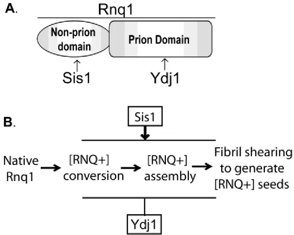
2.5 Selective recognition of the [RNQ+] prion by opposing Hsp40 activities
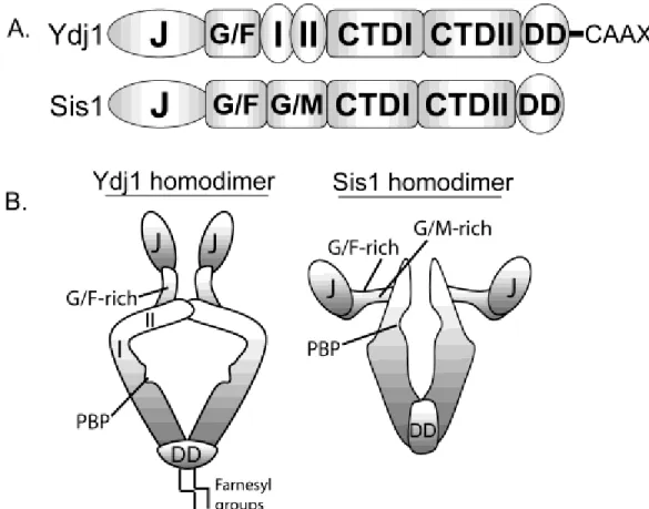
Studies of the yeast prion [RNQ+] have recently revealed novel mechanisms by which Hsp40 co-chaperones bind amyloid-like prion conformers and perhaps regulate prion propagation pathways to distinct endpoints. The yeast prion [RNQ+]/[PIN+] is formed by the yeast protein Rnq1 (rich in asparagines and glutamines) (Fig. 2.2A).(63,64) The [RNQ+] state facilitates the conversion of other prions in yeast(64,65) as well as seeding toxic conformers of an expanded glutamine form of human huntingtin.(66) Rnq1 possesses a C-terminal Gln/Asn-rich prion domain that is sufficient to assemble into amyloid-like fibrils in vitro(67,68) and induce prion formation when fused in place of the Gln/Asn-rich N-terminal domain of Sup35.(63) The N-terminal non-prion domain of Rnq1 appears to regulate [RNQ+] prion propagation though the function of this domain is still unclear.(69) Not long after [RNQ+] was first described, propagation of [RNQ+] prions was shown to be dependent upon Sis1.(70) Deletion of other Hsp40s in yeast has no effect on the [RNQ+] state suggesting this dependency is specific for Sis1.(5) In contrast to Sis1, overexpression of Ydj1 cures yeast of some [RNQ+] prion variants.(71) Thus, similar to other yeast prions discussed above, Sis1 promotes [RNQ+] propagation while Ydj1 can inhibit [RNQ+] assembly perhaps reflecting distinct fundamental functions for these two Hsp40 co-chaperones in the prion assembly pathway (Fig. 2.2B).
22
23
Ydj1 and the Gln/Asn-rich prion domain of Rnq1 is quite surprising because peptide array studies suggest Type I Hsp40s such as Ydj1 prefer substrates enriched in hydrophobic residues.(41,72,73) Furthermore, select binding to the Rnq1 prion domain in the [RNQ+] prion state implies that Ydj1 recognizes the Gln/Asn-rich motifs in a conformation-specific manner. Altogether, two Hsp40s in the cell bind [RNQ+] prions yet target different regions in the Rnq1 protein. The outcome of such binding preferences might (at a rudimentary level) account for the disparate chaperone activities on [RNQ+] prion propagation.
What features in Ydj1 and Sis1 direct these Hsp40 chaperones to bind distinct domains within Rnq1? Interestingly, binding between Ydj1 and the Rnq1 prion domain is dependent upon the Ydj1 ZFLR and farnesylation at its C-terminal CaaX motif.(29) The Ydj1 ZFLR is adjacent to two anti-parallel beta-strands that might bind the beta-rich Rnq1 prion domain through a beta-strand donor mechanism.(74,75) How lipid modification of an Hsp40 co-chaperone contributes to substrate interaction is unclear, although farnesylation of Ydj1 has been implicated in binding to the kinase Ste11.(48) Thus, farnesylation appears required for binding to numerous chaperone substrates including yeast prions. In contrast, Sis1-dependent maintenance of the [RNQ+] prion state requires unique extensions in the G/F-rich region of Sis1.(76) These observations collectively suggest that Hsp40 co-chaperones rely on specialized modules to bind distinct domains in Rnq1 and regulate different aspects of [RNQ+] prion propagation.
24
amyloid-like [RNQ+] assemblies while overexpression of Hsp70 and Hsp104 does not result in such an increase.(12)
In contrast to Sis1, Ydj1 might cap the exposed ends of [RNQ+] prion assemblies and thereby inhibit fibril elongation by sterically hindering contacts between exposed Gln/Asn-rich motifs in the [RNQ+] prion conformer. In addition, Ydj1 might bind a [RNQ+] prion assembly intermediate and cooperate with Hsp70 to refold the Rnq1 protein into its native conformation or partition this protein conformer into an alternative off-pathway assembly that is subsequently remodeled by another chaperone complex.(77) The net result of either mechanism would be solubilization of assembled Rnq1 and loss of the [RNQ+] prion trait. Importantly, Sis1 activity must normally out-compete Ydj1 to promote efficient [RNQ+] propagation. This might occur because Sis1-binding to Rnq1 is stoichiometric(76) and Ydj1-binding motifs in the Rnq1 prion domain are buried within most [RNQ+] assemblies. Furthermore, some but not all [RNQ+] prion variants are sensitive to Ydj1 overexpression(71) suggesting that Ydj1 may differentially recognize Rnq1 prion domain surfaces exposed in specific [RNQ+] prion variants.
25 2.6 Hsp40s protect cells from toxic prion conformers
The study of Hsp40 action in [RNQ+] assembly has further revealed that Sis1 and Ydj1 protect the cell from the accumulation of cytotoxic protein conformers. Overexpression of Rnq1 is toxic to yeast in the presence of pre-existing [RNQ+] prion.(12) Importantly, overexpression of Sis1 suppresses cytotoxicity caused by excess Rnq1, an effect that correlates with enhanced [RNQ+] prion assembly into SDS-insoluble aggregates and a decrease in the pool of SDS-soluble, Rnq1 protein species. Furthermore, mutating the Sis1-binding site in the Rnq1 non-prion domain decreases the efficiency of [RNQ+] prion assembly and exacerbates toxicity.(12) Thus, chaperone-mediated [RNQ+] assembly appears protective although the specific nature of the cytotoxic protein conformer is still unclear.
26 2.7 Concluding Remarks and Future Directions
27 2.3 References
1. Tuite, M. F., and Cox, B. S. (2003) Nat Rev Mol Cell Biol 4, 878-890
2. Jones, G. W., and Tuite, M. F. (2005) Bioessays 27, 823-832
3. Rikhvanov, E. G., Romanova, N. V., and Chernoff, Y. O. (2007) Prion 1, 217-222
4. Chernoff, Y. O., Lindquist, S. L., Ono, B., Inge-Vechtomov, S. G., and Liebman, S. W. (1995) Science 268, 880-884
5. Higurashi, T., Hines, J. K., Sahi, C., Aron, R., and Craig, E. A. (2008) Proc Natl Acad Sci U S A 105, 16596-16601
6. Kushnirov, V. V., Kryndushkin, D. S., Boguta, M., Smirnov, V. N., and Ter-Avanesyan, M. D. (2000) Curr Biol 10, 1443-1446
7. Schwimmer, C., and Masison, D. C. (2002) Mol Cell Biol 22, 3590-3598
8. Carrell, R. W., and Lomas, D. A. (1997) Lancet 350, 134-138
9. Sipe, J. D., and Cohen, A. S. (2000) J Struct Biol 130, 88-98
10. Chiti, F., and Dobson, C. M. (2006) Annu Rev Biochem 75, 333-366
11. Kayed, R., Head, E., Thompson, J. L., McIntire, T. M., Milton, S. C., Cotman, C. W., and Glabe, C. G. (2003) Science 300, 486-489
12. Douglas, P. M., Treusch, S., Ren, H. Y., Halfmann, R., Duennwald, M. L., Lindquist, S., and Cyr, D. M. (2008) Proc Natl Acad Sci U S A
13. Ross, E. D., Minton, A., and Wickner, R. B. (2005) Nat Cell Biol 7, 1039-1044
14. Williams, A. J., and Paulson, H. L. (2008) Trends Neurosci 31, 521-528
28
16. Moriyama, H., Edskes, H. K., and Wickner, R. B. (2000) Mol Cell Biol 20, 8916-8922
17. Derkatch, I. L., Bradley, M. E., Zhou, P., Chernoff, Y. O., and Liebman, S. W. (1997) Genetics 147, 507-519
18. Paushkin, S. V., Kushnirov, V. V., Smirnov, V. N., and Ter-Avanesyan, M. D. (1996) Embo J 15, 3127-3134
19. Kryndushkin, D. S., Alexandrov, I. M., Ter-Avanesyan, M. D., and Kushnirov, V. V. (2003) J Biol Chem 278, 49636-49643
20. Shorter, J., and Lindquist, S. (2004) Science 304, 1793-1797
21. Wegrzyn, R. D., Bapat, K., Newnam, G. P., Zink, A. D., and Chernoff, Y. O. (2001) Mol Cell Biol 21, 4656-4669
22. Newnam, G. P., Wegrzyn, R. D., Lindquist, S. L., and Chernoff, Y. O. (1999) Mol Cell Biol 19, 1325-1333
23. Jung, G., Jones, G., Wegrzyn, R. D., and Masison, D. C. (2000) Genetics 156, 559-570
24. Allen, K. D., Wegrzyn, R. D., Chernova, T. A., Muller, S., Newnam, G. P., Winslett, P. A., Wittich, K. B., Wilkinson, K. D., and Chernoff, Y. O. (2005) Genetics 169, 1227-1242
25. Chernoff, Y. O., Newnam, G. P., Kumar, J., Allen, K., and Zink, A. D. (1999) Mol Cell Biol 19, 8103-8112
26. Chacinska, A., Szczesniak, B., Kochneva-Pervukhova, N. V., Kushnirov, V. V., Ter-Avanesyan, M. D., and Boguta, M. (2001) Curr Genet 39, 62-67
27. Glover, J. R., and Lindquist, S. (1998) Cell 94, 73-82
28. Aron, R., Higurashi, T., Sahi, C., and Craig, E. A. (2007) Embo J 26, 3794-3803
29. Summers, D. W., Douglas, P. M., Ren, H. Y., and Cyr, D. M. (2009) J Biol Chem 284, 3628-3639
29
31. Shorter, J., and Lindquist, S. (2008) Embo J 27, 2712-2724
32. Walsh, P., Bursac, D., Law, Y. C., Cyr, D., and Lithgow, T. (2004) EMBO Rep 5, 567-571
33. Szabo, A., Langer, T., Schroder, H., Flanagan, J., Bukau, B., and Hartl, F. U. (1994) Proc Natl Acad Sci U S A 91, 10345-10349
34. Langer, T., Lu, C., Echols, H., Flanagan, J., Hayer, M. K., and Hartl, F. U. (1992) Nature 356, 683-689
35. Cyr, D. M., Lu, X., and Douglas, M. G. (1992) J Biol Chem 267, 20927-20931
36. Cyr, D. M. (2008) Cell 133, 945-947
37. Sahi, C., and Craig, E. A. (2007) Proc Natl Acad Sci U S A 104, 7163-7168
38. Qiu, X. B., Shao, Y. M., Miao, S., and Wang, L. (2006) Cell Mol Life Sci 63, 2560-2570
39. Lu, Z., and Cyr, D. M. (1998) J Biol Chem 273, 5970-5978
40. Fan, C. Y., Ren, H. Y., Lee, P., Caplan, A. J., and Cyr, D. M. (2005) J Biol Chem 280, 695-702
41. Fan, C. Y., Lee, S., Ren, H. Y., and Cyr, D. M. (2004) Mol Biol Cell 15, 761-773
42. Ramos, C. H., Oliveira, C. L., Fan, C. Y., Torriani, I. L., and Cyr, D. M. (2008) J Mol Biol 383, 155-166
43. Holstein, S. E., Ungewickell, H., and Ungewickell, E. (1996) J Cell Biol 135, 925-937
44. Yan, W., Schilke, B., Pfund, C., Walter, W., Kim, S., and Craig, E. A. (1998) Embo J 17, 4809-4817
45. Lu, Z., and Cyr, D. M. (1998) J Biol Chem 273, 27824-27830
30
47. Caplan, A. J., Tsai, J., Casey, P. J., and Douglas, M. G. (1992) J Biol Chem 267, 18890-18895
48. Flom, G. A., Lemieszek, M., Fortunato, E. A., and Johnson, J. L. (2008) Mol Biol Cell 19, 5249-5258
49. Sha, B., Lee, S., and Cyr, D. M. (2000) Structure 8, 799-807
50. Luke, M. M., Sutton, A., and Arndt, K. T. (1991) J Cell Biol 114, 623-638
51. Yan, W., and Craig, E. A. (1999) Mol Cell Biol 19, 7751-7758
52. Caplan, A. J., and Douglas, M. G. (1991) J Cell Biol 114, 609-621
53. Johnson, J. L., and Craig, E. A. (2001) J Cell Biol 152, 851-856
54. Glover, J. R., Kowal, A. S., Schirmer, E. C., Patino, M. M., Liu, J. J., and Lindquist, S. (1997) Cell 89, 811-819
55. Bagriantsev, S. N., Gracheva, E. O., Richmond, J. E., and Liebman, S. W. (2008) Mol Biol Cell
56. Kryndushkin, D. S., Smirnov, V. N., Ter-Avanesyan, M. D., and Kushnirov, V. V. (2002) J Biol Chem 277, 23702-23708
57. Krzewska, J., and Melki, R. (2006) Embo J 25, 822-833
58. Sweeny, E. A., and Shorter, J. (2008) Prion 2, 1-6
59. Coschigano, P. W., and Magasanik, B. (1991) Mol Cell Biol 11, 822-832
60. Lian, H. Y., Zhang, H., Zhang, Z. R., Loovers, H. M., Jones, G. W., Rowling, P. J., Itzhaki, L. S., Zhou, J. M., and Perrett, S. (2007) J Biol Chem
61. Savistchenko, J., Krzewska, J., Fay, N., and Melki, R. (2008) J Biol Chem
31
63. Sondheimer, N., and Lindquist, S. (2000) Mol Cell 5, 163-172
64. Derkatch, I. L., Bradley, M. E., Hong, J. Y., and Liebman, S. W. (2001) Cell 106, 171-182
65. Derkatch, I. L., Uptain, S. M., Outeiro, T. F., Krishnan, R., Lindquist, S. L., and Liebman, S. W. (2004) Proc Natl Acad Sci U S A 101, 12934-12939
66. Meriin, A. B., Zhang, X., He, X., Newnam, G. P., Chernoff, Y. O., and Sherman, M. Y. (2002) J Cell Biol 157, 997-1004
67. Vitrenko, Y. A., Gracheva, E. O., Richmond, J. E., and Liebman, S. W. (2007) J Biol Chem 282, 1779-1787
68. Wickner, R. B., Dyda, F., and Tycko, R. (2008) Proc Natl Acad Sci U S A 105, 2403-2408
69. Kurahashi, H., Ishiwata, M., Shibata, S., and Nakamura, Y. (2008) Mol Cell Biol 28, 3313-3323
70. Sondheimer, N., Lopez, N., Craig, E. A., and Lindquist, S. (2001) Embo J 20, 2435-2442
71. Bradley, M. E., Edskes, H. K., Hong, J. Y., Wickner, R. B., and Liebman, S. W. (2002) Proc Natl Acad Sci U S A 99 Suppl 4, 16392-16399
72. Li, J., and Sha, B. (2004) Biol Proced Online 6, 204-208
73. Rudiger, S., Schneider-Mergener, J., and Bukau, B. (2001) Embo J 20, 1042-1050
74. Sauer, F. G., Futterer, K., Pinkner, J. S., Dodson, K. W., Hultgren, S. J., and Waksman, G. (1999) Science 285, 1058-1061
75. Choudhury, D., Thompson, A., Stojanoff, V., Langermann, S., Pinkner, J., Hultgren, S. J., and Knight, S. D. (1999) Science 285, 1061-1066
76. Lopez, N., Aron, R., and Craig, E. A. (2003) Mol Biol Cell 14, 1172-1181
32
78. Gokhale, K. C., Newnam, G. P., Sherman, M. Y., and Chernoff, Y. O. (2005) J Biol Chem 280, 22809-22818
79. Douglas, P. M., Summers, D. W., and Cyr, D. M. (2009) Prion 3
80. Fiala, J. C. (2007) Acta Neuropathol 114, 551-571
81. Balch, W. E., Morimoto, R. I., Dillin, A., and Kelly, J. W. (2008) Science 319, 916-919
82. Chai, Y., Koppenhafer, S. L., Bonini, N. M., and Paulson, H. L. (1999) J Neurosci 19, 10338-10347
83. Chan, H. Y., Warrick, J. M., Gray-Board, G. L., Paulson, H. L., and Bonini, N. M. (2000) Hum Mol Genet 9, 2811-2820
84. Cummings, C. J., Mancini, M. A., Antalffy, B., DeFranco, D. B., Orr, H. T., and Zoghbi, H. Y. (1998) Nat Genet 19, 148-154
85. Cohen, E., Bieschke, J., Perciavalle, R. M., Kelly, J. W., and Dillin, A. (2006) Science 313, 1604-1610
86. Cheng, I. H., Scearce-Levie, K., Legleiter, J., Palop, J. J., Gerstein, H., Bien-Ly, N., Puolivali, J., Lesne, S., Ashe, K. H., Muchowski, P. J., and Mucke, L. (2007) J Biol Chem 282, 23818-23828
87. Wyttenbach, A., Carmichael, J., Swartz, J., Furlong, R. A., Narain, Y., Rankin, J., and Rubinsztein, D. C. (2000) Proc Natl Acad Sci U S A 97, 2898-2903
Chapter Three
Use of Yeast as a System to Study Amyloid Toxicity
34 3.1 Abstract
35 3.2. Introduction
An extensive group of neurodegenerative disorders are characterized by the formation of amyloid-like fibrils, including Alzheimer’s disease, Huntington’s disease, and Creutzfeldt-Jakob disease (1). Amyloid-like fibrils are composed of β-sheet-rich conformers of a non-native protein that are assembled in a unique structure called a cross-β spine (2).This structure confers several unique properties that distinguish amyloid-like fibrils from amorphous, or disordered aggregates including resistance to proteases and insolubility in ionic detergents (3). While the accumulation of amyloid deposits is a diagnostic biomarker of amyloidosis, the connection between the assembly of amyloid-like fibrils and neuronal cell death is currently unclear (4-6). Furthermore, specific neuronal subpopulations are selectively susceptible to misfolding and aggregation of particular disease proteins suggesting there are complex cellular pathways that influence amyloid assembly and proteotoxicity. Our understanding of this process is still rudimentary and many questions remain to be answered.
There are numerous model organisms available to study amyloid-linked diseases. While every model organism has unique benefits and limitations, studies in the budding yeast Saccharomyces cerevisiae have provided substantial insight on mechanisms underlying the assembly
36
of this role in prion induction although yeast strains isolated in the wild are predominantly in a [RNQ+] state (15) suggesting there may be an important, yet undiscovered function for this prion (16). In addition, expression of the expanded-polyglutamine form of huntingtin is selectively toxic to yeast when Rnq1 is the [RNQ+] prion conformation (17). Thus, the [RNQ+] prion influences aggregation and toxicity of other glutamine-enriched proteins in yeast although the mechanism underlying this relationship is unclear.
37 3.3 Basic methods for culturing yeast
Methods for culturing and manipulating yeast are well-established and readily available (19). As such, this section will focus primarily on central guidelines for growing yeast cultures to study amyloid assembly and toxicity. All experiments described herein were performed at 30°C although the specific experimental temperature may vary depending upon the optimal growth conditions for the yeast strain background. Plasmids expressing aggregation-prone proteins can easily be transformed into yeast and maintained under selection by utilizing auxotrophic markers (ex. amino acids). Individual colonies are used to inoculate liquid cultures that can be expanded into liter-size volumes as long as cultures are properly aerated and nutrient-rich. The ability to collect large quantities of cells is one of the distinct advantages of using budding yeast over most other model systems. The most important consideration when culturing yeast is to maintain the cells in log-phase growth by consistently diluting cells in fresh media. However, yeast cells will undergo physiological changes similar to aging after repetitive diluting or indefinite time in stationary phase (20). As a result, exogenous plasmids should be freshly transformed to improve reproducibility between experiments.
One additional advantage to working with budding yeast is the availability of diverse promoters (both inducible and constitutive) that provide extensive flexibility in the time and level of protein expression. Inducible promoters are particularly important when studying proteotoxicity because the expression of a toxic protein needs to be tightly controlled during an experiment. One of the more common promoters for this kind of analysis is the GAL1 promoter (21). As described below, this promoter is often used to examine how high level expression of a disease protein impacts cell viability by spotting assays or growth curves. There are several advantages to using the GAL1 promoter for this kind of analysis. Expression from this promoter is repressed in the presence of glucose permitting very robust control of protein expression. To facilitate rapid expression from the GAL1 promoter, yeast cells are initially grown in media containing raffinose as the carbon source
38
expression from the GAL1 promoter cannot be titrated by varying the levels of galactose. Furthermore, some yeast strains are unable to unable to metabolize galactose and are thus inviable when galactose is the sole carbon source.
In contrast to the GAL1 promoter, the CUP1 promoter can be tightly controlled by varying levels of copper sulfate added to the media. Expressing a toxic protein such as Rnq1 from the CUP1 promoter can inhibit cell growth (22); however, caution should be exercised if levels of copper sulfate cause growth defects independent o f protein expression. This promoter is also used to constitutively express a protein of choice using low levels of copper sulfate (ex. 50μM or lower). Long-term, low level expression of an aggregation-prone protein such as Rnq1 allows the exogenous protein to reach steady state equilibrium with cellular pathways that regulate assembly of amyloid-like aggregates. As such, Rnq1 aggregation can be studied under conditions when this protein is not toxic to yeast and independent of changes in cell physiology that might accompany cytotoxicity. Constitutive, high level expression can also be achieved using promoters from housekeeping enzymes such as glyceraldehyde-3-phosphate dehydrogenase (GPD) or alcohol dehydrogenase (ADH). These promoters are powerful tools to analyze how overexpression of various cellular factors affects amyloid assembly and toxicity.
39
In the context of studying prion biogenesis, budding yeast offer one unique advantage over other model systems. Yeast strains can be “cured” of the prion state. For example, Rnq1 is soluble
40 3.4 Growth and viability assays
Spotting Assay
The most basic tool for studying amyloid toxicity in yeast is to monitor cell growth under conditions where an amyloidogenic protein is expressed compared to conditions when this protein either is not expressed or does not assemble into amyloid-like fibrils (ex. a cured prion strain). This method is readily applicable to high through-put screening of genetic factors that influence toxicity. Defects in cell growth can most easily be assessed by spotting yeast cells on solid agar media and comparing growth between strain expressing a toxic protein and a control strain. As shown in Figure 3.1, expressing Rnq1 from the GAL1 promoter is toxic to yeast in a [RNQ+] dependent-manner (18). To perform this assay, single colonies from freshly transformed yeast are used to inoculate a liquid culture and when yeast cells have reach mid-log phase, equal concentrations of cells (~0.5 OD600) are
serially diluted onto solid selective media containing 2% galactose as the carbon source and incubated for 2-4 days at 30°C depending upon the strain. As a control, strains are simultaneously spotted on selective media containing glucose to show that changes in growth rates are dependent upon expression of the aggregation-prone protein.
Growth Curve
If differences in growth are too slight to detect by the spotting assay then toxicity can be quantitatively assessed by measuring growth curves in liquid media. The procedure begins as described above, except liquid cultures are induced with galactose at a very low density (OD600
0.01-0.05) and OD600 measured approximately every 2 hours until strains reach stationary phase.
41
to repress the GAL1 promoter. If cell viability is indeed reduced upon protein expression, then yeast cells will not grow on media with glucose while yeast cells would be expected to reenter the cell cycle when the proteotoxic insult is removed.
Figure 3.1. Expression of amyloid-forming proteins is toxic to yeast. Yeast cells (strain BY4741-[RNQ+] or [rnq-]) were transformed with an empty pRS416 vector or pRS416(GAL1-RNQ1). Liquid cultures were inoculated with freshly transformed yeast and incubated for one day at 30°C. Equal quantities of cells in mid-log phase (0.5 OD600) were serially diluted (1:5) onto selective media
42 3.5 Morphological analysis of protein aggregation Fluorescence microscopy
A rapid method to analyze protein aggregation is fluorescently-tagging a protein of interest and visualizing intracellular localization via fluorescence microscopy. For example, Rnq1-GFP forms punctuate structures in [RNQ+] cells while is predominantly diffuse in [rnq-] cells (Fig. 3.2). If tagged constructs are expressed from inducible promoters, then a time-course can be performed to determine the kinetics of aggregation. To freeze cells at specific timepoints, cells in mid-log phase are treated with 0.1M phosphate buffer (pH6.8) and 3.7% formaldehyde then incubated at 25°C for 30 minutes to 1 hour. Cells are then washed once in phosphate buffer (pH7.4) and resuspended in phosphate buffer (pH 7.4) plus 1.2M sorbitol. Cells can be stored in this solution for several days at 4°C and further processed for immunofluorescent detection of other proteins or stained with commercially available dyes to identify organelles. Alternatively, fluorophore-conjugated proteins can be visualized by live cell imaging to characterize the dynamics of protein aggregation on the order of seconds to minutes.
43
Figure 3. 2. Visualization of protein aggregates by fluorescence microscopy. (A)BY4741 cells ([RNQ+] or [rnq-]) in mid-log phase expressing Rnq1-GFP from the CUP1 promoter were induced with 50uM CuSO4 for 2 hours and live cells visualized with a FITC filter set.
Thioflavin T Staining of Amyloid-like Fibrils in Yeast
44 3.6. Biochemical analysis of amyloid-like aggregates
The structure of amyloid-like aggregates renders these assemblies insoluble in ionic detergents such as sodium dodecyl sulfate (SDS). There are several assays that exploit this unique property to biochemically distinguish large, insoluble assemblies from unassembled, SDS-soluble protein species in yeast cell extracts. As mentioned above, maintaining cells in mid-log phase is critical when performing these experiments. Additionally, if the aggregate-prone protein is expressed from an inducible promoter, a time-course experiment should be performed to characterize assembly kinetics. Once the basic dynamics of protein assembly into SDS-resistant aggregates is established, then these assays can be performed under conditions where select cellular factors are overexpressed or deleted to determine how specific cellular pathways influence the assembly of amyloid-like aggregates.
Differential high speed centrifugation
The most simple approach to characterizing the formation of amyloid-like aggregates is to separate SDS-insoluble aggregates by high speed centrifugation. This technique allows direct comparison between soluble and insoluble pools of an amyloid-forming protein. However, this technique is relatively insensitive to slight changes in the size of amyloid-like oligomers or fibrils. As shown in Figure 3.3A, excess Rnq1-YFP partitions predominantly in the SDS-insoluble fraction yet a pool also remains in the SDS-soluble supernatant. In contrast, excess Rnq1-YFP resolves exclusively in the SDS-soluble fraction in [rnq-] cells (18).
45
Figure 3.3. Biochemical analysis of SDS-insoluble, amyoid-like aggregates. (A) Separation of SDS-insoluble Rnq1-GFP aggregates by high-speed differential centrifugation. BY4741 cells ([RNQ+] or [rnq-] transformed with pGAL-RNQ1YFP were induced for 2 hours with 2% galactose. Cells were processed as described in the main text and equal volumes of total, supernatant, and pellet fractions were analyzed by SDS-PAGE and western immunoblotting for GFP (Roche) and Pgk1 (Invitrogen). (B) SDD-AGE analysis of Rnq1-GFP SDS-insoluble aggregates. BY4741 cells ([RNQ+] or [rnq-] transformed with pCUP-RNQ1GFP were induced with 50uM CuSO4 and samples were
46
specific protein but these conditions appear sufficient for a majority of endogenous Rnq1 to resolve in the insoluble pellet in [RNQ+] cells. An aliquot from the lysate is saved prior to the spin represent the input (or Total) fraction. After the spin, an aliquot from the supernatant is saved and the pellet is resupended in lysis buffer (in a volume equivalent to the original volume used for the spin). Each fraction (total, supernatant, pellet) is mixed with a equal volume of 2X sample buffer (125mM Tris-HCl pH6.8, 4% SDS, 4mM EDTA, and 20% glycerol, 8% beta-mercaptoethanol) and samples are analyzed by SDS-polyacrylamide gel electrophoresis (SDS-PAGE).
Semi-denaturing detergent agarose gel electrophoresis (SDD-AGE)
47
minutes and at least 100μg loaded into a 1.5% agarose gel (made with 1xTAE buffer + 0.1%SDS).
Lysates are run at ~70 volts for approximately 2-3 hours. The gel is transferred onto a PVDF membrane at 24 volts for 1.5 hours in standard Tris-based buffer (alternatively you can transfer for 8 hours at 12 volts).The transfer step generates a large amount of heat so the buffer and transfer apparatus should be chilled at 4°C prior to use. The transfer step is inefficient and requires practice and some optimization. The PVDF membrane is analyzed by western immunoblotting for the protein of interest using standard methods.
Filter Trap
A filter trap assay is another common and rapid method for analyzing the formation of large, SDS-insoluble aggregates. Initially, this technique was used to analyze aggregation of the polyglutamine-expanded form of huntingtin from cell and tissue extracts (31). However, this assay can also be applied to the analysis of large, SDS-insoluble aggregates formed by yeast prions (32,33). In contrast to the assays described above only the large, SDS-insoluble pool is observed and cannot be directly compared to the SDS-soluble pool that is lost through the cellulose acetate membrane. However, this technique is applicable to large scale analysis, for example, assessing changes in SDS-insoluble aggregate formation under a variety of environmental conditions or genetic backgrounds.
To perform this assay, yeast lysates are generated as described above for SDD-AGE analysis. After protein concentrations are standardized between samples, lysates are applied to a cellulose acetate membrane (0.2μM pore size -Beckman) that is previously equilibrated with lysis buffer and
48
If there is a poor signal, then the amount of SDS can be reduced or the protein induction time increased to accumulate more of the SDS-insoluble aggregate pool.
Size-exclusion chromatography
The assays described above separate amyloid-like particles into a soluble and SDS-insoluble protein species. However, there are times when more detailed resolution is required to characterize assembly intermediates that are solubilized by the presence of SDS. In addition, protein-protein interactions are typically disrupted by the presence of SDS and lost in these assays. To bypass this complication, amyloid-like assemblies can be resolved by size exclusion chromatography. We have previously used this technique to distinguish the ratio of assembled, high molecular weight pools of Rnq1 from unassembled, low molecular protein species (22,32,33). Resolving yeast cell extracts by size-exclusion chromatography requires a large quantity of yeast (>100 OD600) because
49
If lysates are separated on the appropriate resin then the relative ratio of assembled to unassembled protein can be directly compared. One important consideration is that this approach does not distinguish SDS-insoluble, amyloid-like particles from disordered aggregates or large protein complexes because denaturing buffers are not always compatible with resins in size-exclusion columns. To overcome this restriction, elution fractions can be applied to the assays described above to identify fractions that contain SDS-insoluble aggregates. Furthermore, the elution profile of Rnq1 in a [RNQ+] strain can be compared back to the elution profile in a [rnq-] strain when these proteins do not assemble into amyloid-like conformers (17,18,32).
50 3.7. Final Conclusions
51 3.8. References
1. Carrell, R. W., and Lomas, D. A. (1997) Lancet 350, 134-138
2. Nelson, R., Sawaya, M. R., Balbirnie, M., Madsen, A. O., Riekel, C., Grothe, R., and Eisenberg, D. (2005) Nature 435, 773-778
3. Chiti, F., and Dobson, C. M. (2006) Annu Rev Biochem 75, 333-366
4. Haass, C., and Selkoe, D. J. (2007) Nat Rev Mol Cell Biol 8, 101-112
5. Treusch, S., Cyr, D. M., and Lindquist, S. (2009) Cell Cycle 8, 1668-1674
6. Robakis, N. K. (2010) Neurodegener Dis 7, 32-37
7. Khurana, V., and Lindquist, S. (2010) Nat Rev Neurosci 11, 436-449
8. Shorter, J., and Lindquist, S. (2005) Nat Rev Genet 6, 435-450
9. Jones, G. W., and Tuite, M. F. (2005) Bioessays 27, 823-832
10. Sondheimer, N., and Lindquist, S. (2000) Mol Cell 5, 163-172
11. Derkatch, I. L., Bradley, M. E., Hong, J. Y., and Liebman, S. W. (2001) Cell 106, 171-182
12. Wickner, R. B., Dyda, F., and Tycko, R. (2008) Proc Natl Acad Sci U S A 105, 2403-2408
13. Patel, B. K., and Liebman, S. W. (2007) J Mol Biol 365, 773-782
14. Vitrenko, Y. A., Pavon, M. E., Stone, S. I., and Liebman, S. W. (2007) Curr Genet 51, 309-319
15. Resende, C. G., Outeiro, T. F., Sands, L., Lindquist, S., and Tuite, M. F. (2003) Mol Microbiol 49, 1005-1017
52
17. Meriin, A. B., Zhang, X., He, X., Newnam, G. P., Chernoff, Y. O., and Sherman, M. Y. (2002) J Cell Biol 157, 997-1004
18. Douglas, P. M., Treusch, S., Ren, H. Y., Halfmann, R., Duennwald, M. L., Lindquist, S., and Cyr, D. M. (2008) Proc Natl Acad Sci U S A
19. Guthrie, C., and Fink, G. R. (2002) Guide to yeast genetics and molecular and cell biology. Part B. Methods in enzymology, Academic Press, San Diego, Calif.
20. Steinkraus, K. A., Kaeberlein, M., and Kennedy, B. K. (2008) Annu Rev Cell Dev Biol 24, 29-54
21. St John, T. P., Scherer, S., McDonell, M. W., and Davis, R. W. (1981) J Mol Biol 152, 317-334
22. Douglas, P. M., Treusch, S., Ren, H. Y., Halfmann, R., Duennwald, M. L., Lindquist, S., and Cyr, D. M. (2008) Proc Natl Acad Sci U S A 105, 7206-7211
23. Jung, G., and Masison, D. C. (2001) Curr Microbiol 43, 7-10
24. Ferreira, P. C., Ness, F., Edwards, S. R., Cox, B. S., and Tuite, M. F. (2001) Mol Microbiol 40, 1357-1369
25. Cox, B. S., Byrne, L. J., and Tuite, M. F. (2007) Prion 1, 170-178
26. Kaganovich, D., Kopito, R., and Frydman, J. (2008) Nature 454, 1088-1095
27. Sipe, J. D., and Cohen, A. S. (2000) J Struct Biol 130, 88-98
28. Kawai-Noma, S., Pack, C. G., Kojidani, T., Asakawa, H., Hiraoka, Y., Kinjo, M., Haraguchi, T., Taguchi, H., and Hirata, A. (2010) J Cell Biol 190, 223-231
29. Kimura, Y., Koitabashi, S., and Fujita, T. (2003) Cell Struct Funct 28, 187-193
53
31. Scherzinger, E., Lurz, R., Turmaine, M., Mangiarini, L., Hollenbach, B., Hasenbank, R., Bates, G. P., Davies, S. W., Lehrach, H., and Wanker, E. E. (1997) Cell 90, 549-558
32. Douglas, P. M., Summers, D. W., Ren, H. Y., and Cyr, D. M. (2009) Mol Biol Cell 20, 4162-4173
Chapter Four
The type I Hsp40 Ydj1 utilizes a farnesyl moiety and zinc finger-like region to suppress prion toxicity
55 4.1 Abstract
Type I Hsp40s are molecular chaperones that protect neurons from degeneration by
modulating the aggregation state of amyloid-forming proteins. How Type I Hsp40s recognize -rich, amyloid-like substrates is currently unknown. Thus, we examined the mechanism for binding between the Type I Hsp40 Ydj1 and the yeast prion [RNQ+]. Ydj1 recognized the Gln/Asn-rich prion domain from Rnq1 specifically when it assembled into the amyloid-like [RNQ+] prion state. Upon deletion of YDJ1, overexpression of the Rnq1 prion domain killed yeast. Surprisingly, binding and suppression of prion domain toxicity by Ydj1 was dependent upon farnesylation of its C-terminal CAAX box and action of a zinc-finger-like region. In contrast, folding of luciferase was independent of farnesylation, yet required Ydj1's zinc finger-like region and a conserved hydrophobic peptide-binding pocket. Type I Hsp40s contain at least three different domains that that work in concert to bind different protein conformers. The combined action of a farnesyl moiety and zinc finger-like region enable Type I Hsp40s to
56 4.2 Introduction
Protein misfolding and aggregation are common themes in neurodegenerative maladies termed conformational diseases. A subset of these disorders including Alzheimer’s disease and
the transmissible spongiform encephalopathies (prion diseases) are characterized by the accumulation of a stable, ß-sheet rich fibrils called amyloid (1). Criteria that distinguish amyloid-like fibrils from amorphous aggregates include resistance to SDS-solubilization and binding of the dye thioflavin T (2). The direct connection between amyloid accumulation and neuropathology is still a matter of debate (3-5). Yet, the flux of proteins through amyloid forming pathways correlates well with disease (3-5).
Hsp70 molecular chaperones protect against neurodegeneration associated with conformational disease via suppression of protein aggregation or conversion of toxic species into non-toxic aggregates or amyloid (6,7). Hsp70 has broad substrate selectivity and co-chaperones in the Hsp40/DnaJ family specify Hsp70 targets. Upon delivery of substrate, Hsp40s stimulate Hsp70 ATPase activity through a conserved J-domain and thereby stabilize Hsp70:polypeptide complexes. Nucleotide exchange factors (NEFs) convert Hsp70-ADP to Hsp70-ATP releasing non-native substrate for further rounds of folding or degradation (8,9). Escape of disease-related proteins from the action of Hsp70/Hsp40 and other quality control machinery leads to the accumulation of toxic protein species (10).
57
of some Type I Hsp40 substrates (15-17). Type I Hsp40s can independently bind non-native polypeptides and also cooperate with Hsp70 to suppress protein aggregation (18,19). Importantly, human and yeast Type I Hsp40s are highly conserved and functionally interchangeable (12).
Interestingly, Type I Hsp40s such as human DnaJ 2 (HDJ-2) and yeast DnaJ 1 (Ydj1) are unique in that they contain a C-terminal CAAX box that is post-translationally modified by farnesyl (20,21). Farnesylation helps localize a pool of Type I Hsp40s to the cytoplasmic face of the endoplasmic reticulum (ER) and is required for cells to survive heat stress (22). One function of ER-localized Type I Hsp40s is the folding of polytopic membrane proteins (21). However, even though the entire pool of Ydj1 is farnesylated a large portion is found in the cytosol (22). Thus, it is conceivable that the farnesyl moiety of Ydj1/Hdj-2 has additional roles in cytoprotection including regulation of stress response or assistance with polypeptide binding.
58
59 4.3 Results
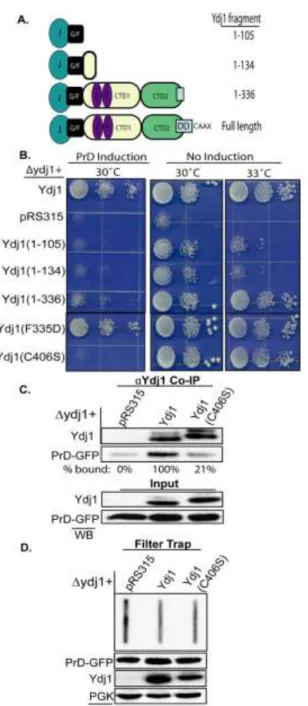
The Rnq1 PrD forms benign intracellular aggregates. Overexpression of Rnq1 is toxic to yeast when endogenous Rnq1 is assembled in its [RNQ+] conformation (4). Cell death is thought to occur due to inefficient conversion of overexpressed Rnq1 into amyloid-like [RNQ+] prions and the accumulation of a templated toxic species. Furthermore, defects in Sis1-binding exacerbate Rnq1 toxicity by decreasing the efficiency of [RNQ+] assembly. The nature of the toxic Rnq1 species is not clear and how defects in chaperone binding lead to its accumulation are unknown. Thus, we investigated whether removal of the entire non-prion domain and elimination of the Sis1 binding site would enhance Rnq1 proteotoxicity. However, while overexpression of Rnq1 was toxic to [RNQ+] cells, overexpression of the PrD was not (Fig. 4.1A & B). Overexpression of Rnq1 or PrD did not alter cell growth in the absence of prion seeds ([rnq-] background). Thus, the presence of the non-prion domain on Rnq1 somehow leads to the accumulation of a toxic Rnq1 species.To gain insight into the nature of the toxic Rnq1 species we characterized intermediates on the pathway for conversion of native Rnq1 and PrD into amyloid-like [RNQ+] prion. Rnq1-GFP and PrD-GFP each formed intracellular aggregates in a [RNQ+] background that were morphologically indistinguishable by fluorescence microscopy (Fig. 4.1C). Rnq1 was not observed to coalesce in a [rnq-] background though a small population of cells (<5%) expressing PrD-GFP contained non-toxic aggregates. In a [RNQ+] background, Rnq1-GFP partitioned predominantly in the Triton-insoluble pellet of yeast extracts, though a significant population was present in the Triton-soluble supernatant (Fig. 4.1D). In contrast, PrD-GFP fractionated exclusively in the Triton-insoluble pellet in a [RNQ+] background. In a [rnq-] state, Rnq1-GFP was predominantly soluble while PrD-GFP was present in both Triton-soluble and insoluble appear toxic.
60
61
62
thyroglobulin (669kDa), but still in the included volume. In addition, a pool of Rnq1-GFP eluted similar to a monomeric form. PrD-GFP also formed two high molecular weight pools, yet in contrast to Rnq1; no low molecular weight species was detected. The material in the void volume was predominantly SDS-soluble while Rnq1/PrD-GFP species in the broad included peak were insoluble in SDS (data not shown). In a [rnq-] background, Rnq1-GFP resided exclusively in the low molecular weight pool while PrD-GFP eluted in the void volume as well as a low molecular weight pool (Fig. 4.1E).
These observations suggest deletion of the non-prion domain predisposes the PrD to spontaneous aggregation in either a [RNQ+] or [rnq-] background. However, PrD aggregates in a [rnq-] background are not amyloid-like because the PrD only forms thioflavin T-positive, SDS-resistant aggregates in a [RNQ+] background (Fig. 4.2). Thus, the presence of the non-prion domain predisposes a small portion of Rnq1 to accumulate in a soluble pool, which could result from inefficient assembly or increased shearing of [RNQ+] prions into [RNQ+] seeds by Hsp104 (27). Observations that PrD overexpression is not toxic and it's assembly into amyloid-like prions is not accompanied by the accumulation of a soluble PrD pool supports the notion that accumulation of soluble Rnq1 in a [RNQ+] background leads to death (4).
Ydj1 binds the Gln/Asn-rich PrD in its [RNQ+] conformation. Deletion of the Rnq1 non-prion domain and the Sis1-binding motif (4), renders the PrD prone to form benign aggregates in [RNQ+] and [rnq-] cells. The combined ability of the cell to package the PrD into amyloid-like assemblies as well as SDS-sensitive aggregates may account for the benign consequences of PrD
overexpression. Chaperones facilitate the formation of benign, non-amyloid aggregates of A1-42
(30). Therefore, we speculated that an Hsp40 other than Sis1 may recognize the Gln/Asn-rich PrD and prevent the formation of a toxic PrD species by facilitating the conversion of soluble, unassembled PrD species into benign SDS-sensitive aggregates.
63
64
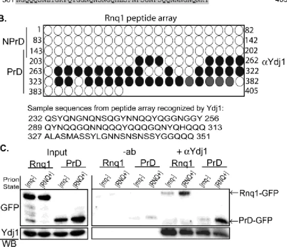
Each peptide in the array was 25 amino acids in length and shared 22 amino acids with the adjacent peptide such that small binding motifs could be identified. Ydj1 bound numerous peptides from the PrD, but no peptides from the non-prion domain (Fig. 2.3B). Ydj1 bound peptides that were typically composed of Gln/Asn-rich motifs interrupted by aliphatic or aromatic residues (Fig. 4.3B). These hydrophobic residues may facilitate binding with the Ydj1 hydrophobic peptide-binding pocket (31). Consistent with this possibility, Ydj1 did not bind a glutamine/glycine repeat at the beginning of the PrD. However, numerous hydrophobic residues are present in the non-prion domain and the terminal 50 amino acids of Rnq1 that were not bound by Ydj1. As such, the arrangement of Gln and Asn amino acids along with hydrophobic residues appears critical for Ydj1 to specifically recognize PrD peptides.
Ydj1 binds proteins with polyglutamine repeats in a manner that is dependent upon the expansion length and regulates their assembly into higher order aggregates (32,33). Thus, Ydj1 may be able to recognize substrates enriched in b-structure. To address whether conversion of Rnq1 or the PrD into a b-rich conformation is required for Ydj1 binding, we assessed Ydj1’s interaction with the native or amyloid-like prion forms of Rnq1 and the PrD. Ydj1 co-immunoprecipitated Rnq1-GFP and PrD-GFP from [RNQ+] lysates, yet not from [rnq-] lysates suggesting Ydj1 prefers the [RNQ+] prion conformation of these proteins (Fig. 4.3C). In addition, Ydj1 did not co-immunoprecipitate the non-prion domain alone and the PrD was a poor substrate for Sis1 in [RNQ+] lysates (Fig. 4.4). The PrD from Rnq1 assembles into thioflavin T-positive,
amyloid-like fibrils (34,35). Therefore Ydj1, but not Sis1, recognizes the PrD of Rnq1 in its -sheet rich, amyloid-like conformation.
Overexpressing the PrD is toxic to yeast in the absence of YDJ1. If Ydj1 binds the PrD in its amyloid-like conformation, then Ydj1 may act analogous to Sis1 in suppressing Rnq1 toxicity by facilitating assembly of the PrD into a benign conformation. To test this hypothesis, the PrD was
65
66
Figure 4.5 Deletion of YDJ1 sensitizes yeast to overexpression of the PrD. (A) Cells in a
67
overexpression was highly toxic to yeast in a ydj1 background (Fig. 4.5A). Cell viability was
rescued by Ydj1 expression from a low copy plasmid from its own promoter. Even though PrD
expression from the GAL1 promoter was toxic, PrD protein levels were much lower in the ydj1
strain compared to the YDJ1-rescued strain (Fig 4.5A; lower panel). This was not surprising because YDJ1 is required for efficient nucleosomal remodeling that is required for activation of the GAL1 promoter (36).
In contrast to what is observed with [RNQ+] assembly when Sis1 is depleted (4,27), the PrD still formed intracellular aggregates and did not accumulate as a soluble species when Ydj1 was deleted (Fig. 4.5B-D) In fact, the only difference we observed was a close to 2-fold increase in
the levels of SDS-resistant PrD-GFP in the ydj1 strain compared to wild type background (Fig.
4.5E). Therefore, Ydj1 appears to enable yeast to tolerate PrD expression by limiting the pool of amyloid-like PrD assemblies. Consistent with this hypothesis, overexpression of Ydj1 decreased the level SDS-resistant PrD-GFP (Fig. 4.5F). Thus, PrD toxicity appears to differ from Rnq1 toxicity in that the accumulation of SDS-resistant forms of PrD, and not a low molecular weight, detergent-soluble species correlates with cell death.
The mechanism via which Ydj1 modulates the accumulation of SDS-resistant PrD and suppresses PrD toxicity is unclear. Ydj1 may cap the exposed ends of elongating PrD amyloid or interact with Hsp104 (37) to maintain the level of this species within a tolerable range. Alternatively, Ydj1 may coat PrD particles to prevent non-specific protein:protein interactions that titrate essential cellular factors. We favor the former model because while Ydj1 co-
68

![Figure 4.1 The Rnq1 PrD assembles into benign [RNQ + ] prion. (A) Domain boundaries of Rnq1 from S](https://thumb-us.123doks.com/thumbv2/123dok_us/8279329.2192598/71.918.144.789.110.773/figure-rnq-prd-assembles-benign-prion-domain-boundaries.webp)
![Figure 4.2 The Rnq1 PrD forms SDS-insoluble, amyloid-like [RNQ + ] prion. (A) In wildtype cells, PrD-RFP was expressed overnight from a copper inducible promoter in [RNQ + ] and [rnq - ] cells](https://thumb-us.123doks.com/thumbv2/123dok_us/8279329.2192598/72.918.168.759.102.788/figure-insoluble-amyloid-wildtype-expressed-overnight-inducible-promoter.webp)
![Figure 4.4 Ydj1 specifically binds the PrD of Rnq1. (A) Ydj1 co-immunoprecipitated Rnq1- Rnq1-YFP, PrD-Rnq1-YFP, yet not NPrD-YFP from [RNQ + ] lysates (right panel)](https://thumb-us.123doks.com/thumbv2/123dok_us/8279329.2192598/76.918.214.702.101.671/figure-specifically-binds-immunoprecipitated-nprd-lysates-right-panel.webp)