RESEARCH
Multivariable analysis to determine
if HIV-1 Tat dicysteine motif is associated
with neurodevelopmental delay in HIV-infected
children in Malawi
Jasmeen Dara
1, Anna Dow
2, Elizabeth Cromwell
2, Christa Buckheit Sturdevant
3, Macpherson Mallewa
4,
Ronald Swanstrom
3, Annelies Van Rie
2*†and Vinayaka R. Prasad
5*†Abstract
Background: HIV-1 Tat protein is implicated in HIV-neuropathogenesis. Tat C31S polymorphism (TatCS) has been
associated with milder neuropathology in vitro and in animal models but this has not been addressed in a cohort of HIV-infected adults or children.
Methods: HIV viral load (VL) in plasma and cerebrospinal fluid (CSF) were determined and plasma HIV tat gene was sequenced. Neurodevelopmental assessment was performed using Bayley Scales of Infant Development III (BSID-III), with scores standardized to Malawian norms. The association between TatCS and BSID-III scores was evaluated using
multivariate linear regression.
Results: Neurodevelopmental assessment and HIV tat genotyping were available for 33 children. Mean age was 19.4 (SD 7.1) months, mean log VL was 5.9 copies/mL (SD 0.1) in plasma and 3.9 copies/mL (SD 0.9) in CSF. The prevalence of TatCC was 27 %. Z-scores for BSID-III subtests ranged from
−1.3 to −3.9. TatCC was not associated with higher BSID-III
z-scores.
Conclusions: The hypothesis of milder neuropathology in individuals infected with HIV TatCS was not confirmed
in this small cohort of Malawian children. Future studies of tat genotype and neurocognitive disorder should be performed using larger sample sizes and investigate if this finding is due to differences in HIV neuropathogenesis between children and adults.
Keywords: HIV-1, HIV-1 subtype C, Tat, Dicysteine motif, Encephalopathy, Neurodevelopment
© 2015 Dara et al. This article is distributed under the terms of the Creative Commons Attribution 4.0 International License (http://creativecommons.org/licenses/by/4.0/), which permits unrestricted use, distribution, and reproduction in any medium, provided you give appropriate credit to the original author(s) and the source, provide a link to the Creative Commons license, and indicate if changes were made. The Creative Commons Public Domain Dedication waiver (http://creativecommons.org/ publicdomain/zero/1.0/) applies to the data made available in this article, unless otherwise stated.
Background
Although prevention of mother-to-child transmission (PMTCT) of HIV has significantly decreased the inci-dence of pediatric HIV, approximately 240,000 children were newly infected with HIV in 2013 [1]. HIV infection
negatively impacts the neurological development of children, with symptoms ranging from mild neurode-velopmental delays to severe neurocognitive and motor impairments [2–4]. While early initiation of anti-ret-roviral therapy (ART) can prevent or delay the onset of HIV-associated neurological disorders (HAND) [5, 6], only one in every seven children living with HIV in sub-Saharan Africa receive ART [7].
Virologic factors that contribute to neurodevelopment are poorly understood, particularly in children. HIV-1 Tat protein has been implicated in both directly [8] and indi-rectly [9] inducing monocyte chemotaxis, damaging the blood brain barrier, direct neurotoxicity and augmenting
Open Access
*Correspondence: [email protected]; vinayaka.prasad@einstein. yu.edu
†Annelies Van Rie and Vinayaka R. Prasad contributed equally 2 UNC School of Public Health, University of North Carolina, Chapel Hill, NC, USA
5 Department of Microbiology and Immunology, Albert Einstein College of Medicine, Bronx, NY, USA
monocyte infiltration across the blood–brain barrier. Tat is released from infected cells and causes direct damage to the central nervous system (CNS) white matter [10,
11] and promotes indirect damage through the induc-tion of chemokines and cytokines [12]. A key molecular determinant of neuropathogenesis in HIV-1 Tat is the C30C31 dicysteine motif (TatCC) and in a given
individ-ual’s plasma, nearly all viruses display the same genotype. For example, Tat in HIV-1 subtype B isolates contains a highly conserved (99 %) C30C31 motif, which mediates its ability to induce β chemokines and promote monocyte chemotaxis [13]—features that play a key role in HAND. In contrast, approximately 90 % of subtype C HIV-1 iso-lates worldwide have a C31S (TatCS) polymorphism [13],
which has been linked with defective monocyte chemo-taxis. We have previously shown that an Indian subtype C HIV-1 isolate with TatCS, when compared to US
sub-type B isolate, displayed decreased in vitro chemokine induction, decreased monocyte recruitment by infected macrophages, and in a mouse model for HIV encephali-tis [14], caused milder neurodegeneration and decreased cognitive impairment following intracranial injection of HIV-infected monocyte-derived macrophages [15]. Furthermore, subtype C HIV TatCS causes decreased
N-methyl d-aspartate (NMDA) receptor induced neuro-toxicity [16] and neuronal cell death [17] compared with subtype B HIV Tat.
The importance of Tat dicysteine motif in HAND is further underscored by the geographic variability of HIV-associated dementia (HAD) in adults in regions where subtype C predominates. In India, where subtype C infec-tion is predominant, HAD prevalence of 2–6 % has been reported [18, 19]. In contrast, South Africa and Botswana, also countries with predominantly subtype C infections, have reported a 25–30 % prevalence of HAD [20, 21]. The higher incidence of HAD correlates with a higher preva-lence of intact Tat dicysteine motif in Southern African isolates (11–22 % of subtype C HIV isolates from Bot-swana and South Africa), while the lower incidence of HAD in India correlates with a lower prevalence of HIV-1 isolates with Tat C30C31 motif [22]. Thus, the variation in the prevalence of HAND between different regions with subtype C HIV epidemics appears to correlate with regional differences in the frequency of the subtype C HIV-1 isolates bearing Tat with an intact dicysteine motif [22]. This hypothesis has been further validated through in vitro experiments and mouse models of HIV encepha-litis using a Zambian HIV-1 subtype C isolate with an intact dicysteine motif [22]. However, the tat genotype and HAD prevalence in the same cohort has not been confirmed so far. A recent study in adults in South Africa showed no significant cognitive differences between adults with subtype C HIV-1 with TatC31S as compared
to those with the dicysteine motif [23]. However, to date, viral genotypic differences related to pediatric HIV neu-rodevelopmental delay have not been evaluated.
We evaluated the relationship between the HIV-1 Tat dicysteine motif and neurodevelopmental delay in HIV-infected children in Malawi using the Bayley Scales of Infant Development III (BSID-III). We hypothesized that HIV-1 Tat dicysteine motif is associated with neurode-velopmental delay in children infected with HIV-1 sub-type C.
Methods Study population
Children were eligible to participate if HIV-infected, age 2–36 months, ART-naïve, but eligible for ART and if their primary caregiver had no chronic illnesses other than HIV that might impede the daily activities or neurode-velopment of the child. Children were recruited through either the pediatric HIV clinic or the emergency depart-ment at Queen Elizabeth Central Hospital in Blantyre, Malawi between June 2009 and June 2010. Children with a history of severe birth asphyxia, cerebral malaria, men-ingitis, a congenital malformation or chronic medical that may interfere with neurodevelopment were excluded.
Children recruited through the HIV clinic had to dem-onstrate delay for one or more neurodevelopmental mile-stones on a parental screening questionnaire, whereas those recruited through the emergency department had to have a clinical indication for a lumbar puncture.
Laboratory and neurodevelopmental evaluations
Participating children underwent neurodevelopmental testing with the BSID-III, to assess motor (fine and gross), cognitive, and language (expressive and receptive commu-nication) development [24]. Neurodevelopmental assess-ment was performed at enrolassess-ment for children recruited through the HIV clinic and at 1-month follow-up visit among children recruited through the emergency depart-ment, to ensure that the acute illness did not interfere with the neurodevelopmental assessment. The BSID-III was administered in the local language of Chichewa by a study pediatrician or nurse trained in administration of the BSID-III. Scores were interpreted using normative data from Malawi [25]. Neurological development for each child was classified as normal (between mean and ±1 SD), mild delay (between −1 and −2 SD), severely delayed (>−2 SD), above normal (between +1 and +2 SD) or advanced (>+2 SD above mean) for each of the five subtests.
sequences (nucleotides 1–214, corresponding to codons 1–70), which includes the codons for amino acids 30 and 31 were determined. The amino acid at Tat codon 31, predicted using the nucleotide sequence, was used in the statistical analysis.
Statistical analysis
Using Stata 12.0 (College Station, TX), we created sepa-rate linear regression models to assess the association between the outcomes of fine motor, gross motor, cog-nitive, expressive communication and receptive com-munication z-scores with TatCC as the primary exposure
variable of interest. Potential covariates included mater-nal ART use (dichotomous), exposure to PMTCT (dichotomous), breastfeeding >6 months of life (dichot-omous), breastfeeding at enrollment (dichot(dichot-omous), gender, log plasma VL (continuous), log CSF VL (con-tinuous), CSF-to-plasma VL ratio (con(con-tinuous), and maternal age (continuous). Frequency distributions were calculated for these covariates among subjects with TatCS
and TatCC. Covariates were examined for their
relation-ship with neurodevelopmental z-scores using t-test with unequal variances, Mann–Whitney U-test, Pearson’s cor-relation, and Spearman’s corcor-relation, as appropriate.
Separate multivariable models were created for each neurodevelopmental z-score using an a priori approach in which variables were chosen based on biological plausibil-ity of confounding the relationship between exposure and outcome. In addition, covariates with missing values could not be included in the model as this lead to small effective sample sizes. Based on these criteria, the variables chosen to include were age, log plasma VL, and log CSF VL. To assess for normality of the linear regression model, the standardized residuals were plotted on a histogram and p–p plot. Outliers with a Cook’s distance greater than 4/n were excluded. Additional models using robust regression diagnostics were created by excluding outliers with Cook’s distance >1. The assumptions of equal variances and line-arity were tested using a residuals-versus-fitted plot. Mul-ticollinearity was tested by using variance inflation factors (VIF) with a cut-off of >10.
We also performed a sensitivity analysis to determine if the exclusion of the six subjects lacking Tat sequenc-ing information affected the estimate of the association between exposure and outcome.
Ethics statement
Institutional Review Board approval was received from the College of Medicine in Blantyre, Malawi, the Uni-versity of North Carolina in Chapel Hill, NC, and Albert Einstein College of Medicine in Bronx, NY. Written informed consent was obtained from all parents or guardians.
Results
Characteristics of study population
Of the 46 HIV-infected children enrolled, 39 completed neurodevelopmental assessments, and 33 had isolates available for genetic sequencing to determine the pre-dicted amino acid sequence at Tat codon 31 (Fig. 1). Of the 33 children included in the analysis, 32 were recruited through the HIV clinic, and one through the emergency department. No HIV-negative children were enrolled. The mean age was 19.4 (SD 7.1) months, mean log plasma VL was 5.9 (SD 0.1) copies/mL, and mean log CSF VL was 3.9 (SD 0.9) copies/mL, and 44.8 % of all mothers in the study received single-dose nevirapine (sdNVP) for PMTCT. Compared to children with TatCS,
children with TatCC were more likely to have received
sdNVP for PMTCT (p = 0.041), have higher log plasma VL (p = 0.008) and tended to have higher log CSF VL (p = 0.139) (Table 1). There was no difference in socioec-onomic conditions such as ownership of goods, housing materials and access to running water between the two groups (data not shown).
Exposure (HIV Tat motif) and outcome (neurodevelopment) in 33 Malawian children
The prevalence of the TatCC was 24 % among all (n = 41)
pediatric isolates available and 27 % among those who also completed BSID-III evaluations (n = 33) (see Table 1).
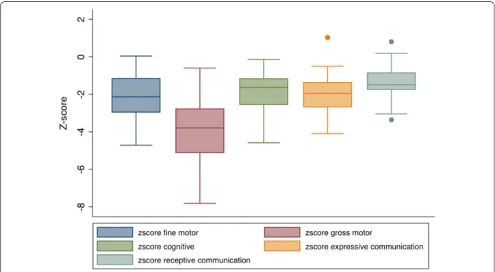
All children had moderate to severe delay in at least one domain: 88 % (n = 29) for cognitive development, 88 % (n = 29) for expressive communication, 60 % (n = 20) for receptive communication, 78 % (n = 26) for fine motor and 97 % (n = 32) for gross motor development (not shown). Unadjusted neurodevelopmental z-scores for each domain were plotted on a box and whiskers plot and shown in Fig. 2. Compared to the Malawian norm, the HIV infected children enrolled in the study had delayed development with z-scores on BSID-III ranging from
−3.9 (SD 1.6) for gross motor development to −1.3 (SD 0.9) for receptive communication (Table 1; Fig. 2).
Association between HIV Tat motif and outcome (neurodevelopment)
The z-scores for the five subtests of the BSID-III did not differ between children infected with TatCC and
TatCS HIV virus. In multivariate analyses, adjusting for
age, log plasma VL, and log CSF VL in the final model, there was no significant association between TatCC and
Fig. 1 Study enrollment flow chart. Subjects were recruited either with neurodevelopmental delays through the cotrimoxazole (COT) HIV clinic or with acute neurological deficit requiring a lumbar puncture through the emergency department. Seven subjects died or were lost to follow-up. Thirty subjects completed full or partial Bayley III assessments and 33 of those subjects had isolates available for genetic sequencing
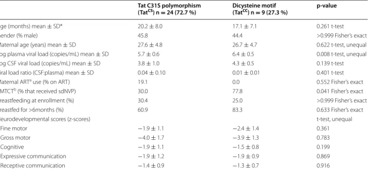
Table 1 Demographic data
* Sample size: age, gender, log viral load, log CSF viral load, viral load ratio, (n = 33); maternal age (n = 30); maternal ART use, PMTCT, breastfed for >6months (n = 29); breastfeeding at enrollment (n = 31)
a ART—antiretroviral therapy
b PMTCT—prevention of mother to child transmission; the use of antepartum antiretroviral therapy to prevent the peripartum transmission of HIV; all mothers that
received PMTCT, received single-dose nevirapine (sdNVP)
Tat C31S polymorphism
(TatCS) n = 24 (72.7 %) Dicysteine motif (TatCC) n = 9 (27.3 %) p-value
Age (months) mean ± SD* 20.2 ± 8.0 17.1 ± 7.1 0.261 t-test
Gender (% male) 45.8 44.4 >0.999 Fisher’s exact
Maternal age (years) mean ± SD 27.6 ± 4.8 26.7 ± 4.7 0.622 t-test, unequal Log plasma viral load (copies/mL) mean ± SD 5.7 ± 0.6 6.4 ± 0.5 0.008 t-test, unequal Log CSF viral load (copies/mL) mean ± SD 3.8 ± 1.0 4.3 ± 0.5 0.139 t-test Viral load ratio (CSF:plasma) mean ± SD 0.04 ± 0.10 0.01 ± 0.01 0.401 t-test
Maternal ARTa use (% on ART) 19.1 0.0 0.552 Fisher’s exact
PMTCTb (% that received sdNVP) 30.0 77.8 0.041 Fisher’s exact
Breastfeeding at enrollment (%) 30.4 25.0 >0.999 Fisher’s exact
Breastfed for >6months (%) 60.9 83.3 0.633 Fisher’s exact
Neurodevelopmental scores (z-scores) t-test, unequal
Fine motor −1.9 ± 1.1 −2.4 ± 1.4 0.361
Gross motor −4.0 ± 1.7 −3.9 ± 1.3 0.783
Cognitive −1.9 ± 1.1 −1.5 ± 0.8 0.199
Expressive communication −1.9 ± 1.2 −1.9 ± 0.9 0.869
illustrates the adjusted point estimates and 95 % confi-dence intervals for the association between TatCC and
the five domains of neurological development measured by the BSID-III.
Children excluded because of lack of Tat sequenc-ing information were on average 5 months younger, and had higher gross motor and expressive communication z-scores compared to children with Tat information. In a sensitivity analysis to assess a potential bias introduced by the exclusion of these children, we found that assign-ing children with missassign-ing Tat information either as all TatCC or as all TatCS did not change our final result of a
lack of association observed between Tat sequence and neurodevelopmental delay.
Discussion
In this cross-sectional study of HIV-infected children in Malawi, we found, contradictory to our hypothesis, the presence of TatCC variants was not significantly
associ-ated with neurodevelopmental severity in multivariate analysis. This may be related to (1) differences in HIV-associated neuropathogenesis between children and adults, (2) small sample size or other factors.
It is possible that TatCC variants are associated with the
occurrence of HIV neuropathogenesis but not the sever-ity thereof, an issue we could not conclusively assess as all participating children presented with neurodevel-opmental delay. Underlying differences in neuropathol-ogy may explain the results identified in this study. HIV
Fig. 2 Neurodevelopmental z-scores obtained by the Bayley III Scales of Infant and Toddler Development standardized to pediatric Malawian norms. Box and whiskers plot mean and range for unadjusted neurodevelopmental z-scores, including gross motor, cognitive, expressive communi-cation, and receptive communication
Table 2 P-values for Tat-CC in univariate and multivariable analysis
Adjusted for age, log plasma VL, and log CSF VL
Fine motor Gross motor Cognitive Expressive
communication Receptive communication
Univariate analysis t-test, unequal variances 0.361 0.783 0.199 0.869 0.916
Multivariable analysis 0.846 0.974 0.155 0.848 0.376
neuropathy is a result of chronic HIV infection that results in the infiltration of activated monocytes into the brain, which leads to the production of Tat, gp120, cytokines, and other neurotoxic viral proteins [26]. In children with HIV-associated neuropathy, both the dura-tion of HIV infecdura-tion and the associated inflammadura-tion are shorter when compared with adults with HAND. Additionally, infants and children have rapidly develop-ing brains, which undergo increases in white matter size, maturation of myelination, and increase in the number of neuronal axons [27]. Central nervous system (CNS) pathology of children with AIDS frequently involves global anoxic-ischemic and necrotizing encephalomy-elopathies [28, 29] which differs from the pathology seen in adults consisting of fronto-temporal atrophy, diffuse parenchymal nodules, as well as perivascular mononu-clear infiltrates [30]. Radiographic evaluations support white matter damage, which differs between neona-tal and adult brains in response to ischemic injury [27]. Immunologic differences also distinguish pediatric and adult HIV-associated neuropathy. CNS vascular inflam-mation found in pediatric AIDS consists of transmural and perivascular mononuclear infiltrates consisting pri-marily of CD3+ and CD8+ T-lymphocytes, in contrast to the inflammatory lesions found in adult patients with HIV-1 encephalopathy in which monocytes and mac-rophages are the predominant mononuclear cells [31], suggesting the immunologic and inflammatory basis of disease in children is fundamentally different from that found in adults. A CNS compartmentalization study con-ducted on the same study population showed that only 10–20 % of the children had HIV variants compartmen-talized in the CSF [32] that have evolved to use low levels of CD4, a characteristic of macrophage-tropic isolates. This supports the hypothesis that CNS HIV replication in
the majority of the children occurs primarily in T-cells. Although prior in vitro research has shown that sub-type C HIV-1 Tat with an intact dicysteine motif induces greater CCL-2 induction, monocyte recruitment, and neuropathology, the effects of HIV-1 Tat with an intact dicysteine motif on T-lymphocytes are unknown and merits evaluation. As previously noted, prior clinical data shows that Southern African countries with predomi-nantly subtype C infection show both a higher prevalence of HAD and a higher proportion of subtype C HIV-1 with a TatCC motif [1, 21, 22, 33, 34]; however, all of these
results were obtained from studies in adults. This is the first study to evaluate the neurodevelopmental outcomes in children infected with subtype C HIV-1 with the TatCC
motif.
In the neurocognitive evaluation conducted by Paul et al. South African adult individuals infected with clade C HIV-1 with TatCC were compared to those infected
with clade C HIV-1 with TatCS motif [23]. They found no
differences in the neurocognitive defects between the two groups. This adult study, like the current pediatric study, also relied on plasma-derived HIV-1 sequences to deter-mine the Tat genotype. It is known that HIV-1 can be compartmentalized in the brain [32] which could mean that it may be more directly relevant to correlate the cog-nitive defects to brain- or CSF-derived HIV-1 sequences than to plasma. Furthermore, studies focused on Tat also do not take into account the role of gp120, another source of neuronal dysfunction. Our recent report has shown that while the gp120 of Indian subtype C HIV-1 is non-neurovirulent, that of Southern African subtype C HIV-1 displayed robust neurovirulence [35].
Our findings should be interpreted in light of the study limitations. First, the sample size was small and informa-tion on some covariates was incomplete, which restricted our ability to adjust for confounding variables. Most impor-tantly, even though in bivariate analysis we observed that children with TatCC were more likely to have higher plasma
VL and were more likely to have received sdNVP for PMTCT (p = 0.041) compared to children with TatCS, we
were only able to include plasma VL and not PMTCT in the multivariate model. This may have biased the association to the null and thus contributed to the lack of a statistically significant relationship between HIV-1 TatCC motif and
developmental delay. Second, data on the plasma CD4 or CD8 lymphocyte count was not available. Low plasma CD4 counts have been associated with neurocognitive dysfunc-tion in children [36] and low plasma CD8 counts are asso-ciated with the risk of neurological impairment in children [37]. Third, the cross-sectional study design hindered the ability to infer causation and allowed for the introduction of biases. Survival bias may have occurred and influenced the interpretation of our findings. Although there may be some
Fig. 3 Adjusted coefficients for TatCC. For each of the five subtests,
the adjusted point estimate and 95 % lower confidence limit (LCL) and upper confidence limit (UCL) for the association for TatCC is
bias due to exclusion of children without Tat information, sensitivity analysis suggest that this did not influence our conclusions. In our analysis, children with HIV TatCC
vari-ants had a significantly higher mean plasma VL than HIV TatCS variants (p = 0.008; t-test, unequal variances). It is
possible that HIV-infected children with TatCC variants did
not survive as long as children with TatCS variants. If such
survival bias occurred, it could appear that children with TatCS variants have more severe neurodevelopmental delay.
Finally, we were unable to assess the association between TatCC and the severity of neurodevelopmental delay as all
children included in the analysis presented with neurode-velopmental delay in at least one domain. A control group of HIV-infected children without neurodevelopmental delay could have contributed valuable information but was precluded given ethical and cultural concerns of perform-ing a lumbar puncture in this population.
Conclusions
In this study of 33 Malawian ART-naïve HIV-1 subtype C infected children with neurodevelopmental delay, we observed that the presence of HIV TatCC motif was not
associated with the severity of developmental delay in children with HIV type C infection. This may be due to differences in HIV-1 neuropathogenesis in children as compared to adults but demonstrating this hypothesis will require studies with larger sample size.
Abbreviations
HIV-1: human immunodeficiency virus type 1; VL: viral load; BSID-III: Bayley Scales of Infant Development III; PMTCT: prevention of mother-to-child transmission; ART: anti-retroviral therapy; CNS: central nervous system; HAND: HIV-associated neurocognitive disorders; HAD: HIV-associated dementia; VIF: variance inflation factors; sdNVP: single-dose nevirapine.
Authors’ contributions
JD analyzed the genetic and amino acid sequences of HIV-1 Tat, performed the statistical analysis, and drafted the manuscript. AD participated in study design, collection of data, analysis of data, and edited the manuscript. EC performed statistical analysis necessary to standardize the BSID-III for the Malawian population and edited the manuscript. CBS performed genetic analysis and confirmed the predicted amino acid sequences of HIV-1 Tat. MM participated in study design and collection of samples and data. RW participated in study design, genetic analysis, and editing the manuscript. AVR participated in study design, validation of statistical analysis, and drafting the manuscript. VP participated in study design, assisted in analysis of genetic and amino acid sequences of HIV-1 Tat and drafted the manuscript. All authors read and approved the final manuscript.
Author details
1 Department of Pediatrics, Montefiore Medical Center, Bronx, NY, USA. 2 UNC School of Public Health, University of North Carolina, Chapel Hill, NC, USA. 3 Department of Biochemistry and Biophysics, University of North Carolina, Chapel Hill, NC, USA. 4 Malawi-Liverpool Wellcome Trust and Department of Pediatrics, College of Medicine, Blantyre, Malawi. 5 Department of Microbiol-ogy and ImmunolMicrobiol-ogy, Albert Einstein College of Medicine, Bronx, NY, USA.
Acknowledgements
JD was supported in part by the National Center for Research Resources (NCRR) and the National Center for Advancing Translational Sciences (NCATS),
components of the National Institutes of Health (NIH), through Clinical Trans-lational Science Award (CTSA) Grant Numbers UL1RR025750, KL2RR025749 and TL1RR025748. AVR was supported by the Fogarty International Center and National Institute of Child Health and Human Development (NICHD) Award Number R01HD053216. MLW is supported by a core grant from the Wellcome Trust. VP was supported by NIH Grant MH083579 and Center for AIDS Research at the Albert Einstein College of Medicine and Montefiore Medical Center (NIH AI-051519). RS was supported by NIH Award R01 MH101024, with infrastructure support from the UNC Center For AIDS Research (NIH Award P30 AI50410) and the UNC Lineberger Comprehensive Cancer Center (NIH Award P30 CA16086). Its contents are solely the responsibility of the authors and do not necessarily represent the official views of the NIH. The authors wish to thank Dr. Robert Heyderman of the Malawi-Liverpool Wellcome Trust for use of the MLW infrastructure and supervision of the Malawian investigators, and Dr. Steve Meschnick of UNC Gillings School of Public Health for his collabora-tive efforts with Dr. Mupase that resulted in samples used in the study and Dr. Vasudev Rao for critically reading the manuscript.
Competing interests
The authors declare that they have no competing interests.
Received: 25 March 2015 Accepted: 27 November 2015
References
1. Lawler K, Mosepele M, Ratcliffe S, Seloilwe E, Steele K, Nthobat-sang R, et al. Neurocognitive impairment among HIV-positive individuals in Botswana: a pilot study. J Int AIDS Soc. 2010;13:15. doi:10.1186/1758-2652-13-15.
2. Hittelman J. Neurodevelopmental aspects of HIV infection. In: Karger, editor. Brain in pediatric AIDS. Basel, Switzerland; 1990. p. 64–71. 3. Boivin MJ, Green SD, Davies AG, Giordani B, Mokili JK, Cutting WA. A
preliminary evaluation of the cognitive and motor effects of pediatric HIV infection in Zairian children. Health Psychol. 1995;14(1):13–21.
4. Chase C, Vibbert M, Pelton SI, Coulter DL, Cabral H. Early neurodevelop-mental growth in children with vertically transmitted human immunode-ficiency virus infection. Arch Pediatr Adolesc Med. 1995;149(8):850–5. 5. Husstedt IW, Frohne L, Bockenholt S, Frese A, Rahmann A, Heese C, et al.
Impact of highly active antiretroviral therapy on cognitive processing in HIV infection: cross-sectional and longitudinal studies of event-related potentials. AIDS Res Hum Retroviruses. 2002;18(7):485–90. doi:10.1089/088922202317406628.
6. Winston A, Duncombe C, Li PC, Gill JM, Kerr SJ, Puls R, et al. Does choice of combination antiretroviral therapy (cART) alter changes in cerebral function testing after 48 weeks in treatment-naive, HIV-1-infected indi-viduals commencing cART? A randomized, controlled study. Clin Infect Dis. 2010;50(6):920–9. doi:10.1086/650743.
7. USAID. HIV/AIDS Health Profile—Africa Region. http://www.usaid.gov. Accessed 28 Mar 2013.
8. Albini A, Benelli R, Giunciuglio D, Cai T, Mariani G, Ferrini S, et al. Identifica-tion of a novel domain of HIV tat involved in monocyte chemotaxis. J Biol Chem. 1998;273(26):15895–900.
9. Weiss JM, Nath A, Major EO, Berman JW. HIV-1 Tat induces monocyte chemoattractant protein-1-mediated monocyte transmigration across a model of the human blood-brain barrier and up-regulates CCR5 expres-sion on human monocytes. J Immunol. 1999;163(5):2953–9.
10. Magnuson DS, Knudsen BE, Geiger JD, Brownstone RM, Nath A. Human immunodeficiency virus type 1 tat activates non-N-methyl-d-aspartate excitatory amino acid receptors and causes neurotoxicity. Ann Neurol. 1995;37(3):373–80. doi:10.1002/ana.410370314.
11. Sabatier JM, Vives E, Mabrouk K, Benjouad A, Rochat H, Duval A, et al. Evidence for neurotoxic activity of tat from human immunodeficiency virus type 1. J Virol. 1991;65(2):961–7.
13. Ranga U, Shankarappa R, Siddappa NB, Ramakrishna L, Nagendran R, Mahalingam M, et al. Tat protein of human immunodeficiency virus type 1 subtype C strains is a defective chemokine. J Virol. 2004;78(5):2586–90. 14. Tyor WR, Power C, Gendelman HE, Markham RB. A model of human
immunodeficiency virus encephalitis in scid mice. Proc Natl Acad Sci USA. 1993;90(18):8658–62.
15. Rao VR, Sas AR, Eugenin EA, Siddappa NB, Bimonte-Nelson H, Berman JW, et al. HIV-1 clade-specific differences in the induction of neuropathogene-sis. J Neurosci. 2008;28(40):10010–6. doi:10.1523/JNEUROSCI.2955-08.2008. 16. Li W, Huang Y, Reid R, Steiner J, Malpica-Llanos T, Darden TA, et al. NMDA
receptor activation by HIV-Tat protein is clade dependent. J Neurosci. 2008;28(47):12190–8. doi:10.1523/JNEUROSCI.3019-08.2008.
17. Campbell GR, Watkins JD, Loret EP, Spector SA. Differential induction of rat neuronal excitotoxic cell death by human immunodeficiency virus type 1 clade B and C tat proteins. AIDS Res Hum Retroviruses. 2011;27(6):647–54. doi:10.1089/AID.2010.0192.
18. Gupta JD, Satishchandra P, Gopukumar K, Wilkie F, Waldrop-Valverde D, Ellis R, et al. Neuropsychological deficits in human immunodeficiency virus type 1 clade C-seropositive adults from South India. J Neurovirol. 2007;13(3):195–202. doi:10.1080/13550280701258407.
19. Satishchandra P, Nalini A, Gourie-Devi M, Khanna N, Santosh V, Ravi V, et al. Profile of neurologic disorders associated with HIV/AIDS from Ban-galore, south India (1989–96). Indian J Med Res. 2000;111:14–23. 20. Sacktor N, Lyles RH, Skolasky R, Kleeberger C, Selnes OA, Miller EN, et al.
HIV-associated neurologic disease incidence changes: multicenter AIDS Cohort Study, 1990–1998. Neurology. 2001;56(2):257–60.
21. Joska JA, Westgarth-Taylor J, Myer L, Hoare J, Thomas KG, Combrinck M, et al. Characterization of HIV-associated neurocognitive disorders among individuals starting antiretroviral therapy in South Africa. AIDS Behav. 2011;15(6):1197–203. doi:10.1007/s10461-010-9744-6.
22. Rao VR, Neogi U, Talboom JS, Padilla L, Rahman M, Fritz-French C, et al. Clade C HIV-1 isolates circulating in Southern Africa exhibit a greater fre-quency of dicysteine motif-containing Tat variants than those in South-east Asia and cause increased neurovirulence. Retrovirology. 2013;10:61. doi:10.1186/1742-4690-10-61.
23. Paul RH, Joska JA, Woods C, Seedat S, Engelbrecht S, Hoare J, et al. Impact of the HIV Tat C30C31S dicysteine substitution on neuropsychological function in patients with clade C disease. J Neurovirol. 2014;20(6):627–35. doi:10.1007/s13365-014-0293-z.
24. Bayley N. Bayley Scales of Infant and Toddler Development. 3rd ed. San Antonio: Harcourt Assessment Inc.; 2006.
25. Cromwell EA, Dube Q, Cole SR, Chirambo C, Dow AE, Heyderman RS, et al. Validity of US norms for the Bayley Scales of Infant Development-III in
Malawian children. Eur J Paediatr Neurol. 2014;18(2):223–30. doi:10.1016/j. ejpn.2013.11.011.
26. Gras G, Kaul M. Molecular mechanisms of neuroinvasion by mono-cytes-macrophages in HIV-1 infection. Retrovirology. 2010;7:30. doi:10.1186/1742-4690-7-30.
27. Neil J, Miller J, Mukherjee P, Huppi PS. Diffusion tensor imaging of normal and injured developing human brain—a technical review. NMR Biomed. 2002;15(7–8):543–52. doi:10.1002/nbm.784.
28. Park YD, Belman AL, Kim TS, Kure K, Llena JF, Lantos G, et al. Stroke in pedi-atric acquired immunodeficiency syndrome. Ann Neurol. 1990;28(3):303– 11. doi:10.1002/ana.410280302.
29. Dickson DW, Llena JF, Nelson SJ, Weidenheim KM. Central nervous system pathology in pediatric AIDS. Ann NY Acad Sci. 1993;693:93–106. 30. Budka H. Neuropathology of human immunodeficiency virus infection.
Brain Pathol. 1991;1(3):163–75.
31. Katsetos CD, Fincke JE, Legido A, Lischner HW, de Chadarevian JP, Kaye EM, et al. Angiocentric CD3(+) T-cell infiltrates in human immunodefi-ciency virus type 1-associated central nervous system disease in children. Clin Diagn Lab Immunol. 1999;6(1):105–14.
32. Sturdevant CB, Dow A, Jabara CB, Joseph SB, Schnell G, Takamune N, et al. Central nervous system compartmentalization of HIV-1 subtype C variants early and late in infection in young children. PLoS Pathog. 2012;8(12):e1003094. doi:10.1371/journal.ppat.1003094.
33. Joska JA, Fincham DS, Stein DJ, Paul RH, Seedat S. Clinical correlates of HIV-associated neurocognitive disorders in South Africa. AIDS Behav. 2010;14(2):371–8. doi:10.1007/s10461-009-9538-x.
34. Holguin A, Banda M, Willen EJ, Malama C, Chiyenu KO, Mudenda VC, et al. HIV-1 effects on neuropsychological performance in a resource-limited country, Zambia. AIDS Behav. 2011;15(8):1895–901. doi:10.1007/ s10461-011-9988-9.
35. Rao VR, Neogi U, Eugenin E, Prasad VR. The gp120 protein is a second determinant of decreased neurovirulence of Indian HIV-1C isolates com-pared to southern African HIV-1C isolates. PLoS One. 2014;9(9):e107074. doi:10.1371/journal.pone.0107074.
36. Brouwers P, Tudor-Williams G, DeCarli C, Moss HA, Wolters PL, Civitello LA, et al. Relation between stage of disease and neurobehavioral measures in children with symptomatic HIV disease. AIDS. 1995;9(7):713–20. 37. Pearson DA, McGrath NM, Nozyce M, Nichols SL, Raskino C, Brouwers P,
et al. Predicting HIV disease progression in children using measures of neuropsychological and neurological functioning. Pediatric AIDS clinical trials 152 study team. Pediatrics. 2000;106(6):E76.
• We accept pre-submission inquiries
• Our selector tool helps you to find the most relevant journal
• We provide round the clock customer support
• Convenient online submission
• Thorough peer review
• Inclusion in PubMed and all major indexing services
• Maximum visibility for your research
Submit your manuscript at www.biomedcentral.com/submit