Abstract:
Previous work from our laboratory demonstrated that obese mice have alterations in
viral cytokine gene expression when infected with the influenza virus. Since these cytokines play a major role in the infected cells’ innate immune response, we evaluated the effects of leptin dos-age on expression of several cytokines during a flu infection in human alveolar lung epithelium (A549 cells). Following 48 hours of infection by influenza A/Puerto Rico/8/1934 H1N1, increas-ing leptin dosage resulted in significant decreases in expression of interferon-α (IFNα) and monocyte chemotactic protein-1 (MCP-1); a strong, similar trend was found for IFNβ, but it was not statistically significant. No leptin dosage trends were found for IL-6 or for the amount of viral particles released by cells into the supernatant. These data suggest that leptin contributes to the increased flu severity in obese individuals by weakening the innate immune response.
I. Aims
Obesity is epidemic throughout the United States and is known to increase the risk of a variety of medical complications, including cardiovascular disease, diabetes, and multiple can-cers1. More recently, obesity has been found to be an independent risk factor for severe compli-cations, hospitalizations, and death due to infection with the influenza virus2. As the rates of obe-sity rise, annual deaths from flu infections may increase, and flu pandemics may end up being even more deadly than in the past. This leads us to ask why does obesity lead to increased sus-ceptibility to flu infection? What is it about the obese state that could affect the flu severity and mortality? One possibility is elevated leptin levels.
We, and others, have demonstrated that leptin levels are significantly elevated in the serum of obese animals. However, our lab has uniquely demonstrated that leptin levels are also elevated in lung lavage fluid during flu infection in obese mice, revealing that the lung epithelial cells are bathed in abnormal levels of leptin. This finding raises the question: does leptin only affect the immune response or does it also affect the influenza virus pathology and the hardiness of the lung tissue?
Aim 1: Determine the role of leptin dosage on the innate immune response of human lung tissue (A549 cells) during influenza infection
a. Evaluate the tissue’s ability to recruit leukocytes. We hypothesize that with
ing leptin dosage, human lung tissue will have an impaired expression of MCP-1.
b. Evaluate the tissue’s ability to repel infections. We hypothesize that with increasing
leptin dosage, human lung tissue will have an impaired expression of IFNα and IFNβ.
c. Evaluate the tissue’s release of pro-inflammatory cytokines. We hypothesize that
Aim 2: Determine the role of leptin dosage on the innate immune response of canine kidney tissue (MDCK cells) during influenza infection
a. Evaluate the tissue’s ability to recruit leukocytes. We hypothesize that with
ing leptin dosage, canine kidney tissue will have an impaired expression of MCP-1.
b. Evaluate the tissue’s ability to repel infections. We hypothesize that with increasing
leptin dosage, canine kidney tissue will have an impaired expression of IFNα and IFNβ.
c. Evaluate the tissue’s release of pro-inflammatory cytokines. We hypothesize that
with increasing leptin dosage, canine kidney tissue will have an excessive expression of IL-6.
Aim 3: Determine the role of leptin dosage on viral titers during influenza infection
a. Evaluate the effects of leptin on the replication of the flu virus. We hypothesize that
with increasing leptin dosage, flu virus will be present at greater levels.
The overall objective of this research is to determine the effects of leptin on the flu res-ponse: leptin’s ability to alter the innate immune response to the flu in human epithelial tissue, and the effects on virus titers. Results from this study can help explain why obese individuals are more susceptible to influenza virus infection compared with healthy weight individuals. With the information gained from this study, we can attempt to identify mechanisms that cause increased flu severity in obese populations and find ways to strengthen infected tissues’ response to infec-tion for a proper reacinfec-tion to the flu.
II. Introduction
Leptin, a polypeptide signaling molecule synthesized primarily by adipocytes, is present in the blood in proportion to the amount of adipose tissue in the human body3. Lean humans have leptin concentrations around 10 ng/mL whereas obese humans have leptin concentrations around 35 ng/mL—more than three times the former10. Once thought to be a cure for obesity due to its role in suppressing appetite, leptin has been found to be pleiotropic, having many effects on many tissues in the body. Leptin is a necessary hormone for hunger inhibition and regulation of adiposity, but it also plays a role in immunomodulation, regulatory T-cell suppression, insulin resistance, and pro-inflammatory responses3,4. Furthermore, studies indicate that leptin may have even more functions in the body: leptin receptor mRNA variants have been isolated in a wide range of tissue types, including the spleen, kidneys, testes, liver, and adrenal glands5.
pro-duction was seriously delayed. These data suggest that obesity has an effect on lung epithelial cells, perhaps making them more susceptible to influenza infection.
Lung epithelial cells have leptin receptors on their membranes, evidencing that leptin signaling must occur in the lung tissue. The functions of those receptors and the signals they transduce are currently unknown, though they may play a role in the remodeling of airways and response to inflammation8. In addition, mice with elevated leptin levels in the body exhibit res-piratory distress and other resres-piratory dysfunction, however these could possibly be due to obe-sity itself and not due to the increased leptin levels9. Since obese individuals have elevated circu-lating levels of leptin, which is an immunomoducircu-lating inflammatory molecule, and have in-creased flu-related complications, we predict that the excess leptin found in obese persons is a contributing factor to the increased severity of flu infection in obese individuals. We also predict that leptin will have similar effects in other infected tissues, such as kidney tissue.
III. Methods
Cell lines and leptin exposures
We used two different cell lines for this study. First, we began with A549 cells, human alveolar basal epithelial cells, which were grown in culture and subsequently infected with influenza virus. These cells were separated into three distinct groups and treated as follows: 1) no leptin, 2) leptin concentration 10 ng/mL, and 3) leptin concentration 100 ng/mL. Group 1 represents the controls and group 2 represents lean individuals10. Group 3 represents extreme levels not usually seen in the serum of healthy humans; however these concentrations may occur locally at the cell-ular level. All cells were exposed to purified recombinant human leptin for 24 hours prior to in-fection. This protocol was repeated using Madin-Darby canine kidney (MDCK) epithelial cells in order to see if leptin has similar effects on different types of tissues. Both A549 and MDCK cells are routinely used to study and grow influenza viruses.
Influenza infection and RNA isolation
Following the leptin exposure, cells were infected with influenza A/Puerto Rico/8/1934 H1N1 (A/PR8) virus. After 8 hours of infection at 37ºC, the supernates were removed and stored at -80ºC; the cells were lysed with TRIzol reagent (Invitrogen) and the RNA was isolated. Unin-fected cells will be treated in the same manner as the inUnin-fected cells to use a control for the leptin exposure. The RNA isolated for all samples was transcribed to cDNA with Oligo(dT)-primed reverse transcription using SuperScript II Reverse Transcriptase (Invitrogen).
Gene expression quantitation
were also determined for all samples to normalize gene expression, allowing for a calculation of fold increase based on the cycle number at which the sample passed a threshold value. Fold in-crease was expressed as a fold of uninfected, leptin-free samples at 8 hours of incubation when available. In samples with no 8-hour data, the fold increase was expressed as fold of the unin-fected, leptin-free group for that gene and time-point.
Virus quantification
Influenza A/PR8 virus titers were measured in the supernatant (a measure of released virus) by a modified TCID50 assay with hemagglutination as an endpoint indicator.
Statistical analysis
Measures of significance were analyzed via two-way ANOVA; p values of less than 0.05 were considered statistically significant.
IV. Results
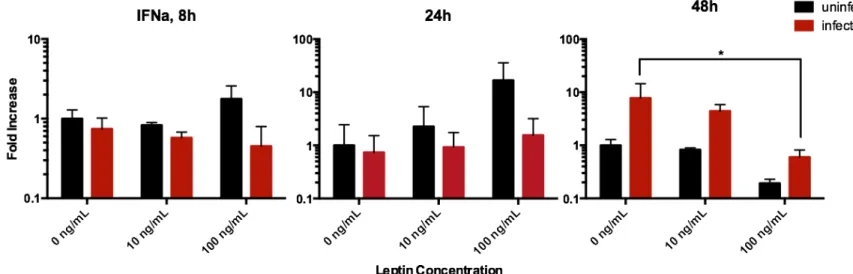
High leptin exposure results in reduced expression of antiviral cytokines
At 48 hours post-infection, leptin exposure resulted in a significant decrease in expression of IFNα mRNA; the decrease in IFNβ is similar but not statistically significant. It should be noted that at 48 hours, the average fold increase in the expression of IFNα for infected 100 ng/mL lep-tin samples were weaker than the average increase 10 ng/mL leplep-tin samples while uninfected
Figure 1. Effects of leptin on human alveolar lung tissue expression of the IFNα gene after 8, 24, and 48 hours of influenza A/PR8 virus infection, measured by quantitative RT-PCR from total RNA. After 48 hours of infection, the leptin dosage as a significant effect on IFNα mRNA expression (p = 0.0062).
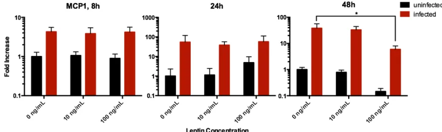
High leptin exposure results in reduced expression of MCP-1
Following 48 hours of A549 tissue infection, there was a significant trend of reduced gene ex-pression as a result of leptin dosage (Figure 3). Infection brought about a 38-fold increase in expression of MCP-1 in the leptin-free samples, a 42-fold increase in the 10 ng/mL leptin sam-ples, and a 41-fold increase in the 100 ng/mL leptin samples. The primary function of the mono-cyte chemotactic protein (MCP-1) is to recruit monomono-cytes to the site of infection so as to clear out virus particles and dead cell debris13—a process crucial to a functional non-specific immune defense. It has already been demonstrated that leptin secretion has a negative effect on regulatory T-cell proliferation14, and it is now evident that leptin negatively affects monocyte recruitment.
Figure 3. Effects of leptin on human alveolar lung tissue expression of the MCP-1 gene after 8, 24, and 48 hours of influenza A/PR8 virus infection, measured by quantitative RT-PCR from to-tal RNA. After 48 hours of infection, the leptin dosage as a significant effect on the increase of MCP-1 mRNA expression (p = 0.0028).
Leptin dosage does not affect IL-6 gene expression after 24 hours of infection
#
Figure 4. Effects of leptin on human alveolar lung tissue expression of the IL-6 gene after 24 hours of influenza A/PR8 H1N1 infection. Infection had a significant effect on IL-6 gene ex-pression; however, leptin dosage had no statistically significant effect on gene expression.
Leptin has no effect on MCP-1 expression in canine kidney cells in the first 48 hours of infection
Leptin had no clear effect on MCP-1 expression in MDCK cells at any point during the first 48 hours of infection (Figure 5). MCP-1 gene transcripts were not detectable after 8 and 24 hours of infection, therefore only the 48-hour data are presented.
#
mRNA transcripts in A549 tissue for IL-6 were detectable only after 24 hours post-infection. The only detectable mRNA samples for MDCK cells were for the MCP-1 gene at 48 hours, which showed no trend.
Leptin dosage has no significant effect on virus titers
The differences between the virus titers of the control (0 ng/mL leptin) and obese (100 ng/mL leptin) samples were not statistically significant, so we can rule out virus dosage as the cause of differences in gene expression for cytokines tested. It should be noted that viral titer does not necessarily correlate with pathogenesis, as obese and lean mice infected with the same dosage of influenza virus can have similar lung virus titers, yet the obese mice will exhibit severe lung pathology and high mortality rates compared to the lean animals.
V. Discussion
As leptin was discovered only 20 years ago, we still have much to learn about its roles
in the body. It was once thought to simply be a bodyweight-regulating hormone, but then it was shown to have immunomodulating properties, and we believe that its roles are even broader than have been shown thus far. High leptin exposure results in the weakening of the cell’s natural abi-lity to ward off infection (reduced interferons alpha and beta expression) and the cell’s abiabi-lity to recruit immune cells to clear the virus (reduced MCP-1 expression).
Now that we have identified leptin as an enhancer of viral replication by diminishing
the anti-viral protective mechanisms used by the lung’s epithelial cells, we can explain, in part, why obesity is associated with more severe influenza lung pathology. This project is the first to show that leptin can directly alter the lung epithelial cell response to influenza infection in hu-mans. Such findings are of great clinical significance; it has been shown that leptin anti-bodies can improve survival of obese mice when infected with flu14, and this project provides further evidence to why that may be. As around 5-20% of the US population becomes infected with the flu each year13, the public health implications of this research can have a profound im-pact on how the disease and other infections are treated in obese persons in the future.
Future studies should look at the expression of other cytokines involved in innate
immunity, such as IL-1 or TNFα; other tissues infected by the influenza virus, such as nasal epi-thelia or bronchial epiepi-thelia; and different concentrations of leptin not used in this study. In addi-tion, different strains of flu at different doses can be tested to see if certain strains or doses pro-duce different effects. These studies should also use tissues that express cytokines at significant levels in order to gather measurable results.
To answer our question of whether leptin affects only the immune response or if
VI. References
1. Pi-Sunyer, F. X. (2002). The obesity epidemic: pathophysiology and consequences of obesity. Obesity Research 10(Suppl 2): 97S–104S.
2. Morgan et al. (2010). Morbid obesity as a risk factor for hospitalization and death due to 2009 pandemic influenza A (H1N1) disease. PLoS ONE 5(3): e9694. doi:10.1371/ journal.pone.0009694
3. La Cava, A., G. Matarese. (2004). The weight of leptin in immunity. National Review of Immunology4: 371–379.
4. Fantuzzi, Giamila. (2005)."Adipose tissue, adipokines, and inflammation." Journal of Allergy and Clinical Immunology,115(5), 911-919.
5. Hoggard et al. (1997). Localization of leptin receptor mRNA splice variants in murine peripheral tissues by RT-PCR and in situ hybridization. Biochemical and Biophysical Research Communications, 232, 383-387.
6. Karlsson, E., Sheridan, P., & Beck, M. (2010). Diet-induced obesity impairs the T cell memory response to influenza virus infection. The Journal of Immunology, 184(6), 3127-3133.
7. Smith, A., Sheridan, P., Harp, J., Beck, M. (2007). Diet-induced obese mice have increased mortality and altered immune responses when infected with influenza virus.
The Journal of Nutrition, 137, 1236-1243.
8. Malli et al. (2010). The role of leptin in the respiratory system: an overview. Respiratory Research. 11:152 http://respiratory-research.com/content/11/1/152
9. Bruno et al. (2009). Leptin and leptin receptor expression in asthma. The Journal of Allergy and Clinical Immunology, 124(2), 230-237.
10. al Maskari, M., & Alnaqdy, A. (2006). Correlation between serum leptin levels, body mass index and obesity in omanis. Sultan Qaboos Medical University Journal, 6(2), 27-31. doi: PMC3074914
11. Biron, C. "Role of Early Cytokines, including Alpha and Beta Interferons (IFN-α/β), in Innate and Adaptive Immune Responses to Viral Infections." Seminars in Immunology
10.5 (1998): 383-90. Print.
12. Deshmane, Satish L., Sergey Kremlev, Shohreh Amini, and Bassel E. Sawaya. "Monocyte Chemoattractant Protein-1 (MCP-1): An Overview." Journal of Interferon & Cytokine Research 29.6 (2009): 313-26. Print.
13. "Seasonal Influenza Q&A." Centers for Disease Control and Prevention. Centers for Disease Control and Prevention, 14 Aug. 2014. Web. 14 Mar. 2015. <http://www.cdc.gov/ flu/about/qa/disease.htm>.
14. De Rosa, Veronica, Claudio Procaccini, Gaetano Calì, Giuseppe Pirozzi, Silvia Fontana, Serafino Zappacosta, Antonio La Cava, and Giuseppe Matarese. "A Key Role of Leptin in the Control of Regulatory T Cell Proliferation." Immunity 26.2 (2007): 241-55. Print. 15. Trujillo, M. E. "Interleukin-6 Regulates Human Adipose Tissue Lipid Metabolism and