Big
Idea
Investigation 7 S83
3
INVESTIGATION
7
CELL DIVISION:
MITOSIS AND MEIOSIS
How do eukaryotic cells divide to produce
genetically identical cells or to produce gametes
with half the normal DNA?
■
BACKGROUND
One of the characteristics of living things is the ability to replicate and pass on genetic information to the next generation. Cell division in individual bacteria and archaea usually occurs by binary fission. Mitochondria and chloroplasts also replicate by binary fission, which is evidence of the evolutionary relationship between these organelles and prokaryotes.
Cell division in eukaryotes is more complex. It requires the cell to manage a complicated process of duplicating the nucleus, other organelles, and multiple
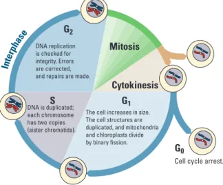
chromosomes. This process, called the cell cycle, is divided into three parts: interphase, mitosis, and cytokinesis (Figure 1). Interphase is separated into three functionally distinct stages. In the first growth phase (G1), the cell grows and prepares to duplicate its
DNA. In synthesis (S), the chromosomes are replicated; this stage is between G1 and the
second growth phase (G2). In G2, the cell prepares to divide. In mitosis, the duplicated chromosomes are separated into two nuclei. In most cases, mitosis is followed by cytokinesis, when the cytoplasm divides and organelles separate into daughter cells. This type of cell division is asexual and important for growth, renewal, and repair of multicellular organisms.
Bio_T_Lab07_01 QU: OK to add callout ?Cyclebegins.
NOTE: Art with callouts requires greater size than specified. Mitosis
Cytokinesis G1 G2
S
G0 The cell increases in size. The cell structures are duplicated, and mitochondria and chloroplasts divide by binary fission. DNA is duplicated;
each chromosome has two copies (sister chromatids).
DNA replication is checked for integrity. Errors are corrected, and repairs are made.
Cell cycle arrest. Inte
rpha se
Figure 1. The Cell Cycle Showing G1, S, and G2 Phases, Mitosis, and Cytokinesis
Genetics and Information Transfer
S84 Investigation 7
Cell division is tightly controlled by complexes made of several specific proteins. These complexes contain enzymes called cyclin-dependent kinases (CDKs), which turn on or off the various processes that take place in cell division. CDK partners with a family of proteins called cyclins. One such complex is mitosis-promoting factor (MPF), sometimes called maturation-promoting factor, which contains cyclin A or B and cyclin-dependent kinase (CDK). (See Figure 2a.) CDK is activated when it is bound to cyclin, interacting with various other proteins that, in this case, allow the cell to proceed from G2 into mitosis. The levels of cyclin change during the cell cycle (Figure 2b). In
most cases, cytokinesis follows mitosis.
Bio_T_Lab07_02ab
NOTE: To keep proper relationships among elements, and to keep circles concentric, a little more depth was needed than specified.
G1
M S G2 M G1 S G2 M MPF activity
Cyclin
Degraded cyclin
MPF
Cdk Cdk
Cyclin Time
Relative concentration
a
b
4
3
2
1
G2 checkpoint
New cyclin builds up
G1 S
G2 M
Figure 2. MPF Production During the Cell Cycle
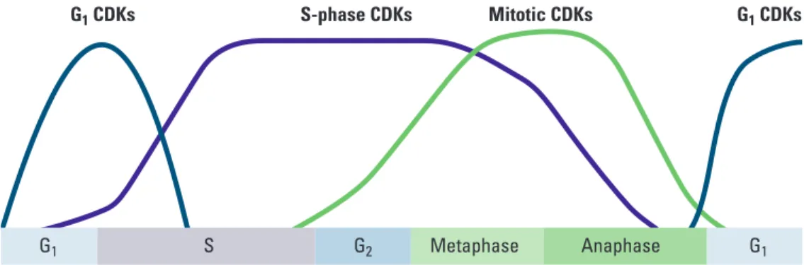
As shown in Figure 3, different CDKs are produced during the phases. The cyclins determine which processes in cell division are turned on or off and in what order by CDK. As each cyclin is turned on or off, CDK causes the cell to move through the stages in the cell cycle.
Figure 3. Levels of CDKs During the Cell Cycle
G1 S G2 Metaphase Anaphase G1
Investigation 7 S85 Cyclins and CDKs do not allow the cell to progress through its cycle automatically.
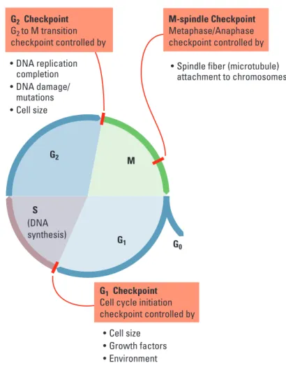
There are three checkpoints a cell must pass through: the G1 checkpoint, G2 checkpoint, and the M-spindle checkpoint (Figure 4). At each of the checkpoints, the cell checks that it has completed all of the tasks needed and is ready to proceed to the next step in its cycle. Cells pass the G1 checkpoint when they are stimulated by appropriate external growth factors; for example, platelet-derived growth factor (PDGF) stimulates cells near a wound to divide so that they can repair the injury. The G2 checkpoint checks
for damage after DNA is replicated, and if there is damage, it prevents the cell from going into mitosis. The M-spindle (metaphase) checkpoint assures that the mitotic spindles or microtubules are properly attached to the kinetochores (anchor sites on the chromosomes). If the spindles are not anchored properly, the cell does not continue on through mitosis. The cell cycle is regulated very precisely. Mutations in cell cycle genes that interfere with proper cell cycle control are found very often in cancer cells.
Bio_T_Lab07_04
QU: What does green circle at upper left represent? Can it be omitted? NOTE: Less depth was needed than specified.
NOTE: We designed this figure to be consistent with Bio_T_Lab07_01.
M G1
G2
G2 Checkpoint G2to M transition
checkpoint controlled by
tDNA replication completion
t%/"EBNBHF
mutations
t$FMMTJ[F
t$FMMTJ[F t(SPXUIGBDUPST t&OWJSPONFOU
tSpindle fiber (microtubule) attachment to chromosomes
M-spindle Checkpoint
.FUBQIBTF"OBQIBTF
checkpoint controlled by
G1 Checkpoint
$FMMDZDMFJOJUJBUJPO
checkpoint controlled by
S
G0 (DNA
synthesis)
■
Learning Objectives
• To describe the events in the cell cycle and how these events are controlled
• To explain how DNA is transmitted to the next generation via mitosis
• To explain how DNA is transmitted to the next generation via meiosis followed by fertilization
• To understand how meiosis and crossing over leads to increased genetic diversity, which is necessary for evolution
■
General Safety Precautions
You must be careful when preparing specimens for viewing under the compound microscope. Always cover the cover slip with a scientific cleaning wipe, such as a Kimwipe, and press down using a pencil eraser.
You should wear safety goggles or glasses and disposable gloves when handling the chemicals and razor blades in Parts 2 and 5. All materials should be disposed of properly as per your teacher’s instructions.
■
THE INVESTIGATIONS
■
Getting Started
These questions are designed to see how well you understand and can explain the key concepts related to cell division before you begin your investigations.
1. How did you develop from a single-celled zygote to an organism with trillions of
cells? How many mitotic cell divisions would it take for one zygote to grow into an organism with 100 trillion cells?
2. How is cell division important to a single-celled organism?
3. What must happen to ensure successful cell division?
4. How does the genetic information in one of your body cells compare to that found
in other body cells?
5. What are some advantages of asexual reproduction in plants?
6. Why is it important for DNA to be replicated prior to cell division?
7. How do chromosomes move inside a cell during cell division?
Investigation 7 S87
■
Procedures
■
Part 1:
Modeling Mitosis
You will investigate mitosis using models. Your teacher will give you sockosomes, clay chromosomes, or pipe-cleaner chromosomes.
Review chromosome duplication and movement using these model chromosomes. Think about these questions as you review the cell cycle and mitosis.
• If a cell contains a set of duplicated chromosomes, does it contain any more genetic information than the cell before the chromosomes were duplicated?
• What is the significance of the fact that chromosomes condense before they are moved?
• How are the chromosome copies, called sister chromatids, separated from each other?
• What would happen if the sister chromatids failed to separate?
■
Part 2:
Effects of Environment on Mitosis
Scientists reported that a fungal pathogen, may negatively affect the growth of soybeans
(Glycine max). Soybean growth decreased during three years of high rainfall, and the soybean roots were poorly developed. Close relatives of R. anaerobis are plant pathogens and grow in the soil. A lectin-like protein was found in the soil around the soybean roots. This protein may have been secreted by the fungus. Lectins induce mitosis in some root apical meristem tissues. In many instances, rapid cell divisions weaken plant tissues.
You have been asked to investigate whether the fungal pathogen lectin affects the number of cells undergoing mitosis in a different plant, using root tips.
• What is your experimental hypothesis? Your null hypothesis? Are these the same?
• How would you design an experiment with onion bulbs to test whether lectins increase the number of cells in mitosis?
• What would you measure, and how would you measure it?
• What would be an appropriate control for your experiment?
Your teacher will provide you with untreated and lectin-exposed roots. You should be comfortable identifying cells in mitosis or in interphase before you begin examining the chromosome squashes.
Preparing Chromosome Squashes
1. Place the onion root tip in 12 M HCl for 4 minutes.
2. Transfer the tip to Carnoy’s fixative for 4 minutes.
3. Remove the slide from Coplin jar containing 70% ethanol, dry with a scientific
cleaning wipe, and label it.
4. Place the tip on the slide and cut off the distal 2 mm portion of the tip; discard the
remainder of the tip.
5. Cover the root tip piece with carbol-fuschin stain for 2 minutes.
6. Blot off excess stain and cover the tip with 1–2 drops of H2O.
7. Place the cover slip over the tip and cover the cover slip with a scientific
cleaning wipe.
8. Firmly press down on the cover slip with the eraser end of a pencil. Do not twist the
slide, and be careful not to break the cover slip.
Counting Cells and Analyzing Data
1. Observe the cells at high magnification (400–500 X).
2. Look for well-stained, distinct cells.
3. Within the field of view, count the cells in each phase. Repeat the counts in two other
root tips.
4. Collect the class data for each group, and calculate the mean and standard deviation
for each group. You must make a table in your notebook for the class data.
5. Compare the number of cells from each group in interphase and in mitosis.
Table 1. Onion Root Tip Cell Phase Data; Treatment Group___________
Tip
Number of Cells
Interphase Mitotic Total
1 2 3 Total
Investigation 7 S89
1. For this experiment, the number of treated cells in interphase and mitosis will be the
observed (o) values.
2. To find out what your expected values are, complete the following steps:
a. Calculate the percentage of cells in interphase and mitosis in the control group from Table 1.
b. Multiply the percentages by the total number of cells in the treated group; this will give the expected numbers (e).
3. Calculate the chi-square (χ2) value for the test.
4. Compare this value to the critical value in Table 2.
Table 2. Critical Values of the Chi-Square Distribution
Degrees of Freedom (DF)
Probability 1 2 3 4 5
0.05 3.84 5.99 7.82 9.49 11.1
0.01 6.64 9.21 11.3 13.2 15.1
0.001 10.8 13.8 16.3 18.5 20.5
1. The degrees of freedom (df) equals the number of groups minus one. In this case,
there are two groups, interphase and mitosis; therefore, df = 2-1, or 1.
2. The p value is 0.05, and the critical value is 3.84. If the calculated chi-square value
is greater than or equal to this critical value, then the null hypothesis is rejected. If the calculated chi-square value is less than this critical value, the null hypothesis is not rejected. In terms of this part of the investigation, what does it mean if your null hypothesis is rejected?
■
Postlab Review
• What was the importance of collecting the class data?
• Was there a significant difference between the groups?
• Did the fungal pathogen lectin increase the number of root tip cells in mitosis?
• What other experiments should you perform to verify your findings?
• Does an increased number of cells in mitosis mean that these cells are dividing faster than the cells in the roots with a lower number of cells in mitosis?
• What other way could you determine how fast the rate of mitosis is occurring in root tips?
■
DESIGNING AND CONDUCTING YOUR INVESTIGATION
Now that you have worked with the root tip model system, design and conduct an investigation to determine what biotic or abiotic factors or substances in the environment might increase or decrease the rate of mitosis in roots. For instance, what factors in the soil might affect the rate of root growth and development? Consider, for example, abiotic soil factors such as salinity and pH or biotic factors, including roundworms, that might alter root growth.■
Part 3:
Loss of Cell Cycle Control in Cancer
Many of us have family members who have or have had cancer. Cancer can occur when cells lose control of their cell cycle and divide abnormally. This happens when tumor-suppressor genes, such as p53 or Rb (retinoblastoma), are mutated. There are many questions you should consider before beginning your investigation.
■
Review from Part 1
• How is the cell cycle controlled in normal cells?
• What are cyclins and cyclin-dependent kinases? What do these proteins do in a cell?
■
Prelab Questions for Part 3
• How are normal cells and cancer cells different from each other?
• What are the main causes of cancer?
• What goes wrong during the cell cycle in cancer cells?
• What makes some genes responsible for an increased risk of certain cancers?
• Do you think that the chromosomes might be different between normal and cancer cells?
The last question is the focus of this part of the lab. With your group, form a hypothesis as to how the chromosomes of a cancer cell might appear in comparison to a normal cell and how those differences are related to the behavior of the cancer cell.
For each of the following cases, look at pictures of the chromosomes (karyotype) from normal human cells. Compare them to pictures of the chromosomes from cancer cells. For each case, count the number of chromosomes in each type of cell, and discuss their appearance. Then answer the following questions.
• Do your observations support your hypothesis?
• If not, what type of information might you need to know in order to understand your observations?
Investigation 7 S91
■
Case 1: HeLa cells
HeLa cells are cervical cancer cells isolated from a woman named Henrietta Lacks. Her cells have been cultured since 1951 and used in numerous scientific experiments. Henrietta Lacks died from her cancer not long after her cells were isolated. Lacks’s cancer cells contain remnants of human papillomavirus (HPV), which we now know increases the risk of cervical cancer.
• From your observations, what went wrong in Henrietta Lacks’s cervical cells that made them cancerous?
• How does infection with human papillomavirus virus (HPV) increase the risk of cervical cancer?
Your teacher may ask you to read The Immortal Life of Henrietta Lacks by Rebecca Skloot. As you read it, think about the following questions:
• Should tissue be removed from a patient without his or her consent for research?
• How was the HeLa cell line cultured?
• What virus infected Henrietta Lacks and may have caused her cervical cancer? What cellular process is affected by this virus?
• Was there bias in the way Henrietta Lacks was treated at Johns Hopkins?
• Put the use of HeLa cells on trial. Debate what is more important: an individual’s rights to his/her own body tissues or the medical knowledge gained by studying a patient’s tissues?
• Should Henrietta Lacks’s family be compensated for the discoveries made using her cells?
• Do companies or universities have the right to patent discoveries made using a patient’s tissues or genes without consulting the patient?
• What other legal and ethical questions are raised in this book?
■
Case 2: Philadelphia Chromosomes
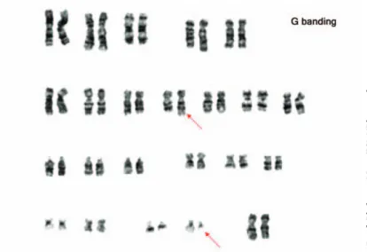
In normal cells, mitosis usually is blocked if there is DNA damage. Sometimes, though, DNA damage makes cells divide more often. Certain forms of leukemia have a unique feature called a Philadelphia chromosome. Look at the karyotype of leukemia cells in Figure 5, and answer the following questions:
• What happens in a normal cell if the DNA has mutations?
• What would happen if cells with mutated DNA replicated?
• How do cells monitor DNA integrity?
• How are the chromosomes different in the cancer cells compared to normal cells?
Use the model chromosomes from Part 1 to explain meiosis and crossing-over events. During your investigation, answer the following questions:
• When is the DNA replicated during meiosis?
• Are homologous pairs of chromosomes exact copies of each other?
• What is crossing over?
• What physical constraints control crossover frequencies?
• What is meant by independent assortment?
• How can you calculate the possible number of different kinds of gametes?
• What happens if a homologous pair of chromosomes fails to separate, and how might this contribute to genetic disorders such as Down syndrome and cri du chat syndrome?
• How are mitosis and meiosis fundamentally different?
Figure 6. Meiotic Cell Division Emphasizing Chromosome Movement
■
Part 5:
Meiosis and Crossing Over in Sordaria
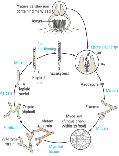
The fungus Sordaria fimicola exchanges genetic material when two mycelia meet and fuse. The resulting zygote undergoes meiosis to produce asci; each ascus contains eight haploid spores. A single gene determines the spore color.
©P
ro
f. P
hi
lip
pe V
ag
o/IS
M/P
ho
to
ta
ke
Figure 5. Karyotype of a Patient with Chronic Myelogenous Leukemia Indicating Chromosomal Deformity
■
Part 4:
Modeling Meiosis
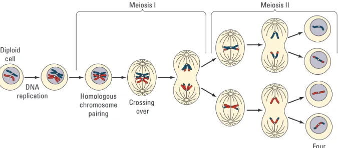
Meiosis resembles mitosis but serves a very different purpose. Meiosis is a cell division resulting in the halving, or reduction, of chromosome number in each cell. A diploid organism has two sets of chromosomes (2n), while a haploid cell or organism has one set (1n). Meiosis produces gametes (ova and sperm) in animals and spores in fungi, plants, and protists. Three other important characteristics of meiosis are the exchange of genetic material (“crossing over”) between homologous chromosomes, the independent assortment of the chromosomes, and the separation of alleles of the same gene (Figure 6). These characteristics, along with random fertilization, increase the genetic variability in the offspring. These mechanisms are essential to our understanding of genetics and evolution in sexually reproducing organisms.
The hallmark of sexual reproduction is the great diversity seen in the gametes and in the resulting offspring produced by fertilization. Meiosis is integral to this process because this type of cell division produces the sex cells, gametes. Before you begin the modeling exercise, your teacher will ask you to discuss these questions.
• How do sexually reproducing organisms produce gametes from diploid progenitors?
• How does the process increase gamete diversity?
• What are the outcomes from independent assortment and crossing over?
• How does the distance between two genes or a gene and a centromere affect crossover frequencies?
Investigation 7 S93 Use the model chromosomes from Part 1 to explain meiosis and crossing-over events.
During your investigation, answer the following questions:
• When is the DNA replicated during meiosis?
• Are homologous pairs of chromosomes exact copies of each other?
• What is crossing over?
• What physical constraints control crossover frequencies?
• What is meant by independent assortment?
• How can you calculate the possible number of different kinds of gametes?
• What happens if a homologous pair of chromosomes fails to separate, and how might this contribute to genetic disorders such as Down syndrome and cri du chat syndrome?
• How are mitosis and meiosis fundamentally different?
Bio_S_Lab07_06 Diploid
cell
Four haploid cells Meiosis I
DNA
replication Homologous
chromosome pairing
Crossing over
Meiosis II
Figure 6. Meiotic Cell Division Emphasizing Chromosome Movement
■
Part 5:
Meiosis and Crossing Over in Sordaria
The fungus Sordaria fimicola exchanges genetic material when two mycelia meet and fuse. The resulting zygote undergoes meiosis to produce asci; each ascus contains eight haploid spores. A single gene determines the spore color.
Mature perithecium containing many asci
8 Ascospores
Mycelium (fungus grows within its food) 8
Haploid nuclei
Cell partitioning Mitosis
Mitosis Mitosis Spore discharge
Filament Ascospore Meiosis
Fertilization
Mycelial fusion 4
Haploid nuclei
Zygote (diploid)
Wild-type strain
Mutant strain Ascus
Figure 7. Sordaria Life Cycle
(tn)
(tn) (+) (+)
Investigation 7 S95 A cross was made between wild type (+; black) and tan (tn) strains. The resulting zygote
produces either parental type asci, which have four black and four tan spores in a row (4:4 pattern), or recombinant asci, which do not have this pattern.
• How do you explain the differences between the recombinant asci and the parental types?
• What meiotic event can account for this difference?
• Using the model chromosomes from Part 4, predict the possible meiotic outcomes.
1. Place a drop of water onto the microscope slide.
2. Gently scrape some perithecia from the agar plate near where the two strains meet.
3. Place a cover slip over the perithecia and put a scientific cleaning wipe over the cover
slip.
4. Gently press down on the cover slip using the eraser end of a pencil.
5. Count at least 50 asci, and score them as either parental or recombinant (crossing
over).
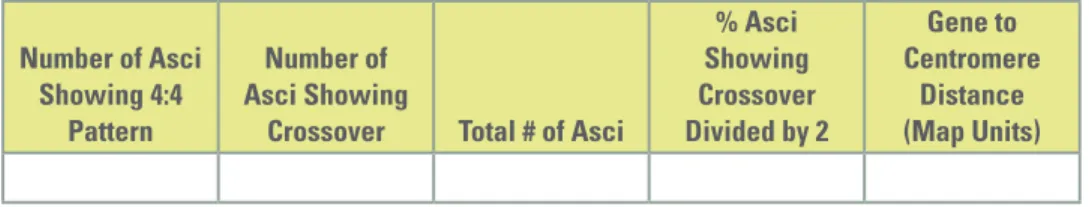
6. Enter the data in Table 3 and make the calculations. One map unit equals one
recombinant per 100 total events. The percentage of asci showing crossover divided by 2 equals the map units in this activity. This is done because each spore produced by meiosis undergoes a mitotic division.
Table 3. Analysis of Results Number of Asci
Showing 4:4 Pattern
Number of Asci Showing
Crossover Total # of Asci
% Asci Showing Crossover Divided by 2
Gene to Centromere
Distance (Map Units)
■
Evaluating Results
1. Why did you divide the percentage of asci showing crossover (recombinant) by 2?
2. The published map distance between the spore color gene and the centromere is 26
map units. How did the class data compare with this distance?
3. How can you account for any disparities between the class data and the published
data?
4. Illustrate what happened during meiosis to produce the results you found.
5. Do you think the Philadelphia chromosome is a result of crossing over as seen in
this part of the investigation or some other chromosomal abnormality? Explain your answer.
6. Do you think the cell cycle described for mitosis could be applied to meiosis as well?
Explain your answer.
■
Where Can You Go from Here?
1. Can the same (or any) environmental factors you tested above affect the amount of
crossing over that occurs in Sordaria? How would you set up an experiment to test this? For example, how does humidity or pH affect the crossover frequency?
2. Revisit the learning objectives stated earlier. Do you better understand mitosis and
meiosis? Could you teach this to another class?
3. How do the mechanisms of cell replication affect genetic diversity and evolution?
Consider the mechanisms such as crossing over, independent assortment, segregation, nondisjunction, and random fertilization.
4. Prepare a video or write and produce a play about the process of chromosome
movement.
5. Investigate how growth factors affect the cell cycle. This will help you review cell
communication.
6. Research what tumor suppressors do in the cell cycle and which types of cancers
may be caused by mutations in tumor suppressor genes. Specific examples include human papillomavirus (HPV), retinoblastoma protein (Rb), BRCA1 and BRCA2, and p53.