Original Article
Effect of Different Irrigation Solutions on the Colour Stability of
Three Calcium Silicate-Based Materials
Sobhnamayan Fa, Adl Ab,Ghanbaran Sa*
aDepartment of Endodontics, School of Dentistry, Shiraz University of Medical Sciences, Shiraz, Iran
bDepartment of Endodontics and Biomaterials Research Center, School of Dentistry, Shiraz University of Medical Sciences, Shiraz, Iran
ARTICLE INFO Abstract
Article History:
Received: 20 April 2017 Accepted: 30 May 2017
Statement of Problem: Previous studies have shown discoloration of mineral trioxide aggregate (MTA) in contact with root canal irrigation solutions. However, there are limited data on colour stability of other calcium silicate– based materials (CSMs).
Objectives: This in vitro study aimed to evaluate the colour stability of three CSMs in contact with different irrigation solutions.
Materials and Methods: Three CSMs including White MTA (wMTA) Angelus, calcium enriched mixture (CEM), and Biodentine were assessed in this study.
Forty five samples of each material were mixed according to the manufactures’
instructions and then placed in silicone tubes. After 24 hours, the materials were removed from the moulds and 9 samples of each material left dry or immersed in normal saline, 5% sodium hypochlorite (NaOCL), 2% chlorhexidinegluco-nate (CHX), or 17%EDTA for 24 hours. Colour changes were measured with a spectrophotometer. Data were evaluated with 2-way analysis of variance, one way analysis of variance and Tukey post hoc tests.
Results: The highest discoloration of all materials was observed after contact
with CHX. In the MTA Angelus and CEM cement groups, significant differences
were observed between CHX and NaOCl and also between these two irrigants with the other three irrigants (p < 0.05).
In the Biodentine group, CHX created statistically significant discoloration
compared to other irrigants (p < 0.05). Only wMTA Angelus showed a
significantly higher discoloration in contact with EDTA compared to normal
saline and dry condition (p < 0.05).
wMTA Angelus showed a significantly higher colour change compared with
CEM cement and Biodentine after contact with NaOCl, CHX, and EDTA (p < 0.05).
Conclusions: The contact of wMTA, CEM cement, and Biodentine with CHX should be avoided because this leads to severe discoloration. Contact with so-dium hypochlorite also leads to discoloration of wMTA and CEM cements. Among of the three tested materials, wMTA showed the highest discoloration after contact with NaOCl, CHX, and EDTA.
Key words:
Biodentine
Calcium Enriched Mixture Discoloration
Mineral Trioxide Aggregate Spectrophotometric Analysis
Corresponding Author:
Sahar Ghanbaran
Department of Endodontics, School of Dentistry, Shiraz University of Medical Sciences, Shiraz, Iran Email: [email protected] Tel: +98-917-3072535 Fax: +98-71-36285276
Cite this article as: Sobhnamayan F, Adl A, Ghanbaran S. Effect of Different Irrigation Solutions on the Colour Stability of Three Calcium Silicate-Based Materials. J Dent Biomater, 2017;4(2):373-378.
Introduction
Nowadays, aesthetics is an important concern to both patient and dentist. Tooth discoloration after end-odontic treatment is a common problem that damages the quality of treatment, resulting in dissatisfaction in 31.6%-57% of the patients [1,2].
Many materials used in endodontics may cause tooth discoloration [1]. Therefore, colour stability of endodontic materials is considered as an important factor for clinical success [3]. A progressive discol-oration is proposed to be the result of penetration of materials into dentinal tubules [4].
Endodontic procedures such as direct pulp capping, Cvek pulpotomy, perforation repair, and regenerative endodontics lead to the placement of some biomaterials in the coronal part of the tooth, which may cause discoloration [5,6].
Mineral trioxide aggregate (MTA) was first introduced in a grey form (gMTA). After reports of tooth discoloration caused by gray MTA [5,7], wMTA (wMTA) with a reduction ofAl2O3, MgO, and FeO was developed with an off-white colourto overcome this shortcoming [8]. However, several studies reported tooth discoloration induced even bywMTA[1,3,8-12].
In recent years, other new calcium silicate-based materials (CSMs) such as Biodentine (Septodent, Saint Maura des Fosses, France) and Calcium-Enriched Mixture (CEM)(BioniqueDent, Tehran, Iran)have been introduced to overcome the shortcoming of MTA [13-16].
CEM cement is a biomaterial [17] with different chemical composition but similar clinical applica-tions to those of MTA [18-22]. According to the result of a recent study, CEM cement did not induce tooth discoloration after six months. Therefore, it can be used in the vital pulp therapy of aesthetically sensitive teeth [23]. Biodentine is a dentin restorative material which has indications similar to MTA in addition to the advantage of a shorter setting time(13). High colour stability of Biodentine has been reported in previous studies [11,24,25].
A recent study showed that endodontic irrigation solutions; sodium hypochlorite (NaOCl) and chlorohexidine (CHX) caused clinically perceptible colour changes in Pro Root wMTA(DENTSPLY, Tulsa Dental), wMTA Angelus, Bioaggregate, and Biodentine. However, Pro Root MTAshowed morecolour change which was attributed to the higher amounts of bismuth oxide in its composition [24].
To the best of our knowledge, there is no published study on the colour stability of CEM cement in contact with different irrigation solutions. In addi-tion, the effect of ethylene diamine tetra acetic acid (EDTA) on the colour stability of CSMs has not been yet investigated. Therefore, the aim of this in vitro study was to evaluate colour stability of three CSMs (MTA Angelus (Angelus Solucoes Odontologicas, Londrina, Brazil), Biodentine (Septodent, Saint Maura des Fosses, France), and CEM (BioniqueDent, Tehran, Iran) in contact with different irrigation solutions (EDTA, NaOCl, CHX, Normal Saline). Materials and Methods
Sample Preparation
Forty five specimens of each material to be tested (MTA, Biodentine, CEM cement were mixed according to the manufactures’ instructions and then placed in 15 mm diameter and 2mm high silicone tubes. The specimens were stored at 37ºC and 100% humidity during the setting to reach their optimal mechanical properties [25]. After 24 hours, the materials were removed from the moulds and 9 samples from each group were left dry ( as control) or immersed in the following 4 irrigation solutions for 24 hours:
Normal saline(Darou pakhsh, Tehran, Iran), 5% NaOCl (Cerkamed, Pawłowski, Poland), 0.2% CHX (Cerkamed, Pawłowski, Poland), EDTA (Cerkamed, Pawłowski, Poland).
Spectrophotometric Analysis
Measurement of colour was performed and repeated 3 times with a spectrophotometer (VITA Easy shade Advance 4.0 Germany) under constant laboratory light by the same operator. The device was calibrated before each measurement. The Commission Internationale de l’eclairage (CIE) system was used to calculate the differences in colour. The colour differences (∆E) of the samples were calculated using the following formula:
ΔE = [(Δa)2 + (Δb)2 + (ΔL)2]1/2
where ΔL* = (L*1 _ L*0), Δa* = (a*1 _ a*0), and Δb* = (b*1 _ b*0).
∆L shows the change in luminosity, ∆a is the change in the red-green parameter, and ∆b shows the change in the yellow-blue parameter. Images of the samples were taken before and after immersion,
using a digital camera (Cybershot DSC-W220; Sony, Tokyo, Japan).
Statistical Analysis
The data were analyzed with two-way ANOVA using SPSS software (PASW Statistics 20; SPSS Inc, Chicago, IL). There was an interaction effect between the material and irrigation soloution. (p ≤ 0.05). Therefore, one-way ANOVA and Tukey post hoc tests were used to perform multiple comparison tests. The level of statistical significance was set at p < 0.05. Results
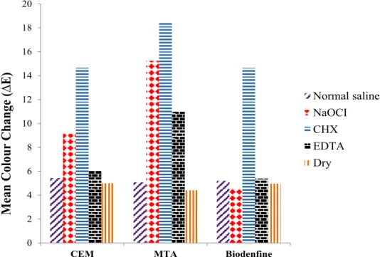
Table 1 shows the mean value and standard deviation of colour changes in different groups. The mean values for changes in the colour of materials are shown in Figure 1. All materials showed the highest discoloration after contact with CHX.
In the MTA Angelus and CEM cement groups, significant differences were observed between CHX and NaOCl and also between these two irrigants with the other three irrigants (p < 0.05).
In the Biodentine group, CHX showed statistically
significant differences with the other irrigants (p < 0.05). Only wMTA Angelus showed a significantly higher discoloration in contact with EDTA compared to normal saline and dry condition (p < 0.05)
There were no significant differences in the ∆E values of three materials in the presence of normal saline and in the dry condition (p = 0.715, p = 0.298, respectively).
White MTA Angelus showed a significantly higher ∆E value compared with CEM cement and Biodentine after contact with NaOCl, CHX, and EDTA (p < 0.05).
Discussion
Tooth discoloration has been reported with the use of a number of endodontic materials; therefore, material selection should not rely solely on biological and functional criteria but also aesthetic considerations should be taken into account [26]. Colour changes in different types of dental materials has been measured using spectrophotometer [27-29]. In the current study, a spectrophotometer was used to evaluate the colour change of three dental cements after contact
0 2 4 6 8 10 12 14 16 18 20
CEM MTA Biodenfine
M
ea
n Colo
ur
Chang
e (∆
E)
Normal saline NaOCI CHX EDTA Dry
Figure 1: Mean values of colour change ((∆E) of all materials
Table 1: Mean value and standard deviation (± SD) of colour changes (∆E) in different groups
Normal saline Naocl CHX EDTA Dry
CEM 5.4 ± 0.78Ad 9.1 ± 0.40Bb 14.6 ± 0.88Ba 6.0 ± 0.52Bc 4.9 ± 1.10Ad MTA 5.1 ± 1.02Ad 15.2 ± 1.11Ab 18.4 ± 1.17Aa 10.9 ± 0.61Ac 4.4 ± 0.48Ad Biodentin 5.2 ± 0.93Ab 4.5 ± 0.98Cb 14.6 ± 1.31Ba 5.3 ± 0.64Ab 4.9 ± 0.87Ab
with different irrigation solution. Spectrophotometric analysis was applied because of its sensitivity to small changes in colour, repeatability, and objectivity [30].
This study showed that MTA Angelus, CEM cement, and Biodentinerevealed the most severe discoloration in contact with CHX. MTA Angelus and CEM cement also showed severe discoloration in contact with NaOCl, while Biodentine was not affected by NaOCl. This finding is in the same line with those of Keskinet a. who reported that Bioden-tine exhibited more discoloration when immersed in CHX compared with NaOCl [24].
Chlorhexidine has been shown to cause extrinsic discoloration of silicate filling materials [31] and dental tissues at varying concentrations by affecting dental pellicle or plaque [32]. However, the exact mechanism of discoloration of dental cements in the presence of CHX is not well defined [24]. The current study has also shown that wMTA and CEM cement exhibited higher discoloration in contact with NaOCl when compared with dry condition, normal saline, and EDTA.
Previously, it had been shown that immersion of wMTA in sodium hypochlorite resulted in the formation of a dark brown discoloration. It has been speculated that the discoloration was attributed to the reaction of sodium hypochlorite with bismuth oxide which is a part of MTA [26].
In contrast to MTA, CEM cement has no bismuth oxide in its content [11]; therefore, the reason of its discoloration in contact with NaOCl is not yet understood.
The current study also evaluated the effect of EDTA on the colour stability of wMTA, CEM cement, and Biodentine. EDTA is normally used for removal of the mineralized portion of smear layer. Recently, EDTA was introduced to regenerative endodontic procedures as the only irrigant in the second visit based on its ability in releasing growth factors from the dentin and also inducing cell attachment and differentiation [33,34].
According to the results, CEM cement and Biodentine exhibited colour stability in contact with EDTA while wMTA Angelus showed higher discoloration compared to normal saline and dry condition. It should be noted that 2 samples of Biodentine were eroded in contact with EDTA and deleted from the study. Therefore, the effect of EDTA on the microstructure of Biodentine should be investigated.
In the dry condition and in the presence of normal
saline, no differences were found between three cements; however, in contact with NaOCl and CHX, wMTA Angelus showed significantly more discol -oration than the other materials. This is somehow in contrast to a previous study reporting no statistically significant difference between discoloration of wMTA Angelus and Biodentine in contact with NaOCl and CHX[24] .
It is well defined that contact of bismuth oxide– containing products with NaOCl and CHX leads to material discoloration [24].Therefore, the higher discoloration of wMTA Angelus which was found in this study may be attributed to the fact that neither CEM cement [11] nor Biodentine [35] contains bismuth oxide as radiopacifire [35].
Conclusions
Under the limitation of this study, contact of wMTA, CEM cement, and Biodentine with CHX should be avoided because this leads to severe discoloration. Contact with sodium hypochlorite also leads to dis-coloration of wMTA and CEM cements. White MTA showed significantly higher discoloration compared with other materials after contact with NaOCl, CHX, and EDTA.
Conflict of Interest: None declared. References
1. Lenherr P, Allgayer N, Weiger R, et al. Tooth discoloration induced by endodontic materials: a laboratory study. Int Endod J. 2012;45:942-949. 2. Thomson AD, Athanassiadis B, Kahler B, et
al. Tooth discolouration: staining effects of various sealers and medicaments. Aust Endod J. 2012;38:2-9.
3. Eghbal MJ, Torabzadeh H, Bagheban AA, et al. Color stability of mineral trioxide aggregate and calcium enriched mixture cement. J Invest Clin Dent. 2016;7:341-346.
4. Van der Burgt T, Mullaney T, Plasschaert A. Tooth discoloration induced by endodontic sealers. Oral Surg Oral Med Oral Pathol. 1986;61:84-89. 5. Karabucak B, Li D, Lim J, et al. Vital pulp
therapy with mineral trioxide aggregate. Dent Traumatol. 2005;21:240-243.
6. Belobrov I, Parashos P. Treatment of tooth discoloration after the use of white mineral trioxide aggregate. J Endod. 2011;37:1017-1020.
7. Bortoluzzi EA, Araújo GS, Guerreiro Tanomaru JM, et al. Marginal gingiva discoloration by gray MTA: a case report. J Endod. 2007;33:325-327. 8. Akbari M, Rouhani A, Samiee S, et al. Effect of
dentin bonding agent on the prevention of tooth discoloration produced by mineral trioxide ag-gregate. Int J Dent. 2011;2012.
9. Moore A, Howley MF, O’Connell AC. Treatment of open apex teeth using two types of white mineral trioxide aggregate after initial dressing with calcium hydroxide in children. Dent Traumatol. 2011;27:166-173.
10. Felman D, Parashos P. Coronal tooth discol-oration and white mineral trioxide aggregate. J Endod. 2013;39:484-487.
11. Kohli MR, Yamaguchi M, Setzer FC, et al. Spectrophotometric analysis of coronal tooth discoloration induced by various bioceramic cements and other endodontic materials. J Endod. 2015;41:1862-1866.
12. Ioannidis K, Mistakidis I, Beltes P, et al. Spectro-photometric analysis of coronal discolouration induced by grey and white MTA. Int Endod J. 2013;46:137-144.
13. Laurent P, Camps J, De Méo M, et al. Induction of specific cell responses to a Ca 3 SiO 5-based posterior restorative material. Dent Mater. 2008;24:1486-494.
14. Koubi G, Colon P, Franquin JC, et al. Clinical evaluation of the performance and safety of a new dentine substitute, Biodentine, in the restoration of posterior teeth-a prospective study. Clinl Oral Investig. 2013;17:243-249.
15. Pradelle-Plasse N, Tran X, Colon P, et al. Biocompatibility or cytotoxic effects of dental composites. 2009.
16. Shokouhinejad N, Nekoofar MH, Pirmoazen S, et al. Evaluation and Comparison of Occurrence of Tooth Discoloration after the Application of Various Calcium Silicate–based Cements: An Ex Vivo Study. J Endod. 2016;42:140-144.
17. Asgary S, Shahabi S, Jafarzadeh T, et al. The properties of a new endodontic material. J Endod. 2008;34:990-993.
18. Asgary S, Eghbal MJ, Parirokh M. Sealing ability of a novel endodontic cement as a root-end filling material. J Biomed Mater Res A. 2008;87:706-709.
19. Malekafzali B, Shekarchi F, Asgary S. Treat-ment outcomes of pulpotomy in primary molars using two endodontic biomaterials. A 2-year
randomised clinical trial. Eur J Paediatr Dent. 2011;12:189-193.
20. Asgary S, Eghbal MJ, Ghoddusi J. Two-year results of vital pulp therapy in permanent molars with irreversible pulpitis: an ongoing multicenter randomized clinical trial. Clin Oral Investig. 2014;18:635-641.
21. Haghgoo R, Arfa S, Asgary S. Microleakage of CEM cement and ProRoot MTA as furcal perforation repair materials in primary teeth. Iran Endod J. 2013;8:187-190.
22. Guler AU, Kurt S, Kulunk T. Effects of various finishing procedures on the staining of provi -sional restorative materials. J Prosthet Dent. 2005;93:453-458.
23. Rouhani A, Akbari M, Farhadi-faz A. Comparison of tooth discoloration induced by calcium-enriched mixture and mineral trioxide aggregate. Iran Endod J. 2016;11:175-178. 24. Keskin C, Demiryurek EO, Ozyurek T. Color
Stabilities of Calcium Silicate–based Materials in Contact with Different Irrigation Solutions. J Endod. 2015;41:409-411.
25. Vallés M, Mercadé M, Duran-Sindreu F, et al. Influence of light and oxygen on the color stability of five calcium silicate–based materials. J Endod. 2013;39:525-528.
26. Camilleri J. Color stability of white mineral trioxide aggregate in contact with hypochlorite solution. J Endod. 2014;40:436-440.
27. Deljoo Z, Sadeghi M, Azar MR, et al. The Effect of Different Polishing Methods and Storage Media on Discoloration of Resin Composites. J Dent Biomater. 2016;3;226-232.
28. Khaledi AAA, Safari A, Adibi A, et al. The Effect of Chlorhexidine Mouth Rinse on the Colour Stability of Porcelain with Three Different Surface Treatments: An in Vitro Study. J Dent Biomater. 2014;1:3-8.
29. Mousavi S, Narimani S, Hekmatfar S, et al. Colour Stability of Various Types of Acrylic Teeth Exposed to Coffee, Tea and Cola. J Dent Biomater. 2016;3:335-340.
30. Khokhar Z, Razzoog M, Yaman P. Color stability of restorative resins. Quintessence Int. 1991;22;733-777.
31. Heyden G. Relation between locally high concentration of chlorhexidine and staining as seen in the clinic. J Periodontal Res Suppl. 1973;12:76-80.
Evaluation of chlorhexidine gluconate mouth-rinse-induced staining using a digital colorimeter: An in vivo study. Quintessence Int. 2011;42:213-223.
33. Martin DE, De Almeida JF, Henry MA, et al. Concentration-dependent effect of so-dium hypochlorite on stem cells of apical papilla survival and differentiation. J Endod. 2014;40:51-55.
34. Zhao S, Sloan A, Murray P, et al. Ultrastructural localisation of TGF-β exposure in dentine by chemical treatment. Histochem J. 2000;32:489-494.
35. Camilleri J, Kralj P, Veber M, et al. Characteriza-tion and analyses of acid-extractable and leached trace elements in dental cements. Int Endod J. 2012;45:737-743.