Key Concepts
Plasma membranes are made up of selectively permeable bilayers of phospholipids. Phospholipids are amphipathic lipid molecules – they have hydrophobic and hydrophilic regions.
Ions and molecules diffuse spontaneously from regions of higher concentration to regions of lower concentration. Movement of water across a plasma membrane is called osmosis.
In cells, membrane proteins are responsible for the passage of
© 2011 Pearson Education, Inc.
The Importance of Membranes
• The plasma membrane, or cell membrane, separates life from nonlife.
• The plasma membrane separates the cell’s interior from the
external environment. Membranes function to:
– Keep damaging materials out of the cell
– Allow entry of materials needed by the cell
Lipids: What Is a Lipid?
• Lipids are carbon-containing compounds that are found in organisms and that are largely nonpolar and hydrophobic.
• Hydrocarbons are nonpolar molecules that contain only carbon
and hydrogen.
• Lipids do not dissolve in water because they have a major hydrocarbon component called a fatty acid.
– A fatty acid is a hydrocarbon chain bonded to a carboxyl
(—COOH) functional group.
Three Types of Lipids Found in Cells
• Lipid structure varies widely.
• The three most important types of lipids found in cells:
1. Fats are composed of three fatty acids linked to glycerol.
– Also called triacylglycerols or triglycerides
2. Steroids are a family of lipids with a distinctive four-ring structure.
– Cholesterol is an important steroid in mammals.
The Structure of Membrane Lipids
Membrane-forming lipids contain both a polar, hydrophilic region and a nonpolar, hydrophobic region.
• Phospholipids are amphipathic:
– The “head” region, consisting of a glycerol, a phosphate, and a charged group, contains highly polar covalent bonds.
– The “tail” region is comprised of two nonpolar fatty acid or isoprene chains.
• When placed in solution, the phospholipid heads interact with
Phospholipids and Water
• Phospholipids do not dissolve when they are placed in water.
• Water molecules interact with the hydrophilic heads but not with the hydrophobic tails.
– This drives the hydrophobic tails together.
• Upon contact with water phospholipids form either:
– Micelles
© 2011 Pearson Education, Inc.
Phospholipid Bilayers
• Phospholipid bilayers form when two sheets of phospholipid molecules align. The hydrophilic heads in each layer face a
surrounding solution, while the hydrophobic tails face one another inside the bilayer.
© 2011 Pearson Education, Inc.
Selective Permeability of Lipid Bilayers
• The permeability of a structure is its tendency to allow a given substance to pass across it.
Phospholipid bilayers have selective permeability.
– Small or nonpolar molecules move across phospholipid bilayers quickly.
© 2011 Pearson Education, Inc.
Many Factors Affect Membrane Permeability
• Many factors influence the behavior of the membrane:
– Number of double bonds between the carbons in the phospholipid’s hydrophobic tail
– Length of the tail
– Number of cholesterol molecules in the membrane
Bond Saturation and Membrane Permeability
• Double bonds between carbons in a hydrocarbon chain can cause a “kink” in the hydrocarbon chain, preventing the close packing of hydrocarbon tails, and reducing hydrophobic interactions.
– Unsaturated hydrocarbon chains have at least one double
bond.
– Hydrocarbon chains without double bonds are termed saturated.
• Saturated fats have more chemical energy than unsaturated fats.
Other Factors That Affect Permeability
• Hydrophobic interactions become stronger as saturated hydrocarbon tails increase in length.
– Membranes containing phospholipids with longer tails have
reduced permeability.
• Adding cholesterol to membranes increases the density of the hydrophobic section.
– Cholesterol decreases membrane permeability.
• Membrane fluidity decreases with temperature because molecules
in the bilayer move more slowly.
Fluidity of the Membrane
• Individual phospholipids can move laterally throughout the lipid bilayer.
– They rarely flip between layers.
Solute Movement across Lipid Bilayers
• Materials can move across the cell membrane in different ways.
– Passive transport does not require an input of energy.
– Active transport requires energy to move substances across the membrane.
• Small molecules and ions in solution are called solutes, have thermal energy, and are in constant, random motion.
– This random movement is called diffusion.
© 2011 Pearson Education, Inc.
Diffusion along a Concentration Gradient
• A difference in solute concentrations creates a concentration gradient.
Molecules and ions move randomly when a concentration gradient exists, but there is a net movement from high- concentration
regions to low-concentration regions. Diffusion along a
concentration gradient increases entropy and is thus spontaneous.
• Equilibrium is established once the molecules or ions are
randomly distributed throughout a solution.
– Molecules are still moving randomly but there is no more net
© 2011 Pearson Education, Inc.
Osmosis
• Water moves quickly across lipid bilayers.
– The movement of water is a special case of diffusion called osmosis.
• Water moves from regions of low solute concentration to regions of high solute concentration.
– This movement dilutes the higher concentration, thus equalizing the concentration on both sides of the bilayer.
© 2011 Pearson Education, Inc.
Osmosis and Relative Solute Concentration
• The concentration of a solution outside a cell may differ from the concentration inside the cell.
– An outside solution with a higher concentration is said to be
hypertonic to the inside of a cell.
– A solution with a lower concentration is hypotonic to the cell.
– If solute concentrations are equal on the outside and inside of a
Osmosis in Hypertonic, Hypotonic, and Isotonic Solutions
• In a hypertonic solution, water will move out of the cell by osmosis and the cell will shrink.
• In a hypotonic solution, water will move into the cell by osmosis
and the cell will swell.
Short-Distance Transport of Water Across Plasma
Membranes
•
To survive, plants must balance water uptake and
loss
© 2011 Pearson Education, Inc.
•
Water potential
is a measurement that combines
the effects of solute concentration and pressure
•
Water potential determines the direction of
movement of water
•
Water flows from regions of higher water potential
to regions of lower water potential
•
Potential refers to water’s capacity to perform work
The Fluid-Mosaic Model of Membrane Structure
• Although phospholipids provide the basic membrane structure, plasma membranes contain as much protein as phospholipids.
• The fluid-mosaic model of membrane structure suggests that some
© 2011 Pearson Education, Inc.
•
Water potential is abbreviated as Ψ and measured
in a unit of pressure called the
megapascal (MPa)
•
Ψ = 0 MPa for pure water at sea level and at room
temperature
How Solutes and Pressure Affect Water Potential
•
Both pressure and solute concentration affect water
potential
•
This is expressed by the water potential equation:
Ψ
Ψ
S
Ψ
P•
The
solute potential (
Ψ
S)
of a solution is directly
proportional to its molarity
© 2011 Pearson Education, Inc.
•
Pressure potential (
Ψ
P)
is the physical pressure on
a solution
•
Turgor pressure
is the pressure exerted by the
plasma membrane against the cell wall, and the cell
wall against the protoplast
•
The
protoplast
is the living part of the cell, which
also includes the plasma membrane
•
Consider
a U-shaped tube where the two arms are
separated by a membrane permeable only to water
•
Water moves in the direction from higher water
© 2011 Pearson Education, Inc.
Figure 36.8
Solutes have a negative effect on by binding water molecules.
Pure water at equilibrium
H2O
Adding solutes to the right arm makes lower there, resulting in net movement of water to the right arm:
H2O Pure
water
Membrane Solutes
Positive pressure has a positive effect on by pushing water.
Pure water at equilibrium
H2O
H2O
Positive pressure Applying positive
pressure to the right arm makes higher there, resulting in net movement of water to the left arm:
Solutes and positive pressure have opposing effects on water
movement.
Pure water at equilibrium
H2O
H2O
Positive pressure
Solutes In this example, the effect of adding solutes is
offset by positive
pressure, resulting in no net movement of water:
Negative pressure (tension) has a negative effect on by pulling water.
Pure water at equilibrium
H2O
H2O
Negative pressure Applying negative
Figure 36.8a
Solutes have a negative effect on by binding water
molecules.
Pure water at equilibrium
H O
Adding solutes to the right
arm makes lower there,
resulting in net movement of water to the right arm:
H O
Pure water
© 2011 Pearson Education, Inc.
Figure 36.8b
Positive pressure has a positive effect on by pushing water.
Pure water at equilibrium
H2O H2O
Positive pressure
Applying positive pressure to
the right arm makes higher
Solutes and positive pressure have opposing effects on water movement.
Pure water at equilibrium Positivepressure
Solutes
In this example, the effect of adding solutes is offset by positive pressure, resulting in no net movement of water:
© 2011 Pearson Education, Inc.
Negative pressure (tension) has a negative effect on by
pulling water.
Pure water at equilibrium
H2O H2O
Negative pressure
Applying negative pressure
to the right arm makes
lower there, resulting in net movement of water to the right arm:
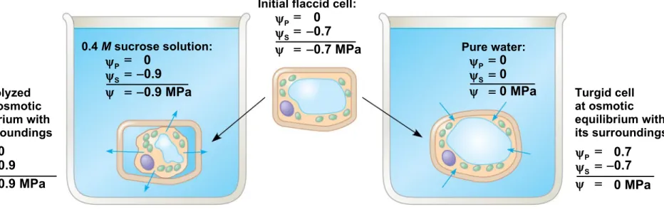
Figure 36.9
Plasmolyzed cell at osmotic equilibrium with its surroundings
0.4 M sucrose solution:
Initial flaccid cell:
Pure water:
Turgid cell at osmotic equilibrium with its surroundings
(a) Initial conditions: cellular environmental (b) Initial conditions: cellular environmental P 0
P 0
S P 0
S
P 0.7
S 0.9
0.9 MPa
S 0.9
0.9 MPa
0.7 MPa
S 0.7
P 0
0 0 MPa
0.7 0 MPa
© 2011 Pearson Education, Inc.
Figure 36.9a
Plasmolyzed cell at osmotic equilibrium with its surroundings
0.4 M sucrose solution:
Initial flaccid cell:
(a) Initial conditions: cellular environmental
0.9 MPa
S 0.9 P 0
P 0
S 0.9
0.9 MPa
P 0
S 0.7
Figure 36.9b Pure water: Turgid cell at osmotic equilibrium with its surroundings
(b) Initial conditions: cellular environmental Initial flaccid cell:
0.7 MPa
P 0
S 0.7
P 0
0 MPa
S 0
P 0.7
S 0.7
© 2011 Pearson Education, Inc.
•
If a flaccid cell is placed in a solution with a lower
solute concentration, the cell will gain water and
become
turgid
•
Turgor loss in plants causes
wilting
, which can be
reversed when the plant is watered
Aquaporins: Facilitating Diffusion of Water
•
Aquaporins
are transport proteins in the cell
membrane that allow the passage of water
•
These affect the rate of water movement across the
© 2011 Pearson Education, Inc.
•
Water potential affects uptake and loss of water by
plant cells
•
If a
flaccid
cell is placed in an environment with a
higher solute concentration, the cell will lose water
and undergo plasmolysis
•
Plasmolysis
occurs when the protoplast shrinks
and pulls away from the cell wall
© 2011 Pearson Education, Inc.
© 2011 Pearson Education, Inc.
Membrane Proteins
• Integral proteins are amphipathic and so can span a membrane, with segments facing both its interior and exterior surfaces.
– Integral proteins that span the membrane are called
transmembrane proteins.
– These proteins are involved in the transport of selected ions and molecules across the plasma membrane.
– Transmembrane proteins can therefore affect membrane permeability.
• Peripheral proteins are found only on one side of the membrane.
Membrane Proteins Affect Ions and Molecules
• The transmembrane proteins that transport molecules are called transport proteins. There are three broad classes of transport proteins, each of which affects membrane permeability:
1. Channels
2. Carrier proteins or transporters
© 2011 Pearson Education, Inc.
Ion Channels and the Electrochemical Gradient
• Ion channels are specialized membrane proteins.
– Ion channels circumvent the plasma membrane’s impermeability to small, charged compounds.
• When ions build up on one side of a plasma membrane, they establish both a concentration gradient and a charge gradient, collectively called the electrochemical gradient.
• Ions diffuse through channels down their electrochemical gradients.
© 2011 Pearson Education, Inc.
Facilitated Diffusion via Channel Proteins
• Cells have many different types of channel proteins in their membranes, each featuring a structure that allows it to admit a particular type of ion or small molecule.
• These channels are responsible for facilitated diffusion: the
© 2011 Pearson Education, Inc.
Facilitated Diffusion via Carrier Proteins
• Facilitated diffusion can occur through channels or through carrier proteins, or transporters, which change shape during the transport process.
• Facilitated diffusion by transporters occurs only down a
concentration gradient, reducing differences between solutions.
• Glucose is a building block for important macromolecules and a major energy source, but lipid bilayers are only moderately
permeable to glucose.
© 2011 Pearson Education, Inc.
Active Transport by Pumps
• Cells can transport molecules or ions against an electrochemical gradient.
– This process requires energy in the form of ATP and is called
active transport.
• Pumps are membrane proteins that provide active transport of molecules across the membrane.
– For example, the sodium-potassium pump, Na+/K+-ATPase,
uses ATP to transport Na+ and K+ against their concentration
© 2011 Pearson Education, Inc.
Secondary Active Transport
• In addition to moving materials against their concentration gradients, pumps set up electrochemical gradients.
• These gradients make it possible for cells to engage in secondary
active transport, or cotransport.
– The gradient provides the potential energy required to power
Summary of Membrane Transport
• There are three mechanisms of membrane transport:
1. Diffusion
2. Facilitated diffusion
3. Active transport
• Diffusion and facilitated diffusion are forms of passive transport and thus move materials down their concentration gradient and do not require an input of energy.
• Active transport moves materials against their concentration
Plasma Membrane and the Intracellular Environment
The selective permeability of the lipid bilayer and the specificity of the proteins involved in passive transport and active transport enable cells to create an internal environment that is much