Spinal cord. Spinal nerves and
reflexes.
Spinal Cord
•
Gross Anatomy of the Spinal Cord
•
About 18 inches (45 cm) long
•
1/2 inch (14 mm) wide
•
Ends between vertebrae L
1and L
2•
Bilateral symmetry
•
Grooves divide the spinal cord into left and right
•
Posterior median sulcus – on posterior side
• Enlargements of the Spinal Cord • Caused by:
• Amount of gray matter in segment
• Involvement with sensory and motor nerves of limbs • Cervical enlargement
• Nerves of shoulders and upper limbs • Lumbar enlargement
• Nerves of pelvis and lower limbs • The distal end
• Conus medullaris
• Thin, conical spinal cord below lumbar enlargement • Filum terminale
• Thin thread of fibrous tissue at end of conus medullaris • Attaches to coccygeal ligament
• Cauda equina
• The Spinal Meninges
• Specialized membranes isolate spinal cord from surroundings
• Functions of the spinal meninges include:
• Protecting spinal cord
• Carrying blood supply
• Continuous with cranial meninges
• Meningitis
• Viral or bacterial infection of meninges
• The Three Meningeal Layers
Dura mater
• Outer layer of spinal cord
Arachnoid mater
• Middle meningeal layer
Pia mater
•
The Dura Mater
• Tough and fibrous • Cranially
• Fuses with periosteum of occipital bone
• Is continuous with cranial dura mater
• Caudally
• Tapers to dense cord of collagen fibers
• Joins filum terminale in coccygeal ligament
• The epidural space
• Between spinal dura mater and walls of vertebral canal
• Contains loose connective and adipose tissue
• The Arachnoid Mater
• Middle meningeal layer • Arachnoid membrane
• Simple squamous epithelia • Covers arachnoid mater • Subdural space
• Between arachnoid mater and dura mater • Subarachnoid space
• Between arachnoid mater and pia mater
• Contains collagen/elastin fiber network (arachnoid trabeculae)
•
The Pia Mater
• Is the innermost meningeal layer
• Is a mesh of collagen and elastic fibers • Is bound to underlying neural tissue
• Blood vessels
• Along surface of spinal pia mater
• Within subarachnoid space
• Paired denticulate ligaments
• Extend from pia mater to dura mater
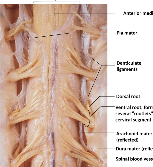
Figure 13-4 The Spinal Cord and Associated Structures.
Anterior median fissure
Pia mater
Denticulate ligaments Spinal cord
Dorsal root
Ventral root, formed by several “rootlets” from one cervical segment
Arachnoid mater (reflected)
• Sectional Anatomy of the Spinal Cord • White matter
• Is superficial
• Contains myelinated and unmyelinated
axons
• Gray matter
• Surrounds the central canal of spinal
cord
• Contains neuron cell bodies, neuroglia,
unmyelinated axons
• Organization of Gray Matter • The gray horns
• Posterior gray horns contain somatic and visceral sensory nuclei • Anterior gray horns contain somatic motor nuclei
• Lateral gray horns are in thoracic and lumbar segments; contain visceral motor
nuclei
• Gray commissures
• Axons that cross from one side of cord to the other before reaching gray matter • Organization of White Matter
• Posterior white columns lie between posterior gray horns and posterior median
sulcus
• Anterior white columns lie between anterior gray horns and anterior median fissure • Anterior white commissure is area where axons cross from one side of spinal
cord to the other
• Lateral white columns located on each side of spinal cord between anterior and
•
Organization of
White Matter
• Tracts or fasciculi • In white columns • Bundles of axons
• Relay same information in same direction • Ascending tracts – sensory
• Carry information to brain • Descending tracts - motor
Spinal nerves
• Connect PNS with the spinal cord (part of CNS)
• They exit the spinal column at the intervertebral foramen. • 31 pairs
• Dorsal root : sensory neurons. It has a dorsal ganglion on its path. Connects the receptors with the sensorial nuclei in the spinal cord.
• Anterior root : motor neurons. Connects the motor nuclei with the effectors .
• ANTERIOR AND DORSAL ROOT UNIT TO FORM THE SPINAL NERVE
The spinal nerve branches : DORSAL RAMUS (SMALLER)- CARRIES SENSORY & MOTOR INFO VENTRAL RAMUS- (THICKER)- CARRIES SENSORY & MOTOR INFO
COMMUNICANTES – towards the sympathetic ganglia.
•
Peripheral Nerves
• Interconnecting branches of spinal nerves
• Each spinal nerve:
• Is surrounded by three connective tissue layers • That support structures and contain blood vessels
1. Epineurium
Outer layer, covers the entire nerve Dense network of collagen fibers 2. Perineurium
Middle layer
Divides nerve into fascicles (axon bundles) 3. Endoneurium
Inner layer
•
Nerve Plexuses
• Complex, interwoven networks of nerve fibers
• Formed from blended fibers of ventral rami of adjacent spinal nerves • Control skeletal muscles of the neck and limbs
• The Four Major Plexuses of Ventral Rami
1. Cervical plexus
2. Brachial plexus
3. Lumbar plexus
•
The Cervical Plexus
• Includes ventral rami of spinal nerves C1–C5
• Innervates neck, thoracic cavity, diaphragmatic muscles • Major nerve
• The Brachial Plexus
• Includes ventral rami of spinal nerves C5–T1 • Innervates pectoral girdle and upper limbs
• Major nerves
• Musculocutaneous nerve (lateral cord)
• Median nerve (lateral and medial cords) • Ulnar nerve (medial cord)
• Axillary nerve (posterior cord)
• The Lumbar Plexus
• Includes ventral rami of spinal nerves T12–L4 • Major nerves
• Genitofemoral nerve
•
The Sacral Plexus
• Includes ventral rami of spinal nerves L4–S4 • Major nerves
• Pudendal nerve
• Sciatic nerve
• Two branches of the sciatic nerve
• Spinal Reflexes
• Automatic responses coordinated within spinal cord
• Through interconnected sensory neurons, motor neurons, and interneurons • Produce simple and complex reflexes
•
Neural
Reflexes
• Rapid, automatic responses to specific stimuli • Basic building blocks of neural function
• One neural reflex produces one motor response
• Reflex arc
• The wiring of a single reflex • Beginning at receptor
• Ending at peripheral effector
•
Five Steps in a Neural Reflex
• Step 1: Arrival of stimulus, activation of receptor
• Physical or chemical changes
• Step 2: Activation of sensory neuron
• Graded depolarization
• Step 3: Information processing by postsynaptic cell
• Triggered by neurotransmitters
• Step 4: Activation of motor neuron
• Action potential
• Step 5: Response of peripheral effector