http://www.cmbl.org.pl
DOI: 10.2478/s11658-006-0038-y Received: 17 January 2006
Revised form accepted: 20 June 2006
© 2006 by the University of Wrocław, Poland
*Present address: Department of Endocrinology and Metabolism, University Hospital of Odense, Medical Biotechnology Center, SDU, DK-5000 Odense C, Denmark, tel: +45 65503057, fax: +45 65503950, e-mail: [email protected]
Abbreviations used: ALP – alkaline phosphatase; BMP-2 – Bone morphogenetic protein-2; Cbfa1/Runx2 – Core binding factor, runt domain, alfa subunit 1; Dex – Dexamethasone; GC – glucocorticoid; IGFBPs – insulin like growth factor binding proteins; IGFs – insulin like growth factors; MSC – mesenchymal stem cells; OC – osteocalcin; OPN – osteopontein; RT – reverse transcriptase.
Short communication
OSTEOBLAST DIFFERENTIATION OF NIH3T3 FIBROBLASTS IS ASSOCIATED WITH CHANGES IN THE IGF-I/IGFBP EXPRESSION
PATTERN
BASEM M. ABDALLAH*
Department of Biochemistry, Faculty of Science, Helwan University, Cairo, Egypt
Abstract: Insulin-like growth factors (IGFs) and IGF-binding proteins (IGFBPs) are essential regulators for osteoblast proliferation and differentiation. It has been reported that Dexamethasone (Dex), an active glucocorticoid (GC) analogue, synergizes the stimulatory effect of 1,25-dihydroxyvitamin D3
(1,25(OH)2D3) on osteoblast differentiation in the mouse fibroblastic cell line
NIH3T3. I investigated whether this stimulatory effect is associated with changes in the expression pattern of the IGF/IGFBP system. Quantitative real-time PCR technology was used to quantify the gene expression levels of the IGF-system during osteoblast differentiation and in response to 1,25(OH)2D3 or
Dex alone under serum-containing and serum-free culture conditions. Interestingly, NIH3T3 was shown to express high mRNA levels of IGF-I, IGF-II and IGFBP-5, and low levels of both IGFBP-2 and -6. During osteoblast differentiation (days 6-12), IGF-I mRNA was repressed by more than 60%, while the transcript of IGFBP-5 was markedly up-regulated, by more than 50-fold. Similarly, treatment with Dex alone resulted in a dose- and time-dependent increase in the expression of IGFBP-5 and a decrease in IGF-I mRNA. Treatment with 1,25(OH)2D3 alone increased the mRNA levels of IGF-I and
which are regulated by glucocorticoid in the presence of 1,25(OH)2D3.
Modulation of the IGF/IGFBP levels by glucocorticoid might suggest important roles for the IGF-system in mediating the osteoblast differentiation of the NIH3T3 cell line.
Key words: IGFs/IGFBPs, NIH3T3, Gene expression, Osteoblast differentiation
INTRODUCTION
The proliferation and differentiation of osteoblasts are controlled by various local growth factors and cytokines produced in the bones, and by systemic hormones [1]. IGF-I and IGF-II areamong the most abundant growth factors synthesized by osteoblasts. They can be stored locally in the bone matrix. A number of in vitro and in vivo findings demonstrated that IGFs are important regulators of bone formation and that they stimulate the proliferation and differentiation of osteoblasts [2].
The actions of IGFs in bone are mediated via two cell surface IGF-receptors (IGF1R and IGF2R), and positively or negatively regulated by a family of six structural and evolutionary IGF-binding proteins (IGFBP-1 to -6) [3]. Each individual IGFBP either inhibits or potentiates the effects of IGFson osteoblasts. For example, IGFBP-1, IGFBP-3 and IGFBP-4 have been consistentlyshown to inhibit IGF activity.By contrast, exogenous IGFBP-5 enhances the mitogenic potential of IGF-Ior IGF-II in osteoblast culture, and has an important role in the storage of IGF-I in the bone matrix, while IGFBP-6 has a much greater affinity to IGF-II than IGF-I, and acts as a negative regulator of IGF-II activity in osteoblasts. IGFBPs can also act independently as growth modulators for cell growth, migration and apoptosis. For example, IGFBP5 can function as a growth regulator to stimulate bone formation parameters in vitro and in vivo (reviewed in [4]).
The production of the IGF-system in osteoblasts was shown to be regulated by the steroid hormone glucocorticoids (GCs), and the secosteriod hormone (1,25-dihydroxyvitamin D3) [5]. GCs were among the first agents identified to mediate osteogenic differentiation in vitro, where their effect was associated with the regulation of the IGF-system [6]. GCs were shown to suppress the production of IGF-I and -II at the mRNA and protein levels in osteoblastic cell cultures [7] and to have a major effect on the modulation of mRNA expression and the secretion levels of IGFBPs, including IGFBP-3, -4, and -5 [8].
1,25(OH)2D3 plays an important role in maintaining skeletal integrity and
osteoblast functions. 1,25(OH)2D3 stimulates the osteoblasts to produce several
non-collagenous proteins, including osteocalcin, osteopontin and alkaline phosphatase [9]. Several studies have reported on the regulatory effect of 1,25(OH)2D3 on IGF/IGFBP synthesis and release in bone marrow mesenchymal
stem cells (MSC) and osteoblastic cell lines. 1,25(OH)2D3 was shown to
[10] and IGFBP-3 in human osteoblastic cells [11]. Considering the regulation of the IGF-system by GCs and 1,25(OH)2D3, it is believed that IGF-system can
act as a mediator for the biological function of GCs and the effects of vitamin D3 on osteoblasts.
In the study of osteoblast differentiation, the NIH3T3 cell line has generally been used as a negative control for other mouse osteoblastic-like cell lines, including MC3T3 and C3H10T1/2; however, several studies demonstrated that NIH3T3 cells have some osteoblastic potential when treated with different growth factors and hormones [12]. For example, the treatment of NIH3T3 with BMP-2 resulted in an increase in the alkaline phosphatase (ALP) activity [13], while Dex stimulates the 1,25(OH)2D3-induced osteoblastic differentiation of
NIH3T3 by up-regulating ALP, ostocalcin and osteopontin [12]. Moreover, overexpression of Core-binding factor1 (Cbfa1/Runx2), (the main regulator of osteoblast formation) promotes the activation of osteocalcin in NIH3T3 [14]. In my attempt to understand the molecular mechanism(s) underlying the osteoblast differentiation of NIH3T3 cells, I studied the possible regulation of the IGF/IGFBP expression pattern by 1,25(OH)2D3 and Dex during osteoblast
differentiation. I found that NIH3T3 cells express the mRNA of IGF-I and -II, and BP-2, -5 and -6 at different levels under basal culture conditions. My data demonstrated the regulation of the expression pattern of the IGF-system during the osteoblast differentiation of the NIH3T3 cell line.
MATERIALS AND METHODS
Cell culture
The murine NIH3T3 fibroblast cell line was obtained from the American Type Culture Collection. Cells were maintained in Dulbecco’s modified Eagle’s medium (DMEM) supplemented with 10% fetal calf serum (FCS) and 1% penicillin/streptomycin (Gibco, Denmark) at 37ºC in a humidified atmosphere containing 5% CO2.
For osteoblast differentiation, cells were seeded at a density of 3 × 10⁴ cells/cm2
in 6-well plates. At 70-80% cell confluence, the medium was changed to an osteogenic medium consisting of DMEM supplemented with 10 nM 1,25(OH)2D3 (Vitamin D3) (provided by Leo Pharma, Denmark) and 10 nM
Dexamethasone (Dex) (Sigma-Aldrich, Denmark). The medium was changed every third day throughout the study.
To study the effect of 1,25(OH)2D3 or Dex on IGF/IGFBP expression pattern,
cells were seeded at a density of 3 × 10⁴ cells/cm2 in 6-well plates. At 80-90%
cell confluence, the cells were induced with different concentrations of either 1,25(OH)2D3 or Dex in the presence or absence of fetal calf serum.
Alkaline phosphatase (ALP) activity assay
osteoblastic-induction medium. A colorimetric ALP activity assay was performed on whole cell extracts as described previously [15] using p-nitrophenyl phosphate as a substrate (ABX Pentra ALP CP kit, HORIBA ABX Diagnostic, Montpellier, France). ALP activity was normalized to the total cellular protein assessed via the Bradford assay (Bio-Rad, Hercules,CA, USA) and expressed as units/mg protein. One unit of alkaline phosphatase activity is defined as the enzyme activity that will liberate 1 μM of p-nitrophenol per 30 min at 37ºC.
RNA isolation and RT-PCR analysis
Total RNA was isolated from cultured cells using the single step method with TRIzol according to the manufacturer’s instructions. The integrity and purity of the total RNA was verified spectrophotometrically and by gel-electrophoresis on 0.8% SeaKem agarose (BMA, Hellerup, Denmark). For reverse transcriptase (RT), first strand complementary DNA was synthesized from 5 μg of total RNA using a commercial revertAid H minus first strand cDNA synthesis kit (Fermentas, Copenhagen, Denmark) according to the manual.
Real-time PCR
Quantitative PCR was performed in an iCycler IQ detection system (Bio-Rad, Herlev, Denmark) using SYBR® Green I as a double-strand DNA-specific
binding dye. Thermocycling was performed in a final volume of 20 µL containing3 µL of cDNA sample (diluted 1:30), 20 pmole of each primer, 2mM MgCl2, 0.2 mM dNTP mixture, 1x Taq reaction buffer, 0.5 U HotStart Taq DNA
polymerase (Qiagen, VWR, Denmark), 0.5 µL of a 1:3000 dilution of SYBR Green I (Roch Molecular Biochemicals, Denmark), and 10 nM Flourescein Calibration Dye (Bio Rad) (for collecting the well factor directly from the experimental plate). The quantification of the target gene and ß-actin mRNA was performed in separate tubes using the primers in Tab. 1. The cycle conditions for the iCycler were started by using the experimental plate as a source of collecting well factors according to the Bio-Rad operating instructions, followed by a denaturing step at 95ºC for 10 min and 40 cycles of 95ºC for 30 sec, 60ºC for 30 sec and 72ºC for 1 min. Each reaction was run in triplicate and the fluorescence data was specified for collection at the end of the extension step in every cycle. To ensure specific amplification, a melting curve was done for each PCR reaction by increasing thetemperature from 60ºC to 95ºC at an increment rate of 0.5ºC/10 sec. To verify the melting curve results, all the samples were run in 1.5% agarose gel electrophoresis and visualised with ethidium bromide staining. The relative expression for each target gene was calculated using the comparative Ct method (formula: [(1/(2∆Ct)], where ∆Ct is
Tab. 1. Real-time PCR primer sequences used in this study.
Gene Primer sequence Product size
Reference gene
β-actin 5’-ACGGGGTCACCCACACTGTGC-3’
5’-CCGCTCGTTGCCAATAGTGATGA-3’ F R 292 bp
Target genes
IGF-I 5’-GCTGAGCTGGTGGATGCTCTTCAGTTC-3’
5’-CTTCTGAGTCTTGGGCATGTCAGTGTG-3’ F R 215 bp
IGF-II 5’-GAGCTTGTTGACACGCTTCAGTTTGTC-3’
5’-ACGTTTGGCCTCTCTGAACTCTTTGAG-3’ F R 356 bp
IGFBP-2 5’-CAACTGTGACAAGCATGGCCG-3’
5’-CACCAGTCTCCTGCTGCTCGT-3’ F R 180 bp
IGFBP-5 5’-CAGGAGTTCAAAGCCAGCCCAC-3’
5’-CGAAGGCGTGGCACTGAAAGTC-3’ F R 198 bp
IGFBP-6 5’-TAATGCTGTTGTTCGCTGCG-3’
5’-CACTGCTGCTTGCGGTAGAA-3’ F R 552 bp
ALP 5’-GCCCTCTCCAAGACATATA-3’
5’-CCATGATCACGTCGATATCC-3’ F R 372 bp
OC 5’-CAGACAAGTCCCACACAGCA-3’
5’-CTTTATTTTGGAGCTGCTGT-3’ F R 456 bp
OPN 5’-GAAACTCTTCCAAGCAATTC-3’
5’-GGACTAGCTTGTCCTTGTGG-3’ F R 589 bp
Statistical analysis
All the values are expressed as the means ± SD (standard deviation). The statistical significance of differences between the induced and non-induced cells was calculated via Student’s t-test. P < 0.05 was considered significant.
RESULTS
Osteoblast differentiation of NIH3T3 is associated with the regulation of IGF/IGFBP expression
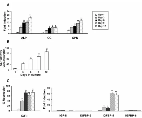
I first examined the osteoblast differentiation potential of the NIH3T3 cell line. As shown in Fig. 1A, the treatment of NIH3T3 cells with a combination of both 1,25(OH)2D3 and Dex was sufficient to stimulate the osteoblast differentiation of
Fig. 1. The regulation of IGFs/IGFBPs during osteoblast differentiation of NIH3T3 cells. (A) NIH3T3 cells were induced to differentiate into osteoblasts over a period of 12 days in an osteogenic medium as described in the Materials and Methods. Total RNA was isolated and the gene expression levels of ALP, OC and OPN were quantified by real-time PCR. (B) ALP activity measurements in NIH3T3 cell extracts during osteoblast differentiation. (C) Real-time PCR analysis of the IGF/IGFBP pattern during osteoblast differentiation of NIH3T3 cells. The expression level of each target gene was normalized for β-actin expression and represented as fold induction over control non-induced cells or % repression of control. The data is the means ± SD of three independent experiments. *p < 0.05, **p < 0.001 vs. control non-induced cells.
IGF-I and IGFBP-5 mRNA expression are mainly regulated by glucocorticoid during the osteoblast differentiation of NIH3T3 cells
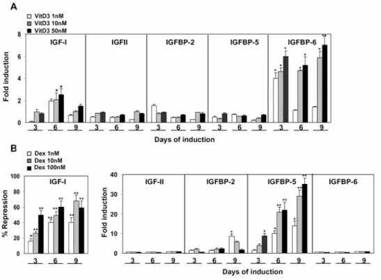
To further identify the main regulator of IGF-I and IGFBP-5 gene expression in NIH3T3, I first analyzed the expression pattern of IGFs/IGFBPs in response to the addition of either 1,25(OH)2D3 or the glucocorticoid, Dex, in the presence of
serum. By contrast to the expression pattern of the IGF-system during osteoblast
differentiation, the treatment of NIH3T3 with 1,25(OH)2D3 alone at
concentrations ranging from 1 to 50 nM was shown to moderately stimulate the expression of IGF-I and IGFBP-6, by 3- and 7-fold, respectively (Fig. 2A). On the other hand, the administration of Dex alone resulted in an IGF-expression pattern similar to that obtained during the osteoblast differentiation of NIH3T3
in the presence of both Dex and 1,25(OH)2D3 (Fig. 1C). As shown in Fig. 2B,
treating NIH3T3 cells with Dex markedly inhibits the expression of IGF-I (by 60%) and stimulates IGFBP-5 mRNA by 30- to 40-fold in a dose- and time-dependent manner.
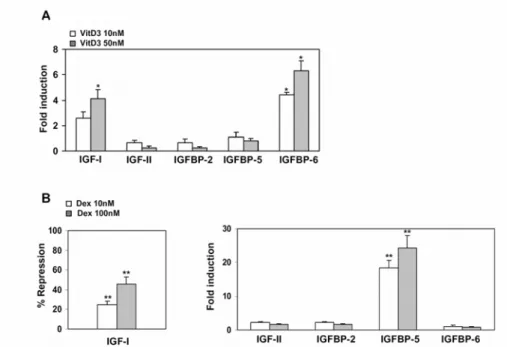
In order to avoid interference from any IGFs or IGFBPs that are normally found in the serum, I analyzed the expression pattern of IGFs/IGFBPs in NIH3T3 cells in response to 1,25(OH)2D3 or Dex in a serum-free medium. As with the expression
pattern of the IGF-system that was obtained in the serum-containing medium, 1,25(OH)2D3 was shown to exert a stimulatory effect on both IGF-I and IGFBP-6,
by 4- and 6-fold, respectively, as assessed via real-time PCR (Fig. 3A). Furthermore, Dex was able to reduce the expression level of IGF-I by 45% and to stimulate the expression of IGFBP-5 by 25-fold in a dose-dependent manner after 48 h of induction (Fig. 3B). This data demonstrated the regulation of the IGF/IGFBP system by Dex during osteoblast differentiation of the NIH3T3 cell line.
DISCUSSION
In this study, I demonstrated that the osteoblast differentiation of the murine fibroblast cell line NIH3T3 is associated with the regulation of the IGF/IGFBP system by glucocorticoid in the presence of 1,25(OH)2D3. This data might
suggest a mechanism by which the stimulatory effect of Dex on 1,25(OH)2D3
-induced osteoblast differentiation in NIH3T3 is mediated at least in part by regulating the IGF-system. These results are of great importance to be considered when using NIH3T3 as a control for any murine osteoblastic cell lines.
IGFs act in diverse patterns, via a combination of endocrine, autocrine, and paracrine modes of action, to regulate the differentiation functions of the osteoblasts. In this study, I demonstrated that the NIH3T3 cell line expresses the mRNA of IGF-I and -II, and that of three of the IGFBPs (-2, -5 and -6) when cultured under basal culture conditions. Among these expressed components of the IGF-system, only IGF-I and IGFBP-5 mRNA were regulated during osteoblast differentiation by glucocorticoid when administered alone or in combination with 1,25(OH)2D3.
My finding that Dex markedly inhibited IGF-I expression is consistent with the known inhibitory effect of Dex on IGF-I expression in several osteoblastic cell models. For example, Dex was observed to inhibit IGF-I expression in cultured rat bone cells [7] and human bone marrow stromal cell cultures [8], and also to have an adverse effect on IGF-I-mediated signaling of osteoblast differentiation in hMSC cells [16]. In addition, by contrast to the anabolic effect of IGF-I on bone, in vivo exposure to excessive glucocorticoids resultsin rapid bone loss with an increase in osteoporotic fracture risk [17]. Therefore, it was suggested that some of the actions of glucocorticoids may be mediated by the down-regulationof osteoblastic IGF [18].
In vitro studies indicate that the effects of GCs on osteoblast differentiation vary with the stage of cell growthand differentiation. GCs were reported to be crucial for the induction of osteoblast differentiation in both human and rat bone marrow stromal cells [6, 8], while they decreased the rate of cell proliferation of mature osteoblasts and osteocytes by inducingapoptosis as well as inhibiting the cell cycle [19]. It has been reported that the administration of Dex alone is not enough to induce osteoblast differentiation in NIH3T3, but it can rather inhibit its cell proliferation [20]. Since IGF-I is known to have a mitogenic effect on many cells [21], it is therefore plausible that the inhibitory effect of Dex on IGF-I expression might represent one of the mechanisms used by Dex to induce the growth arrest of NIH3T3, and hence promote the stimulatory effect of 1,25(OH)2D3 on osteoblast differentiation. In this context, the inhibitory effect of
Dex on cell growth and IGF-I synthesis has been reported for murine osteoblastic cells during their differentiation [22].
cultured cells from human rib marrow [8]. On the other hand, in non-osteoblastic cell types, the regulatory effects of Dex on IGFBP-5 were controversial. Dex was shown to block the induction of IGFBP-5 mRNA by IGF-I in a bovine fibroblast model, while it did not exert any influence on the IGF-I regulation of IGFBP-5 in human fibroblasts [24]. Moreover, Dex decreases the steady state levels of IGFBP-5 mRNA in the pituitary cell line [25]. This data suggests that the regulation of IGFBP-5 by GC treatment may be species and/or cell-type specific.
IGFBP-5 is the most abundant IGFBP produced by the osteoblasts; it is stored in bone, where its binding to the extracellular matrix proteins provides a mechanism of regulating the fixation of IGFs, and hence modulates their bioavailability. In vivo and in vitro data support the notion that IGFBP-5 acts as a growth factor via an IGF-independent mechanism [26]. The addition of IGFBP-5 to mouse osteoblast cells derived from IGF-I knockout or wild type mice increased proliferation, ALP activity and osteocalcin expression. Similarly, local administration of rhIGFBP-5 to the outer periosteum of calvarial bone of IGF-I knockout and wild type mice increased the bone formation parameters and osteoblastic markers of ALP and osteocalcin [27]. Given the independent stimulatory effects of IGFBP-5 on bone cells, it might be suggested that the stimulatory effect of Dex on the vitamin D3-induced osteoblast differentiation of NIH3T3 is mediated, at least in part, by increasing the production of stimulatory IGFBP-5. However, this proposed mechanism needs further investigation, as the in vitro overexpression of IGFBP-5 in the clonal mouse osteoblastic cell line MC3T3 resulted in impaired osteoblast function as assessed by the reduced expression of osteoblastic markers and inhibition of matrix mineralization [28]. In this context, it is better to investigate the biological consequences of the loss of function of endogenous IGFBP-5 using siRNA rather than to study the effect of adding exogenous IGFBP-5, which resulted in broadly variable effects in many cell systems.
The effects of GCs have been studied on other members of the IGF/IGFBP system. For example, it has been reported that Dex can induce IGF-II at both the mRNA and protein levels in human mesenchymal stem cells (hMSC) during osteoblast differentiation [8], and the expression of IGFBP-2 was shown to be regulated by Dex in osteoblastic cell cultures in a positive [29] or negative manner [30]. However, I could not detect any changes in the mRNA expression levels of IGF-II or IGFBP2 in NIH3T3 cells treated either with Dex alone or in combination with 1,25(OH)2D3.
On the other hand, the regulatory effects of 1,25(OH)2D3 on the IGF-system are
controversial based on the cell type, culture condition and the stage of cell differentiation [9]. I found that treating NIH3T3 with 1,25(OH)2D3 alone, leads
to moderate increases in the expression of IGF-I and IGFBP-6 in a dose-and time-dependent manner. Consistent with my findings for IGF-I, 1,25(OH)2D3
IGFBPs including IGFBP-2, -3 and 4 in hMSC and IGFBP-5 in mouse osteoblastic cells [11, 33]. On the other hand, the stimulatory effect of 1, 25(OH)2D3 on IGFBP-6 is unique for NIH3T3 and has not been reported before
for any osteoblastic cell line, and therefore might need further investigation. IGFBP-6 has a much greater affinity for IGF-II than IGF-I, and it exerts an inhibitory effect on IGF-II-induced effects on proliferation and differentiation in osteoblasts and other cell types [34]. For example, exogenous IGFBP-6 inhibits the IGF-II-stimulated DNA synthesis in cultured rat calvarial cells [35]. In addition, IGFBP-6 mediates the inhibitory effect of retinoids on human osteoblast function [36]. Despite this inhibitory effect of IGFBP-6 on osteoblast differentiation in vitro, the effect of IGFBP-6 on bone in vivo remains unclear, since the IGFBP-6 knockout mice do not appear to differ significantly from the controls [37].
In conclusion, my data suggested a role of the IGF-system in osteoblast differentiation of the mouse fibroblastic cell line NIH3T3. The direct implication of the IGF-system in this process still needs further investigation.
NIH3T3 as a clonal and non-tumorogenic cell line that has infinite proliferative capacity in vitro and can differentiate into both adipocyte and osteoblast lineages could provide an excellent in vitro cell model to study the regulation of the IGF-system by several growth factors and hormones that mediate osteoblast or adipocyte differentiation. Such studies will enable an understanding of the direct/indirect involvement of IGFs/IGFBPs in these differentiation processes.
Acknowledgments. Professor Moustapha Kassem is thanked for providing all the research facilities to perform this study at the KMEB laboratory, OUH, Odense, Denmark. Mrs. Tina K. Nielsen is thanked for her excellent technical assistance. Dr. B.M. Abdallah received a research fellowship from the Alfred Benzon foundation.
REFERENCES
1. Kassem, M., Kristiansen, M. and Abdallah, B.M. Mesenchymal stem cells: cell biology and potential use in therapy. Basic Clin. Pharmacol. Toxicol.
95 (2004) 209-214.
2. Langdahl, B.L., Kassem, M., Moller, M.K. and Eriksen, E.F. The effects of IGF-I and IGF-II on proliferation and differentiation of human osteoblasts and interactions with growth hormone. Eur. J. Clin. Invest. 28 (1998) 176-183. 3. Rajaram, S., Baylink, D.J. and Mohan, S. Insulin-like growth factor-binding
proteins in serum and other biological fluids: regulation and functions.
Endocr. Rev. 18 (1997) 801-831.
5. Robson, H., Siebler, T., Shalet, S.M. and Williams, G.R. Interactions between GH, IGF-I, glucocorticoids, and thyroid hormones during skeletal growth. Pediatr. Res. 52 (2002) 137-147.
6. Eijken, M., Koedam, M., van Driel, M., Buurman, C.J., Pols, H.A. and van Leeuwen, J.P. The essential role of glucocorticoids for proper human osteoblast differentiation and matrix mineralization. Mol. Cell Endocrinol.
248 (2006) 87-93.
7. Delany, A.M. and Canalis, E. Transcriptional repression of insulin-like growth factor I by glucocorticoids in rat bone cells. Endocrinology 136 (1995) 4776-4781.
8. Cheng, S.L., Zhang, S.F., Mohan, S., Lecanda, F., Fausto, A., Hunt, A.H., Canalis, E. and Avioli, L.V. Regulation of insulin-like growth factors I and II and their binding proteins in human bone marrow stromal cells by dexamethasone. J. Cell. Biochem. 71 (1998) 449-458.
9. Gurlek, A., Pittelkow, M.R. and Kumar, R. Modulation of growth
factor/cytokine synthesis and signaling by 1alpha,25-dihydroxyvitamin D(3): implications in cell growth and differentiation. Endocr. Rev. 23 (2002) 763-786.
10. Kveiborg, M., Flyvbjerg, A., Eriksen, E.F. and Kassem, M.
1,25-Dihydroxyvitamin D3 stimulates the production of insulin-like growth factor-binding proteins-2, -3 and -4 in human bone marrow stromal cells.
Eur. J. Endocrinol. 144 (2001) 549-557.
11. Nakao, Y., Hilliker, S., Baylink, D.J. and Mohan, S. Studies on the regulation of insulin-like growth factor binding protein 3 secretion in human osteosarcoma cells in vitro. J. Bone Miner. Res. 9 (1994) 865-872.
12. Shui, C. and Scutt, A.M. Mouse embryo-derived NIH3T3 fibroblasts adopt an osteoblast-like phenotype when treated with 1alpha,25-dihydroxyvitamin D(3) and dexamethasone in vitro. J. Cell Physiol. 193 (2002) 164-172. 13. Wang, E.A., Israel, D.I., Kelly, S. and Luxenberg, D.P. Bone morphogenetic
protein-2 causes commitment and differentiation in C3H10T1/2 and 3T3 cells. Growth Factors 9 (1993) 57-71.
14. Xiao, Z.S., Hinson, T.K. and Quarles, L.D. Cbfa1 isoform overexpression upregulates osteocalcin gene expression in non-osteoblastic and pre-osteoblastic cells. J. Cell. Biochem. 74 (1999) 596-605.
15. Abdallah, B.M., Haack-Sorensen, M., Fink, T. and Kassem, M. Inhibition of osteoblast differentiation but not adipocyte differentiation of mesenchymal stem cells by sera obtained from aged females. Bone 39 (2006) 181-188. 16. Koch, H., Jadlowiec, J.A. and Campbell, P.G. Insulin-like growth factor-I
induces early osteoblast gene expression in human mesenchymal stem cells.
Stem Cells Dev. 14 (2005) 621-631.
17. Barnes, P.J. Corticosteroids: the drugs to beat. Eur. J. Pharmacol. 533 (2006) 2-14.
19. Smith, E., Coetzee, G.A. and Frenkel, B. Glucocorticoids inhibit cell cycle progression in differentiating osteoblasts via glycogen synthase kinase-3beta. J. Biol. Chem. 277 (2002) 18191-18197.
20. Jorgensen, N.R., Henriksen, Z., Sorensen, O.H. and Civitelli, R.
Dexamethasone, BMP-2and 1,25-dihydroxyvitamin D enhance a more differentiated osteoblast phenotype: validation of an in vitro model for human bone marrow-derived primary osteoblasts. Steroids 69 (2004) 219-226. 21. Minuto, F., Palermo, C., Arvigo, M. and Barreca, A.M. The IGF system and
bone. J. Endocrinol. Invest. 28 (2005) 8-10.
22. Chen, T.L., Mallory, J.B. and Hintz, R.L. Dexamethasone and 1,25(OH)2 vitamin D3 modulate the synthesis of insulin-like growth factor-I in osteoblast-like cells. Calcif. Tissue Int. 48 (1991) 278-282.
23. Chevalley, T., Strong, D.D., Mohan, S., Baylink, D. and Linkhart, T.A. Evidence for a role for insulin-like growth factor binding proteins in glucocorticoid inhibition of normal human osteoblast-like cell proliferation.
Eur. J. Endocrinol. 134 (1996) 591-601.
24. Conover, C.A., Clarkson, J.T. and Bale, L.K. Effect of glucocorticoid on insulin-like growth factor (IGF) regulation of IGF-binding protein expression in fibroblasts. Endocrinology 136 (1995) 1403-1410.
25. Fielder, P.J., Tauber, J.P., Wilson, K.F., Pham, H.M. and Rosenfeld, R.G. Insulin-like growth factors (IGFs) stimulate and dexamethasone inhibits IGF binding protein (BP)-5 expression in a mouse pituitary cell line. Growth Regul. 3 (1993) 226-234.
26. Firth, S.M. and Baxter, R.C. Cellular actions of the insulin-like growth factor binding proteins. Endocr. Rev. 23 (2002) 824-854.
27. Miyakoshi, N., Richman, C., Kasukawa, Y., Linkhart, T.A., Baylink, D.J. and Mohan, S. Evidence that IGF-binding protein-5 functions as a growth factor. J. Clin. Invest. 107 (2001) 73-81.
28. Durant, D., Pereira, R.M. and Canalis, E. Overexpression of insulin-like growth factor binding protein-5 decreases osteoblastic function in vitro.
Bone 35 (2004) 1256-1262.
29. Jia, D. and Heersche, J.N. Expression of insulin-like growth factor system constituents in differentiating rat osteoblastic cell populations. Growth Horm. IGF. Res. 12 (2002) 399-410.
30. Chen, T.L., Chang, L.Y., Bates, R.L. and Perlman, A.J. Dexamethasone and 1,25-dihydroxyvitamin D3 modulation of insulin-like growth factor-binding proteins in rat osteoblast-like cell cultures. Endocrinology 128 (1991) 73-80. 31. Chenu, C., Valentin-Opran, A., Chavassieux, P., Saez, S., Meunier, P.J. and
Delmas, P.D. Insulin like growth factor I hormonal regulation by growth hormone and by 1,25(OH)2D3 and activity on human osteoblast-like cells in short-term cultures. Bone 11 (1990) 81-86.
parathyroid hormone, transforming growth factor-betaand 1,25-dihydroxyvitamin D3. Endocrinology 128 (1991) 1511-1518.
33. Schmid, C., Schlapfer, I., Gosteli-Peter, M.A., Hauri, C., Froesch, E.R. and Zapf, J. 1 alpha,25-dihydroxyvitamin D3 increases IGF binding protein-5 expression in cultured osteoblasts. FEBS Lett. 392 (1996) 21-24.
34. Kim, E.J., Schaffer, B.S., Kang, Y.H., Macdonald, R.G. and Park, J.H. Decreased production of insulin-like growth factor-binding protein (IGFBP)-6 by transfection of colon cancer cells with an antisense IGFBP-6 cDNA construct leads to stimulation of cell proliferation. J. Gastroenterol. Hepatol. 17 (2002) 563-570.
35. Schmid, C. Insulin-like growth factors. Cell Biol. Int. 19 (1995) 445-457. 36. Yan, T., Wergedal, J., Zhou, Y., Mohan, S., Baylink, D.J. and Strong, D.D.
Inhibition of human osteoblast marker gene expression by retinoids is mediated in part by insulin-like growth factor binding protein-6. Growth Horm. IGF. Res. 11 (2001) 368-377.