R E V I E W
Open Access
Plants and microbes assisted selenium
nanoparticles: characterization and application
Azamal Husen
1*and Khwaja Salahuddin Siddiqi
2Abstract
Selenium is an essential trace element and is an essential component of many enzymes without which they become inactive. The Se nanoparticles of varying shape and size may be synthesized from Se salts especially selenite and selenates in presence of reducing agents such as proteins, phenols, alcohols and amines. These biomolecules can be used to reduce Se salts in vitro but the byproducts released in the environment may be hazardous to flora and fauna. In this review, therefore, we analysed in depth, the biogenic synthesis of Se
nanoparticles, their characterization and transformation into t- Se, m-Se, Se-nanoballs, Se-nanowires and Se-hollow spheres in an innocuous way preventing the environment from pollution. Their shape, size, FTIR, UV–vis, Raman spectra, SEM, TEM images and XRD pattern have been analysed. The weak forces involved in aggregation and transformation of one nano structure into the other have been carefully resolved.
Keywords:Nanotechnology, Selenium, Plant extracts, Microbes, Biofabrication
Introduction
Selenium was known as a notorious element until it was recognized by Schwarz and Foltz in 1957 as an essential trace element for both plants and mammals. Normally Se is available as selenate and selenite oxoanions. The reduction of soluble Se4+and Se6+by microbes to insol-uble non toxic elemental Se is an effective way to re-move it from contaminated soil, water and drainage [1]. Se is one of the chalcogens occurring as selenate SeO42−, selenite SeO32− and selenide Se2− which may be reduced to atomic state by a precursor containing an appropriate reducing agent. Biogenic synthesis of Se nanoparticles is frequently achieved by reduction of selenate/selenite in presence of bacterial proteins or plant extracts containing phenols, flavonoids amines, alcohols, proteins and alde-hydes. The deficiency of Se is known to be associated with over 40 diseases in man [2,3]. At low dosage it can stimu-late the growth of the plant whereas at high dosages it can cause damage to it [4-6]. Se has also been shown to be ef-fective against cancer [7,8]. Their compounds in the form of selenocysteine and selenomethionine are metabolized in biological system [7,8].
A variety of microorganisms, enzymes and fungi, besides plant extracts have been used to synthesise Se nanoparti-cles of different size and morphology. Se itself is used in rectifier, solar cells, photocopier and semiconductor. In addition, they exhibit biological activity owing to their interaction with the proteins and other biomolecules present in the bacterial cells and plant extracts,
contain-ing functional groups such as ›NH, C = O, COO and
C-N [9]. Se-nanobelts have been synthesised on large scale with an approximate diameter of 80 nm and length up to 5μm [10]. Se exists in many crystalline and amorph-ous forms but the shape, size and structure of the nano-particles depend on the concentration, temperature, nature of biomolecules and pH of the reaction mixture. The properties of Se nanoparticles varies with size and shape for instance, Se nanospheres have high biological activity and low toxicity while Se nanowires of t-Se have high photoconductivity [11]. Various methods have been employed to produce large scale Se-nanowires and trigonal selenium (t-Se) [12,13]. Pulse laser ablation, electro-kinetic technique, hydrothermal treatment, vapour deposition methods [10,14-16] generally used for production of Se nanoparticles on large scale require either sophisticated in-struments or specific chemicals which are time consuming and uneconomical. Such methods often employ toxic
* Correspondence:[email protected]
1
Department of Biology, College of Natural and Computational Sciences, University of Gondar, P.O. Box 196, Gondar, Ethiopia
Full list of author information is available at the end of the article
chemicals or high temperature and high pressure which further pollute the environment.
In order to circumvent the effect of toxic chemicals in the fabrication of nanoparticles, biogenic protocol is generally followed [17,18]. Scientists have developed be-nign and harmless methods for the fabrication of nano-particles using plant extracts, bacteria and fungi. For instance,Capsicum annum,Escherichia coliandBacillus subtilis [19-21] have recently been used to produce nanoparticles. Over 16 different species of bacteria and Arechaea have been found to reduce colourless selenate and selenite to red elemental Se of different shape and size [22,23]. Plants and microbes act as producers and protectors of the environment when they are properly used. Pure element in its, atomic state may be produced by many bacteria [24,25] mainly due to the chemicals present in them or protein exuded by them. We have limited knowledge of the mechanism of the formation of Se nanoparticles by microbes and plant extract, never-theless for a better understanding attempts are being made to explore the chemical reactions occurring in these media. Many bacterial strains have been found to reduce selenate/selenite to Se nanoparticles in different environment [26] even in sewage and sludge under both aerobic and anaerobic conditions [27-29]. It has been suggested that substantial quantity of soluble toxic selen-ate/selenite is reduced by bacterial strain to produce non toxic insoluble Se nanoparticles, although in doing so many such microbes would die. The production of Se0/Te0 by two anaerobic bacteriaSulfurospirillum bar-nesiiand Bacillus selenireducenshas been demonstrated by Oremland et al. and Baesman et al. [25,24].
The main objective of this review is to identify the plant extracts and bacterial strains involved in the bio-synthesis of Se nanoparticles. Also the characterization and identification of Se-nanoballs, nanorods, nanowires and hollow spheres have been undertaken with a view to update the nanobiotechnology of Se nanoparticles and their application in diverse areas.
Se nanoparticles from plants, characterization and application
There is a fine line between optimum limit/or deficiency and excess of Se in living system which may cause tox-icity. It is known that the Se nanoparticles prepared from biological material are less toxic than the bulk Se nanoparticles prepared from chemicals. The biomole-cules present in the extract act both as reducing agent and stabilizers of Se nanoparticles. Bacteria, algae, dry fruits and plant extracts are used to produce nanoparti-cles. Green synthesis of selenium nanoparticles from sel-enious acid was achieved by dried extract of raisin (Vitis vinifera) [30]. Like other biological materials, raisin also contains sugar, flavonoids and phenols in addition to
minerals such as iron, potassium and calcium [19,31]. A change from colourless to deeply brick-red colour indi-cated the formation of nanoparticles. The formation of Se-nanoballs was examined at different interval of time. It took nearly 6 min to start conversion of Se ion to Se nanoparticles which was indicated by a decrease in Se ion concentration in the solution. The nature of nano-particles was analysed by TEM images. It showed that the diameter of nanoballs ranges between 3 and 18 nm. They were found to be encapsulated with a thin poly-morphic layer. The formation of Se nanoparticles was confirmed from the energy dispersive x-ray spectros-copy. The Se nanoballs were identified from their char-acteristic absorption peaks at 1.37KeV, 11.22KeV and 12.49KeV [32]. The morphology of Se nanoparticles can be analysed by x-ray diffraction (XRD) analysis. The broad diffraction peak suggests the presence of amorph-ous nature of Se nanoparticles [33]. Their particle size [34] has been found to be of the order of 12 nm.
Sharma et al. [30] have characterized Se nanoballs
fab-ricated from V. vinifera by FTIR spectral studies. The
spectrum exhibited two sharp absorption peaks at
3420 cm−1 attributed to OH and, the second peak at
1620 cm−1to C-H vibration of the organic molecules. A distinct peak at 1375 cm−1has been assigned to phenolic
OH. The other peaks of medium intensity are due to –
CH3and OCH3groups associated with the biopolymers,
present in the V. vinifera extract acting as reducing
agent and stabilizer for the Se nanoballs. Since lignin is a component of all vegetables, fruits and cell wall, it can be extracted from them and the compounds present in them may be identified. In the present work, phenolic group has been identified which generally acts as redu-cing agent and, it is oxidised to ketone during the redox process. However, the extract also contains fairly sub-stantial amount of reducing sugars and therefore, they also help in the reduction and formation of Se nanoballs. These authors have given a flow diagram for Se nanopar-ticles synthesis but it does not reveal the chemical changes which occur as a consequence of redox reac-tions. We now propose the following scheme Figure 1 based on the general synthetic route.
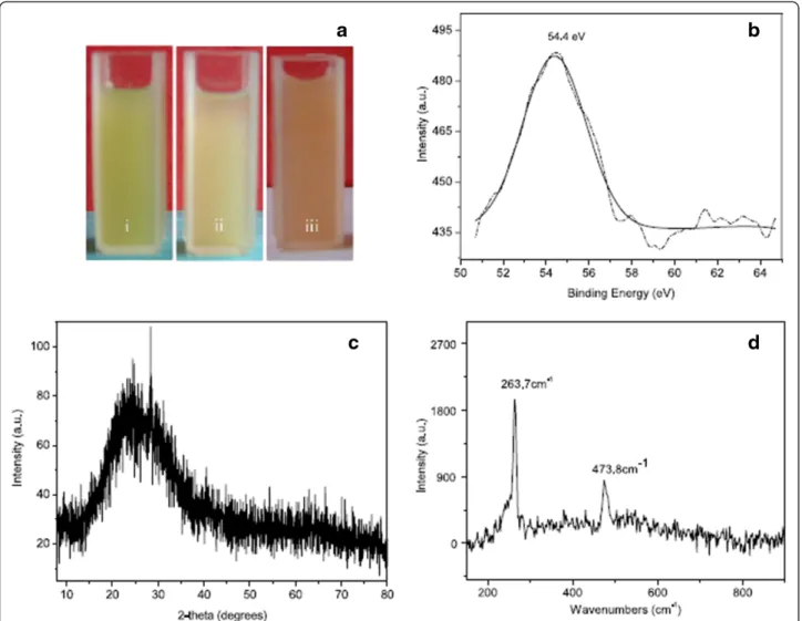
Although, biochemicals may often be used for the synthe-sis of nanomaterials, the biogenic synthetic route is fre-quently applied due to its ease and simplicity and, also because no hazardous and toxic residues are released in the environment [35,36]. In general, a variety of Se nanoparti-cles are produced when H2SeO3is treated with plant ex-tracts for instance,α-Se nanoparticles have been fabricated fromCapsicum annumextract in aqueous medium at low pH and at ambient temperature [19]. The light green extract of C. annum turns pale after 5 h of the addition
of H2SeO3, and then gradually turned red after 12 h
of α-Se in the x-ray photoemission spectroscopy (XPS) which is due to excitation of their surface plasmon vibra-tion [37]. Its XPS spectrum (Figure 2b) shows a sharp peak at 54.4 eV which corresponds to the elemental selenium [38]. The XRD pattern of the Se nanoparticles shows a broad peak at 2θ angles of 15-350 (Figure 2c) which sug-gests that the nanoparticles are not crystalline. Their
Ra-man spectrum displayed a resonance peak at 263.7 cm−1
which (Figure 2d) further confirms the formation of α-Se nanoparticles [39]. An additional peak at 474 cm−1 has been attributed to the protein vibration which is mixed with amorphous Se.
SEM and TEM images of the α-Se nanoparticles
showed that they consist of nanorods and nanoballs laced with C. annum protein which makes them slightly irregular in shape. The length and diameter of rods and
nanoballs range between 1–3 μm and 0.4 μm,
respect-ively. A closer look at the highly magnified field emission scanning electron microscopy (FESEM) image suggests that rod like nanoparticles are actually aggregates of spherical particles with protein coating, making the sur-face rough and uneven. It is quite likely that proteins are held together by hydrogen bonding and Se nanoparticles are held by van der Waals forces.
When the pH of the reaction mixture is lowered to 2 the time to produceα-Se nanoparticles increases. It has been observed from their FESEM images that a variety of polygonal Se nanoparticles are produced with size varying from 200–500 nm. It is of interest to note that some hollow spherical particles were also produced with a pore diameter of 160 nm. Li et al. [19] have hypothesized
that hollow spheres are formed as a consequence of rise in temperature when the reaction product is placed in an electric field. Although, the melting point ofα-Se nanopar-ticles is not very low (~490 K) even then this temperature is seldom achieved in such system, so that it may melt and produce hollow spheres. It is to be noted that even if microwave energy is supplied without rise in temperature only the outer surface ofα-Se nanoballs, made of protein layer would melt, because organic materials have inher-ently lower melting point than metalloid Se. However, if theseα-Se particles also melt with electronic impact even then the hollow sphere would not be produced because the lattice would rupture resulting in the formation of irregular sheets and dot like structures. It is more likely,
that hollow spheres of α-Se are also formed along with
solid nanoballs and polygonal structures during the synthesis of nanoparticles.
A comparison of FTIR spectrum of pure C. annum
extract with the reaction mixture (C. annum extract +
SeO32−) showed many peaks at 1652, 1542 and 1241
cm−1corresponding to amide I, II and III bands owing
to ʋ(C = O), ʋ(N-H) and ʋ(C-N) respectively [36].
These bands slightly shift after the formation of
nano-particles. The UV–vis spectrum of the C. annum
pro-tein (washed with SDS-PAGE gel) with molecular weight of 30 kDa, showed peak (210 nm) corresponding to peptide bonds and amino residues (280 nm). As these are reducing agents they help in the formation of nano-particles. It has also been confirmed from cyclic voltam-mogram that the redox reaction occurs between - 0.7 and 0.9 V [19].
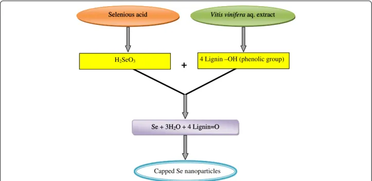
+
Selenious acid Vitis vinifera aq. extract
H2SeO3 4 Lignin –OH (phenolic group)
Se + 3H2O + 4 Lignin=O
Capped Se nanoparticles
+
Selenious acid Vitis vinifera aq. extract
H2SeO3 4 Lignin –OH (phenolic group)
Se + 3H2O + 4 Lignin=O
Capped Se nanoparticles
Inorganic Se (selenite or selenate) also occur as seleno-methionine, selenocysteine, selenocystathione, methyl selenol, dimethyl selenide and selenium methyl seleno-cysteine. Absorption of Se depends on its morphology and solubility in aqueous medium. Sodium salts of Se are generally soluble in water. One form may change into another to suit the basic requirements of binding to certain functional groups such as proteins. Different plants absorb Se in different quantities for instance; wheat accumulates Se proportional to its availability in
the soil while Astragalus grown in the same soil had
manifold excess of the element in it. Broccoli is known as Se accumulator. Finley [40], showed from an experi-ment, on broccoli grown in sodium selenate laden soil,
that it accumulated ~ 103 μg Se/gm dry weight of the
plant tissue. Broccoli is known to contain fairly [41] large quantity of selenium as methyl selenocysteine,
perhaps due to its greater solubility in aqueous medium. However, it is strange that Se from broccoli does not accumulate efficiently in man or rats [42,43] because its major part as selenium methyl selenocysteine is perhaps metabolised to methyl selenol [44]. It has been demon-strated experimentally that, methyl substituted forms of Se is an effective anticancer agent than the other deriva-tives of organo-Se compounds [45]. Garlic with as much
Se as 1000 μg/gm of dry weight has been grown [46]
to induce phase II enzymes [42] is chemoprotective against bladder cancer. Se fed experimental rats in the forms of selenite, selenate, selenomethionine did not acumulate in most rats which means it is either not absorbed or excreted through urine.
Trigonal Se nanowires and nanotubes have been syn-thesized in absolutely ecofriendly way. The Se nanowires
of 70–100 nm width and length in severalμm were
pre-pared in absolute ethanol at room temperature while tri-gonal Se nanotubes of diameter 180–350 nm were obtained in aqueous medium at 85°C (358 K). It was ob-served that amorphous Se nanoparticles were formed in the beginning and subsequently transformed into nano-wires and tubes [51].
Stable Se nanoparticles in colloidal form were prepared fromTerminalia arjunaextract in aqueous medium. They were characterized by UV–vis, energy dispersive X-ray
analysis (EDAX), transmission electron microscopy
(TEM), FTIR and XRD analysis. The colloidal solution had an absorbance maximum at 390 nm. Its IR spectrum showed peaks corresponding to O-H, NH, C = O and C-O stretches suggesting the presence of hydroxyl, amino, ketonic and carbonyl functional groups in the extract which act both as stabilizer and capping agent for the Se nanoparticles [52]. The Se nanoparticles synthesized from fenugreek seed extract in aquous medium at room temperature are between 50–150 nm. They have been found to be active against human breast cancer cells [53].
Se nanoparticles of approximately 35 nm have been synthesized from gum arabic which remain stable in so-lution for about 30 days. The gum arabic was found to be a better stabilizer for Se nanoparticles than the hy-drolysed gum arabic [54]. The Se nanoparticles synthe-sized from lemon leaf extract exhibited an absorption maximum at 395 nm in the UV–vis region. Initially, the mixture of leaf extract and SeO32− remains colourless but after stirring and incubating it for 24 hr at 30°C, it turns red [55]. The photoluminescence spectra exhibited excitation peak at 395 and emission peak at 525 nm (Figure 3). It has been found from TEM image that the size of particles ranges between 60–80 nm. They are polydispersed in colloidal solution but crystalline in na-ture [55]. The FTIR spectra of the samples with and without Se nanoparticles were compared to examine the changes in the functional groups of the biomolecules.
The broad peak at 3415 due to ʋ(NH) shifts to 3418
cm−1 but new peaks appear at 2930 and 3456 cm−1 in
the colloidal solution containing Se nanoparticles. The
region 1500–1800 cm−1
is due to various amide bands which split into some new bands in colloidal solution. However, after reduction of the Na2SeO3to Se nanopar-ticles by the biomolecules in the extract containing func-tional groups such as alcohol, aldehyde, phenol etc., they are oxidized to the following species:
Na2SeO3+ H2O→H2SeO3+ Na2O
H2SeO3⇌SeO32−+ 2H+
Alcohol + SeO32−→Se + Carboxylic acid
Aldehyde + SeO32−→Se + Ketone
Phenol + SeO32−→Se + Phenone
It has also been detected from gel electrophoresis that Se nanoparticles prevented DNA damage when cells were exposed to UVB [55].
Polyphenol gallic acid nanoparticles from plant have been used to fabricate Se nanoparticles since the gallic acid nanoparticles may behave differently than the bulk gallic aicd. Since gallic acid can quantitatively coordinate with the Se ions, another reducing agent dithioerthreitol was added to gallic acid-coated with Se ions. A change in colour was taken as an indication of the formation of nanoparticles which was confirmed by UV–vis and emis-sion spectroscopy [56]. A slightly different method has also been employed by Ingole et al. [34] to prepare Se nanoparticles from glucose. Na2SeSO3prepared from Se powder was treated with glucose solution according to the following chemical reactions:
Se powder + Na2SO3→Na2SeSO3
Na2SeSO3+ H2O→H2SeO4+ Na2S
H2SeO4⇌2H++SeO42−
SeO42−+ Glucose→Se + Gluconic acid
The colourless solution in the beginning becomes yellow then orange and finally turns red which does not change even after heating for over 1 h. These changes have been ascribed to the changes in size of Se nanoparticles.
Se nanoparticles from microbes, characterization and application
Microorganisms reduce the toxic, selenate and selenite oxoanions into non toxic elemental selenium which is insoluble in water. Continuous use of water or edible plants from Se rich soil can cause skin lesion and early hair fall. Effort is therefore, made to reduce Se com-pounds to elemental Se with the help of bacteria. It is a simple process of detoxification of selenites/selenates to Se nanoparticles as the reverse reaction is too slow to produce Se compounds [57].
Fast (forward reaction)
Se(IV)/Se (VI)⇌Se(o)
Slow (backward reaction)
Due to their unique property Se nanoparticles are photovoltaic and semiconductor, antioxidant and chemo-protective agents [58]. Since Se nanoparticles inhibit the
medicine againstS. aureusinfection. Different concentra-tion of Se starting from 65 to 230 mg/L of Se(IV) were allowed to interact with different types of microbes. Ap-pearance of red colour was taken as sign of reduction of Se(IV) to Se(0) as shown in forward reaction above. How-ever, there was no decolouration later, indicating the ab-sence of any species causing oxidation of Se(IV)→Se(VI) (Figure 4). The redox process is time and concentration dependent. When bacterial culture was grown in presence
of 40–100 mg/L selanate, no change in colour was
ob-served even after long time. It appears as if the bacteria are resistant to Se(VI) reduction. However, such bacterial culture may be used to reduce soluble and toxic Se(IV) to
non toxic and insoluble Se nanoparticles. It is also indica-tive of bioremediation of Se from selenites.
Amporphous Se nanoparticles have been synthesized from sodium selenite in presence ofShewanellasp. HN-41 in aqueous medium under anaerobic conditions taking care of reaction time, selenite concentration and biomass of Shewanella [59]. Different types of the Se nanopar-ticles are synthesized using protein, peptides and several other reducing agents [1,60,61]. Nanowires and nanorods
have been fabricated from Rhizobium selenireducens sp.,
Dechlorosomasp.,Pseudomonassp.,Paracoccussp., Enter-obactorsp.,Thaureasp.,Sulfurospirilliumsp.,Desulfovibrio sp., andShewanella sp., [61-63]. It has been reported that the particle size is decreased in presence of O2. It is obvi-ous that oxygen will promote oxidation of Se (forward re-action) as a consequence of which the redox step becomes slower producing smaller Se nanoparticles. Selenite reduc-tase is also helpful in the synthesis of Se nanoparticles. A wide range of selenite concentration starting from 0.01, 0.05, 0.15, 0.25, 0.050, 0.75 and 1 mM were used to study the effect of concentration, size and morphology. Average particle size for all the above concentrations was nearly 103 ± 5.1 nm. For large quantity of nanoparticles the selen-ite concentration not exceeding 0.1 mM is needed. It has been observed from TEM and SAED image that Se nano-particles are amorphous [59]. Extracellular synthesis [64] of fairly smaller particles of the order of 47 nm from the fun-gus,Aspergillus terreuswas done in 60 min.
Figure 4Selenite reduction by the mixed microbial culture isolated from agricultural soil.Selenite reduction at different Se (IV) concentrations(a)and development of red coloration in cultures after 5.5 h(b), 23 h(c)and 48 h(d)of incubation [58].
Microbes like Klebsiella pneumoniae [31] and Pseudo-monas alcaliphila[65] have also been used to synthesize Se nanoparticles in good yield. When Na2SeO3was added to the activated culture of P. alcaliphila the reaction started immediately but completed after 48 h [65]. A gradual colour change with time was observed in the following order:
Grey→Red→Intense Red
0 h 6 h 48 h
The characteristic red colour of Se nanoparticles [37] was detected spectrophotometrically and has been as-cribed to the excitation of the surface plasmon vibration of the monoclinic Se. It has been noticed that particle size is directly proportional to reaction time and it ranges between 50–500 nm [21]. Field emission scan-ning electron microscopic (FESEM) image shows nano-particles of varying size and shape.
The FTIR spectra of the samples with and without Se nanoparticles showed that the intensity of the spectral peaks containing Se nanoparticles is drastically dimin-ished [65] which suggests strong interaction between Se atoms and the protein molecules present in theP. alcali-phila. This is to be noted that the interaction between Se nanoparticles and protein is simply electrostatic be-cause the intensity of sample containing Se atoms was
decreased followed by an increase in ʋ(NH) from 3421
to 3435 cm−1. The Raman spectra also support the for-mation of trigonal selenium (t-Se) and monoclinic selen-ium (m-Se) by the appearance of peaks at 234 and 254 cm−1, respectively (Figure 5). A peak at 235 cm−1is mainly due to chain like structure of t-Se. As the peak at 234 cm−1appears after 48 h of inoculation, it is consid-ered as the transformation of one form of Se into other. The FESEM images which show the accumulation of nanorods on the nanoballs. The size can be controlled by PVP at different time of incubation of nanospheres ranging from 20–200 nm.
On the basis of above studies a possible mechanism for the formation and transformation of Se nanoballs to Se nanorods has been proposed. Zhang et al. [65]
pre-sume that SeO32− is reduced to elemental selenium by
the protein excreted by theP. alcaliphila and, their ag-gregation gives Se nanoparticles of varying size [13]. It is true that protein acts as reducing agent for SeO32− but it is available as excretion fromP. alcaliphilais not con-vincing. The excretion contains toxins, pyrogens and traces of protein but they may not be sufficient for re-duction of selenite. Authors also suggest that large m-Se nanoballs are not stable in solution and they dissolve to form Se atoms. A fraction of dissolved Se atoms crystallize as t-Se forming nanorods [66]. It is not rue because Se in atomic state is not soluble in a solvent but stays in colloidal form. It is a misconception. However,
PVP controls the size of Se nanoparticles. If the Se nano-particles without PVP are left for 2–3 weeks they form aggregates of different shapes and size. Since SeO32− in ionic form is toxic to bacterial culture, the bacteria in selenite solution may therefore, die and the disintegrated protein may then act as reducing agent for selenite. The large m-Se nanoparticle can not dissolve in solution to give Se atom forming Se-nanorods. It is quite likely that they may be segregated and rearranged into nanorods.
Se nanoparticles synthesized from sodium selenite and glutathione in aqueous medium had been tested for its growth inhibition efficacy against Staphylococcus aureus [67]. It was found that growth of S. aureus was inhibited in presence of Se nanoparticles within 3–4 h with as low concentration as 7–15 μg/ml which suggests that Se nanoparticles may be used against bacterial infections.
Biogenic Se nanoparticles fabricated from protein
pro-duced byE. colihave been compared with those
synthe-sized from chemical reaction via redox mechanism [68].
There are specific types of protein produced by E. coli
(AdhP, Idh, OmpC, AceA) which are associated with Se nanoparticles. They are also responsible for their
uniform size and distribution. One of the proteins (AdhP) was found to bind strongly to Se nanoparticles. E. coli was found to produce Se nanoparticles from
2 mM of SeO32−in about 48 h which was distinguished
by the change in colour from colourless to dark red. The amorphous Se nanoparticles thus produced were be-tween 10–90 nm. Since the bacterial growth continued even in presence of selenite, it is confirmed that selenite is not toxic toE. coli at this concentration. The enzymes alcohol dehydrogenase, propanol-preferring (ADHP), ACEP (Isocitrate lyase), ENO, KPYKI, IDH and GLPK require metal ions as cofactor for their activity while the enzymes DCEP, ASTC and TNAA require pyridoxal phosphate as coenzyme without which their activity is lost. The authors have not distinguished between cofac-tor and co-enzyme, they have termed both as metallic cofactor and non-metallic cofactor which is misleading. The cofactors in the enzyme are metal ions bonded through a coordinate covalent bond and can accept lone pair of electrons from the donor atoms in the enzyme into their vacant orbital to form the bond. The Se nano-particle is in the elemental state and no metal in atomic state can bind to protein or any electron donating spe-cies. It is therefore; proper to use the word association of Se nanoparticles to protein rather than bonding. The size of Se nanoparticles produced in presence of the pro-tein, alcohol dehydrogenase propanol-preferring (AdhP) were much smaller than those produced in their
ab-sence. However, since E. coli contains many other
pro-teins than only pAdhP (purified protein), the decrease in Se nanoparticle size may be the cumulative effect of all proteins available inE. coli.
Conclusion
Bioreduction of selenate or selenite from microorganism such as bacteria, fungi and plant extract have become the favourite pursuit of biologist, chemist and engineers. It is expected that in future the metal would be ex-tracted by biomineralization because they produce the purest form of the element. Many raw materials like waste vegetables, fruit peels and leather cuttings may be utilized to produce elemental metal/metalloid from their oxide, halide, nitrate, sulphide and carbonates. Generally, protein, phenol, alcohol, flavonoid or sugar are required for the reduction of SeO3−2, SeO4−2and at least one of the above organic molecules is present in microbes and plant extracts. They may therefore, be exploited for the biotransformation of selenate and selenide to elemental Se of various shape and size. Since the reduced metals or metalloids are insoluble in aqueous medium they can be easily sequestered. Growth inhibition of some of the bacterial stains occurs during the redox process which suggests that selenite/selenate may be used against infec-tion caused by such microbes.
Competing interests
The authors declare that they have no competing interests.
Authors’contributions
AH gathered the research data. AH and KSS analysed these data findings and wrote this review paper. Both authors read and approved the final manuscript.
Acknowledgements
The authors are thankful to the publishers for the permission to adopt their figures for this review.
Author details
1
Department of Biology, College of Natural and Computational Sciences, University of Gondar, P.O. Box 196, Gondar, Ethiopia.2Department of
Chemistry, College of Natural and Computational Sciences, University of Gondar, P.O. Box 196, Gondar, Ethiopia.
Received: 15 May 2014 Accepted: 31 July 2014 Published: 16 August 2014
References
1. Dungan RS, Frankenberger T Jr:Microbial transformations of selenium and the bioremediation of seleniferous environments.Biorem J1999,3:171–188. 2. Tapiero H, Townsend DM, Tew KD:The antioxidant role of selenium and
seleno-compounds.Biom Pharmaco2003,57:134–144.
3. Cox DN, Bastiaans K:Understanding Australian consumers’perceptions of selenium and motivations to consume selenium enriched foods.
Food Qua Pref2007,18:66–76.
4. Turakainen M, Hartikainen H, Seppanen MM:Effects of selenium treatments on potato (Solanum tuberosumL.) growth and concentrations of soluble sugars and starch.J Agric Food Chem2004,
52:5378–5382.
5. Hartikainen H, Xue T, Piironen V:Selenium as an antioxidant and pro-oxidant in ryegrass.Plant Soil2000,225:193–200.
6. Lyons GH, Genc Y, Soole K, Stangoulis JCR, Liu F, Graham RD:Selenium increases seed production inBrassica.Plant Soil2009,318:73–80. 7. Ip C, Thompson HJ, Zhu Z, Ganther HE:In vitro and in vivo studies of
methylseleninic acid: evidence that a monomethylated selenium metabolite is critical for cancer chemoprevention.Cancer Res2000,
60:2882–2886.
8. Miller S, Walker SW, Arthur JR, Nicol F, Pickard K, Lewin MH, Howie AF, Beckett GJ:Selenite protects human endothelial cells from oxidative damage and induces thioredoxin reductase.Clin Sci2001,100:543–550. 9. Zhang Y, Zhang J, Wang HY, Chen HY:Synthesis of selenium
nanoparticles in the presence of polysaccharides.Mater Lett2004,
58:2590–2594.
10. Xie Q, Dai Z, Huang WW, Zhang W, Ma DK, Hu XK, Qian YT:Large-scale synthesis and growth mechanism of single-crystal Se nanobelts.
Crystal Growth Des2006,6:1514–1517.
11. Liu MZ, Zhang SY, Shen YH, Zhang ML:Seleniumnanoparticles prepared from reverse microemulsion process.Chin Chem Lett2004,15:1249. 12. Quintana M, Haro-Poniatowski E, Morales J, Batina N:Synthesis of selenium
nanoparticles by pulsed laser ablation.App Surf Sci2002,195:175–186. 13. Gates B, Mayers B, Cattle B, Xia Y:Synthesis and characterization of
uniform nanowires of trigonal selenium.Adv Fun Mat2002,12:219–227. 14. Wang MCP, Zhang X, Majidi E, Nedelec K, Gates BD:Electrokinetic
assembly of selenium and silver nanowires into macroscopic fibers.ACS Nano2010,4:2607–2614.
15. Yang L, Shen Y, Xie A, Liang J:Oriented attachment growth of three-dimensionally packed trigonal selenium microspheres into large area wire networks.Eur J Inor Chem2007,2007:4438–4444.
16. Filippo E, Manno D, Serra A:Characterization and growth mechanism of selenium microtubes synthesized by a vapor phase deposition route.
Crystal Growth Des2010,10:4890–4897.
17. Husen A, Siddiqi KS:Carbon and fullerene nanomaterials in plant system.
J Nanobiotechno2014,12:16.
19. Li SK, Shen YH, Xie AJ, Yu XR, Zhang XZ, Yang LB, Li CH:Rapid, room-temperature synthesis of amorphous selenium/protein composites usingCapsicum annuum L. extract.Nanotechno2007,18:405101–405109.
20. Gurunathan S, Kalishwaralal K, Vaidyanathan R, Venkataraman D, Pandian SRK, Muniyandi J, Hariharan N, Eom SH:Biosynthesis, purification and characterization of silver nanoparticles usingEscherichia coli.Coll Surf B 2009,74:328–335.
21. Wang T, Yang L, Zhang B, Liu J:Extracellular biosynthesis and transformation of selenium nanoparticles and application in H2O2biosensor.Coll Surf B 2010,80:94–102.
22. Oremland RS, Stolz J:Dissimilatory reduction of selenate and arsenate in nature.InEnvironmental Metal–Microbe Interaction.Edited by Lovley DR. Washington, DC: ASM Press; 2000:25.
23. Stolz JF, Oremland RS:Bacterial respiration of arsenic and selenium.FEMS Micro Rev1999,23:615–627.
24. Baesman SM, Bullen TD, Dewald J, Zhang D, Curran S, Islam FS, Beveridge TJ, Oremland RS:Formation of tellurium nanocrystals during anaerobic growth of bacteria that use Te oxyanions as respiratory electron acceptors.Appl Environ Microbiol2007,73:2135–2143.
25. Oremland RS, Herbel MJ, Blum JS, Langley S, Beveridge TJ, Ajayan PM, Sutto T, Ellis AV, Curran S:Structural and spectral features of selenium nanospheres produced by Se-respiring bacteria.Appl Environ Microbiol 2004,70:52–60.
26. Narasingarao P, Haggblom MM:Identification of anaerobic selenate-respiring bacteria from aquatic sediments.Appl Environ Microbiol2007,
73:3519–3527.
27. Lortie L, Gould WD, Rajan S, McCready RG, Cheng KJ:Reduction of Selenate and Selenite to Elemental Selenium by aPseudomonas stutzeri Isolate.Appl Environ Microbiol1992,58:4042–4044.
28. Oremland RS, Blum JS, Culbertson CW, Visscher PT, Miller LG, Dowdle P, Strohmaier FE:Isolation, growth, and metabolism of an obligately anaerobic, selenate-respiring bacterium, strain SES-3.Appl Environ Microbiol1994,60:3011–3019.
29. Sabaty M, Avazeri C, Pignol D, Vermeglio A:Characterization of the reduction of selenate and tellurite by nitrate reductases.Appl Environ Microbiol2001,67:5122–5126.
30. Sharma G, Sharma AR, Bhavesh R, Park J, Ganbold B, Nam JS, Lee SS:
Biomolecule-mediated synthesis of selenium nanoparticles using dried Vitis vinifera(raisin) extract.Molecules2014,19:2761–2770.
31. Fesharaki PJ, Nazari P, Shakibaie M, Rezaie S, Banoee M, Abdollahi M, Shahverdi AR:Biosynthesis of selenium nanoparticles usingKlebsiella pneumoniaeand their recovery by a simple sterilization process.
Braz J Microbiol2010,41:461–466.
32. Dhanjal S, Cameotra SS:Aerobic biogenesis of selenium nanospheres byBacillus cereusisolated from coalmine soil.Microb Cell Fact2010,
9:52.
33. Ingole AR, Thakare SR, Khati NT, Wankhade AV, Burghate DK:Green synthesis of selenium nanoparticles under ambient condition.
Chalcogenide Lett2010,7:485–489.
34. Klug HP, Alexander LE:X-ray Diffraction Procedures for Polycrystalline and Amorphous Materials.New York, NY, USA: Wiley; 1967.
35. Mukherjee P, Senapati S, Mandal D, Ahmad A, Khan MI, Kumar R, Sastry M:
Extracellular synthesis of gold nanoparticles by the fungusfusarium oxysporum.Chem Bio Chem2002,5:461–463.
36. Mukherjee P, Ahmad A, Sastry M, Kumar R:Bioreduction of AuCl4−ions by the fungus,Verticilliumsp. and surface trapping of the gold
nanoparticles formed.Angew Chem Int Ed2001,40:3585–3588. 37. Lin ZH, Wang CRC:Evidence on the size-dependent absorption spectral
evolution of selenium nanoparticles.Mater Chem Phys2005,92:591–594. 38. Xi GC, Xiong K, Zhao QB, Qian YT:Nucleation–dissolution–recrystallization:
a new growth mechanism for t-Selenium nanotubes.Cryst Growth Des 2006,6:577–582.
39. Cao XB, Xie Y, Zhang SY, Li FQ:Ultra-thin trigonal selenium nanoribbons developed from series-wound beads.Adv Mater2004,16:649–653. 40. Finley J:Selenium from broccoli is metabolized differently than Se from
selenite, selenate or selenomethionine.J Agric Food Chem1998,
46:3702–3707.
41. Roberge MT, Borgerding AJ, Finley JW:Speciation of selenium compounds from high selenium broccoli is affected by the extracting solution.
J Agric Food Chem2003,51:4191–4197.
42. Finley J:The retention and distribution by healthy young men of stable isotopes of selenium consumed as selenite, selenate or hydroponically-grown broccoli are dependent on the chemical form.J Nutr1999,
129:865–871.
43. Finley JW, Davis C, Feng Y:Selenium from high-selenium broccoli protects rats from colon cancer.J Nutr2000,130:2384–2389.
44. Ganther HE, Lawrence JR:Chemical transformations of selenium in living organisms: improved forms of selenium for cancer prevention.
Tetrahedron1997,53:12299–12310.
45. Ip C, Ganther HE:Activity of methylated forms of selenium in cancer prevention.Cancer Res1990,50:1206–1211.
46. Ip C, Lisk D:Characterization of tissue selenium profiles and
anticarcinogenic responses in rats fed natural sources of selenium-rich products.Carcinogen1994,15:573–576.
47. Ip C, Lisk D:Enrichment of selenium in allium vegetables for cancer prevention.Carcinogen1994,9:1881–1885.
48. Ip C, Lisk J, Stoewsand G:Mammary cancer prevention by regular garlic and selenium enriched garlic.Nutr Cancer1992,17:279–286.
49. Ip C, Lisk DJ:Efficacy of cancer prevention by highselenium garlic is primarily dependent on the action of selenium.Carcinogen1995,
16:2649–2652.
50. Lu J, Pei H, Ip C, Lisk DJ, Ganther H, Thompson HJ:Effect of an aqueous extract of selenium-enriched garlic on in vitro markers and in vivo efficacy of cancer prevention.Carcinogen1996,17:1903–1907. 51. Chen H, Shin DW, Nam JG, Kwon KW, Yoo JB:Selenium nanowires and
nanotubes synthesized via a facile template-free solution method.
Mat Res Bull2010,45:699–704.
52. Prasad KS, Selvaraj K:Biogenic synthesis of selenium nanoparticles and their effect on As(III)-induced toxicity on human lymphocytes.Biol Trace Elem Res2014,157:275–283.
53. Ramamurthy C, Sampath KS, Arunkumar P, Kumar MS, Sujatha V, Premkumar K, Thirunavukkarasu C:Green synthesis and characterization of selenium nanoparticles and its augmented cytotoxicity with doxorubicin on cancer cells.Bioprocess Biosyst Eng2013,36:1131–1139.
54. Kong H, Yang J, Zhang Y, Fang Y, Nishinari K, Phillips GO:Synthesis and antioxidant properties of gum arabic-stabilized selenium nanoparticles.
Int J Biol Macromol2014,65:155–162.
55. Prasad KS, Patel H, Patel T, Patel K, Selvaraj K:Biosynthesis of Se nanoparticles and its effect on UV-induced DNA damage.Coll Surf B 2013,103:261–266.
56. Stacey B, Sarker N, Dowdell A, Banerjee I:The spontaneous formation of selenium nanoparticles on gallic acid assemblies and their antioxidant properties.Ford Under Res J2011,1:41–46.
57. Bajaj M, Schmidt S, Winter J:Formation of Se (0) nanoparticles by Duganellasp. andAgrobacteriumsp. isolated from Se-laden soil of North-East Punjab, India.Microbial Cell Fact2012,11:64.
58. Chen T, Wong YS, Zheng W, Bai Y, Huang L:Selenium nanoparticles fabricated inUndaria pinnatifidapolysaccharide solutions induce mitochondria-mediated apoptosis in A375 human melanoma cells.
Coll Surf B2008,67:26–31.
59. Tam K, Ho CT, Lee JH, Lai M, Chang CH, Rheem Y, Chen W, Hur HG:
Growth mechanism of amorphous selenium nanoparticles synthesized byShewanellasp. HN-41.Biosci Biotech Bioch2010,74:696–700. 60. Hunter WJ, Kuykendall LD, Manter DK:Rhizobium selenireducenssp. nov.:
a selenite-reducingα-Proteobacteriaisolated from a bioreactor.
Curr Microbiol2007,55:455–460.
61. Rathgeber C, Yurkova N, Stackebrandt E, Beatty JT, Yurkov V:Isolation of tellurite- and selenite-resistant bacteria from hydrothermal vents of the juan de fuca ridge in the pacific ocean.Appl Environ Microbiol2002,
68:4613–4622.
62. Morita M, Uemoto H, Watanabe A:Reduction of selenium oxyanions in wastewater using two bacterial strains.Eng Life Sci2007,7:235–240. 63. Klonowska A, Heulin T, Vermeglio A:Selenite and tellurite reduction by
Shewanella oneidensis.Appl Environ Microbiol2005,71:5607–5609. 64. Zare B, Babaie S, Setayesh N, Shahverdi AR:Isolation and characterization
of a fungus for extracellular synthesis of small selenium nanoparticles.
Nanomed J2013,1:13–19.
65. Zhang W, Chen Z, Liu H, Zhang L, Gaoa P, Li D:Biosynthesis and structural characteristics of selenium nanoparticles byPseudomonas alcaliphila.
66. Gates B, Yin Y, Xia Y:A solution-phase approach to the synthesis of uniform nanowires of crystalline selenium with lateral dimensions in the range of 10–30 nm.J Am Chem Soc2000,122:12582–12583. 67. Tran PA, Webster TJ:Selenium nanoparticles inhibitStaphylococcus aureus
growth.Int J Nanome2011,6:1553–1558.
68. Dobias J, Suvorova EI, Bernier-Latmani R:Role of proteins in controlling selenium nanoparticle size.Nanotechno2011,22:195605.
doi:10.1186/s12951-014-0028-6
Cite this article as:Husen and Siddiqi:Plants and microbes assisted selenium nanoparticles: characterization and application.Journal of Nanobiotechnology201412:28.
Submit your next manuscript to BioMed Central and take full advantage of:
• Convenient online submission
• Thorough peer review
• No space constraints or color figure charges
• Immediate publication on acceptance
• Inclusion in PubMed, CAS, Scopus and Google Scholar
• Research which is freely available for redistribution
![Figure 3 Photoluminescence spectra of selenium nanoparticles synthesized using leaf extract [55].](https://thumb-us.123doks.com/thumbv2/123dok_us/7885067.2101372/6.892.87.809.683.1057/figure-photoluminescence-spectra-selenium-nanoparticles-synthesized-using-extract.webp)
![Figure 5 Raman scattering spectra of SeNPs trapped at different incubation times: (a) 24 h and (b) 48 h [65].](https://thumb-us.123doks.com/thumbv2/123dok_us/7885067.2101372/7.892.458.807.132.636/figure-raman-scattering-spectra-senps-trapped-different-incubation.webp)
