Original article:
Ethanol Extract of Black Sugarcane Decrease Ischemia Volume and Bax Expression In Rats’ Brain Ety Sari Handayani1, Alfan Nur1,Kuswati1, Zainuri Sabta Nugraha1, Nastiti Widya Ikhsani2,
Faisal Ridho Sakti2, Syaefudin Ali Akhmad3 Abstract:
Background:Black sugarcane contains policosanol, quercetin antioxidant and 14-3-3 protein. These compounds are proven in a pre-clinical test of their ability to reduce brain ischemia. There are no pre-clinic test that prove the neuro-protectant act of black sugarcane extract on ischemia-induced rats, and how its mechanism to protect brain is not yet clear. This study’s purpose is to find the effect of ethanol extract of black sugarcane (BSEE) towards brains’ ischemic volume and to find is there any Bax role on the BSEE’s neuro-protectant mechanism. Methods: This study used fifteen male rats (Wistar). The rats were divided into three groups: group A that had been induced with BCCAO and received BSSE (group A), the rats that had been induced with BCCAO (group B), and sham group (group C). BSEE were given for a week, before bilateral ligation of common carotid artery. The BCCAO duration is 20 minutes, reperfusion periods of 2 hours. Rats’ head decapitation is done two hours after ligation. Brain tissues were colored with TTC. Ischemic brain area were analyzed using Image I software. Bax’ expression scoring was done in the brain preparations with immonohistochemical with Anti-Bax antibody. Anti-Bax proteins were anylized using ALLRED Score, while the statistic analyzing was done with One Way ANOVA. Results: There is a difference of ischemia volume’s mean (p=0,018) and bax protein expression (p=0,013) on the rats brain.Conclusion: BSEE is able to reduce ischemia volume and Bax protein expression on the rats’ brain.
Keywords: Sugarcane, brain’s ischemia volume, bax protein, BCCAO.
Correspondence to: Syaefudin Ali Akhmad, Biochemistry Departement Faculty of Medicine, Univer-sitas Islam Indonesia, Yogyakarta, 55584, Indonesia. E-mail: [email protected]
1. Lecturer, Department of Anatomy, Faculty of Medicine, Universitas Islam Indonesia 2. Student,Faculty of Medicine, Universitas Islam Indonesia
3. Lecturer, Departement of Biochemistry, Faculty of Medicine, Universitas Islam Indonesia Background
The development of rat stroke model bilateral common carotid artery occlusion (BCCAO) is giving scientists the possibility to induce a pre-clinic study about any medications or herbs that might prevent or cure the ischemic brain, including stroke (1). One of the herbs used is black
sugarcane. Black sugarcane contains policosanol, quercetin antioxidant, and protein 14-3-3. Some studies shown that policosanol can reduce the stroke incidences (2). The administration of 200 mg/
kgBBpolicosannol on stroke-induced Mongolian gerbils can lower the cerebral ischemic (3).
Previous studies had proven that boiled water from boiled black sugarcane can lower the ischemia volume of the rats’ brain post-BCCAO (4). The
mechanism of the lowering of ischemia volume itself is not yet known. The antioxidant and
protein in black sugarcane are able to rehabilitate ischemic brain. It is known that protein 14-3-3 in black sugarcane can regulate Bax expression in neuron degeneration 5).
The previous study used water from boiled black sugarcane. Ethanol was used for extraction because it increased policosanol concentration in the extract’s preparation, and policosanol is also long-chained alcohol compound. Until now, the study related to Sugarcane Ethanol Extract’s administration effect on the Bax expression of the ischemic rats is not yet conducted. According to that background, we need to induced a research on the effect of the black sugarcane policosanol extract on the Bax expression in the brain of Post-BCCAO rats.
Methods
This is an experimental study with post-test control
group study design. This study is conducted at GMU Laboratory of Integrated Research and Testing (LPPT UGM) and Physiology Laboratory FK UII.
The study subjects were maleadult wistar strain rats (Rattus norvegicus) that are bred by Research Laboratory Faculty of medicine Islamic University of Indonesia (FKUII) that fulfill the inclusion and exclusion criteria.
Inclusion criteria of this study were healthy male rat with no disabilities, aged three months old, weighed weight of 175-300 grams. The healthy rats were determined according to the physical conditions such as clean fur that are not wet or sticky, move actively; eat, drink, and sleep regularly according their life cycle. The exclusion criteria of this study were sick and dead rats during the studies. Rats sample were determined using resource equation methods (Charan & Kantharia, 2013). E= sum of the animal – amount of group, with E I is in the range of 10-20. Subjects were divided into three groups and each groups consisted of five rats. More details explained below:
1. Group A is BCCAO (Bilateral Common Carotid Artery Occlusion) + Black Sugarcane 2. Group B is BCCAO group
3. Group C is sham operated group. Preparation of Black Sugarcane Extract The sugarcane taken from the same region, so that the variety, planting-time, and demography of the planting location are same. The making of ethanol extract was based on Asikin Protocol (2014) (6)
that had been modified described below:
Each 100 g fresh skin contained 500 mg policosanol. The policosanol needs per rat is 200mg/kgBW/day (7).
One rat needs 40g/kgBW/day fresh black sugarcane skin. The amount of extracted black sugarcane is adjusted with daily needs.
Preparation of The Animal
On the day-1 to the day-7, the animals were placed in 40X30X 20cm3 cage, one cage per animal with
room temperature. The lightning was regulated with dark-light cycle started from 18.00 WIB. Pellets were given everyday from 06.00 WIB. Drinking waters were given ad libitum.
Bilateral Common Carotid Artery Ligation Arteries ligations were done on the day-8. During the operation, the rats were anesthetized with kethamine 8-0-100mg/kgBW IM. The rats
were placed in a sterile platform and the rectal temperature of 37±1oC was maintained. Next step
was disinfection in order to prevent infection using alcohol and betadine solution in the rats’ anterior neck surface. Incision is done vertically in anterior median neck. Neck exploration was done without cutting submandibular gland and phrenicus nerve. When the common carotid arteries are seen, the ligation was performed using micro-clamp for artery. After ligation, the rats were given an analgesic therapy (bupivacaine 0,25% 0,1 mL local) once per day (Analgetic suggested for rat stroke model). The incision is sutured with silk thread, while the area nearby the incision placed was disinfected by betadine.
Sham Operated Group’s Treatment
On the day-8, sham group are given the operation treatment like the control group, but without bilateral common carotid artery ligation.
Tube-Feeding of the Black Sugarcane’s Ethanol Extract
The administration of the ethanol extract from black sugarcane were done for seven days before ligation
Euthanasia and Brain-Taking
2,3,5 Triphenyltetrazholium Chloride (TTC) Staining
The tools used for the making of 0,05% TTC solution is analytic scales (Metler Toledo AL54), 10mL Measurement glass, spatula, 20 mL plastic pots, pH meter, wet cloth, Aluminum foil, and refrigerator. The material used was TTC. The storage was done under 4-8oC temperature in the
refrigerator.
PBS solution was made properly based on SOP P-001 Histology Laboratory, Medical Faculty UGM. TTC 0,05% Solution was done in reagent preparation room, the table were cleaned with wet cloth before the solution was made, the measurement glass were prepared, 5 mg TTC was scaled in closed an alumunium foil-sealed plastic pot that matched the Analytic scale (SOP No. A-001 Histology Laboratory, Medical Faculty, UGM), 10 mL PBS were added to the plastic pot, shaked well. After the TTC was properly mixed, the label contained information such as name, making date, and the maker’s name can be given and the solution can be used. Solution was placed in 4-8oC after used, with maximum storage time
of three days.
The brain that had been taken out from rats’ head were sliced with tissue slicer on 2 mm thickness from frontal to caudal part of the brain. The tissue incissions were inserted in the TTC 0,05% solution for 20 minutes in 37oC degrees (inside
an incubator). After 20 minutes incubation, the brain were put inside the PBS formalin 10%. Next step was recording and documentation of the TTC coloring using camera. After this step, the sliced brain was saved inside PBS formalin 10% for one night. (8)
IHC Staining
Brain tissues were made into paraffin blocks, then sliced 5mcm thick using rotary microtome. One slice was taken, then continued by IHC staining with anti-bax antibody.
IHC staining was started with deparaffinization using xylol and alcohol with reduced concentration. Next, the tissue was incubated with H2O2 3% in 10% methanol for 20 minutes, continued by three times washing with distilled water and PBS for the same repetition. The next step was antigen retrieval with pH 6 citrate buffer in a microwave. Sliced block was heated under a high temperature (100oC) for 10 minutes then continued with the
moderate-low temperature for 20 minutes. Then, let cool the sliced block, and washed it three times
with PBS.
The next step was blocking with background snipper protein for 10 minutes. After this step, a primary antibody (Ab) activate caspase-3 was dropped then the sliced block was incubated for one night under the temperature of 4oC before
being washed three times with PBS. After that, the sliced block was incubated with Trekki Universal Link for 10 minutes and re-washed with PBS for three times. Continued by incubation process with horseradish peroxidase conjugated Streptavidin (SA-HRP complex) for 10 minutes and re-washed in PBS for three times.
The introduction of labelled-Bax cell was performed with 3-3’-diaminobenzidin (1:100) for five minutes. The tissue further then washed five times using distilled water and the counterstained was continued using hematoxylin Meyers for one minutes, then washed with running water/tap water for two minutes. The next step was dehydration with stratified ethanol, from 70%, 80%, 90%, 95%, and 100%; one minutes each. After that, it was cleaned with xylene and cover-slipped with Canada balsam
Sample Observation
Histologic samples were observed under 1000x magnification in Olympus CX21 light microscope with optilab camera that connected to computer. The computer has optilab viewer software to record the picture.
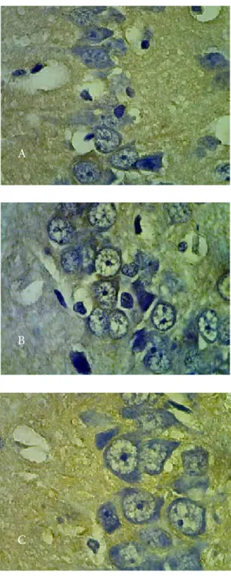
Bax expressions were scored with Allred scoring that considers the proportion of the positive cells on the scale of 0 to 5 and color intensity on the scale of 0 to 3. The sum of the result from both parameters would be used to interpret Bax expression. If the result was 0-2 then the Bax expression was considered negative, while the result of 3-8 considered positive (Picture 1).
Result
The ethical clearancefor this study is registered number 19/Ka.Kom.Et/70/KE/VIII/2017. From Ethics Committee Faculty of Medicine Universitas Islam Indonesia. This study used 15 male wistarstrain rats that fulfilled inclusion and exclusion criteria. There are three group: group A contained of BCCAO-induced rats that received black sugarcane’s ethanol extract from the feed tube; group B consisted of BCCAO-induced rats that were not received black sugarcane’s ethanol extract from the feed tube; group C consisted of sham-operated rats.
The measuring of the rats’ ischemic brain volume was performed using TTC staining. The result of the staining can be seen below:
Picture 2. TTC staining result a. BCCAO rats+Black sugarcane, b. BCCAO rats, c. Sham group
The ischemic part of the brain would be white-colored, while the non-ishcemic were red colored. The ischemic areas from each group were analyzed using Software Image J program. The result of the mean test of the brain ischemia’s volume was analyzed with One Way Anova and post-hoc (table 1)
Table 1. ANNOVA test
Group (mmMean 2) SD ANOVAP Value
Post Hoc
BCCAO +
sugarcane BCCAO sham BCCAO +
sugarcane 2,86 2,44 0,018*
0,016* 0,848
BCCAO 9,40 5,23 0,016* 0,011
Sham 2,40 2,702 0,011 0,848
This result shown that there are differences of the mean of the rats’ ischemic brain between the sham group (2,40 mm2), BCCAO group
(9,40mm2), and BCCAO + black sugarcane group
(2,86 mm2) (p value 0,018). Post-hoc test shown
that there is no difference of the percentage of the ischemic area between the sham group and BCCAO+black sugarcane groups. In other hand, there are significant differences on BCCAO group compared to other groups.
Picture 3. Bax Expression in Hippocampal neurons. A. Ischemia-inducted with black sugarcane treatment B. ischemia-inducted without black sugarcane treatment. C. sham operated
A
B
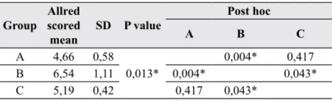
Table 2. Statistic Test for Bax expression
Group Allred scored
mean SD P value
Post hoc
A B C
A 4,66 0,58
0,013* 0,004* 0,417
B 6,54 1,11 0,004* 0,043*
C 5,19 0,42 0,417 0,043*
Discussion
Sugarcane contains carbohydrates, fiber and
minerals, often used as additional nutrients. Phytochemical compounds contained in sugar
cane include alkaloids, saponins, flavonoids,
magnesium minerals, potassium and calcium (11). The policosanol compound is a long chain alcohol compound. This compound is found in several kinds of plants, including rice, olives, and sugarcane. The sugarcane plant (Saccharum
officinarum L.) has various varieties. One of the differentiating varieties is the color of sugar
cane. There are green, yellow, purple to blackish-looking cane. Asikin’s research (2014) states that
the content of policosanol in sugar cane is affected
by cane varieties, structure, extraction techniques, harvest age and demography of the place where the sugarcane is growing(6). A study conducted
in Japan on policosanol content in various cane varieties showed that the Ni-22 variety has the highest policosanol content of 500 mg / 100 g fresh skin weight. Sugarcane skin has the highest level of policosanol (500 mg / 100 g fresh leather weight) compared to other parts such as leaves,
flowers and the cane (355mg / kg stem). The
cane skin coating resembles a candle, the highest source of policosanol. The technique of peeling the skin manually will have higher policosanol content than using a machine. Sugarcane that is 12 months old contains more policosanol compared to sugarcane which is 9 months old (12).
Sugarcane is also a source of antioxidant. This is caused by the high concentration of phenolic (381 mcg/ g drops) in each drops. Inside the sugarcane’s phenolic compound, there are cathecin(16,42 μg/g drops) and quercet in 3-O-glucosyl-xyloxide
(25,27 μg/g drops). Cathecin acts as antioxidant, and quercetin is known of its anti-tumor, anti thrombotic, anti-inflammation, and anti-apoptosis. Quercetin plays active role in cellular mechanism; it inhibits phosphatidylinositol- 3 kinase, C protein kinase, xanthine oxidase and NADPH diaphorase, and it can be said that quercetin has neuro-protective effect (13). The depletion of ROS
level will affect the Bax invasions to mitochondria so apoptosis process can be reduced (14-19).
High concentration of phenolic in sugarcane allows the use of such compounds in prevention and therapy of the disease caused by oxidative stress because sugarcane drops can protect cell, even more protective compared to α-tocopherol(6,20).
Antioxidant activity of phenol compound on the sugarcane process can block an enzyme called lipoperoxidase in rats brain; hence, black sugarcane can be used as neuro-protectant (21).
Sanchez et al (2013) stated that oxidative stress is main key that takes part of causing brain reperfusion injury, after stroke. After the attack, the elevation of reactive oxygen species (ROS) inside the brain is occurred, and so do the damage of the oxidative respiration chain inside the neuron’s mitochondria, and the activation of neural cytoplasm’s oxidation. This ‘mechanism chain’ can not be handled by endogenous antioxidant solo, so the administration of exogenous antioxidant is necessary. Administration of neuro-protective agent vitamin C (500 mg/day, iv) since day one of the ischemic stroke attack can increase the antioxidant in blood, but it fails to improve the stroke patients’ neurologic functions (22).
Another study indicates that sugarcane contains 14-3-3 like proteins. These proteins are similar to protein 14-3-3s that found in neurons’ cytoplasm and mammals’ brain synapse membrane (23). In
cytoplasm, protein 14-3-3s binds with Bax. This stimulation of apoptosis will cause the release of the bindings, so that Bax protein will be translocated to mitochondria. Besides, caspase can cause the release of the bindings between 14-3-3s and Bax. In other hands, the over-increasing of the will block apoptosis process because Bax binds with protein 14-3-3s (24). The increase of protein
14-3-3c can protect brain’s neural cortex when ischemia occurred through Bax-reducing pathway from the interaction of 14-3-3c/p-b-catenin Ser37 inside the neuron’s nucleus (5). The binding of protein 14-3-3
and pro-apoptotis protein Bax, ASK1, BAD, and FOX03, can prevent neural apoptosis(25,26). Protein
14-3-3 plays rol in neurogenesis and neuronal migration(27).
in lowered concentration of SRPK2, FOXO1, and Cdc 25 inside the nucleus, so that the apoptosis process is blocked. Protein 14-3-3 binds Bax in cytoplasma, so mitochondrial Bax is lowered and apoptosis process will be blocked again (25).
This result shows that black sugarcane can act as neuro-protectant through the blocking of Bax protein pathway. Bax expression is regulated with p53 protein that also a transcription factors when activated as part of the cell response on stress and it also regulates plenty of gene targets, one of them is Bax. In other words, p53 can induces apoptosis; in this case it takes role in independent transcription of theapoptosis itself, so that specifically it interacts with Bax, and then promotes activation inside mitochondria, and it increase Bax expression.
The depletion of the bax protein expression on hippocampal part of the rats’ brain with black sugarcane administration is also followed by the depletion of the volume of the brain ischemia as well. This result matches previous study, where the lower number of the brain ischemia was also found in the post-tBCCAOrats’s brain that received boiled sugarcane water (4). The
observation of the ischemia volume of the rat brain is performed with TTC staining. This compound will bind with the dehydrogenase and NAD co-factor in mitochondria (28). The necrotic cells
swell, intra-cell organelles and plasmic membrane break and come out toward plasma, and one of them is Lactate dehydrogenase (LDH) (29). This
enzyme will go to the plasma so the measuring of the plasma-enzyme level can be performed. LDH is more sensitive for ischemic brain incident. This
enzyme can be used to grade stroke incident as well (1).
Conclusion
There is an effect of black sugarcane extract on ischemia volume and Bax expression of rat’s brain post-tBCCAO.
Conflict of interests : No relevant disclosures Acknowledgement
This study was used a grant from UPPM (Unit Penelitian dan Pengabdian Masyarakat), Faculty of medicine, Islamic University of Indonesia. 1. Authors’s contribution:
Data gathering and idea owner of this study: Ety Sari Handayani, Alfan Nur, Kuswati, Zainuri Sabta Nugraha, Nastiti Widya Ikhsani, Faisal Ridho Sakti, Syaefudin Ali Ahmad.
Study design: Ety Sari Handayani, Kuswati, Zainuri Sabta Nugraha
Data gathering: Ety Sari Handayani, Alfan Nur, Kuswati, Zainuri Sabta Nugraha, Nastiti Widya Ikhsani, Faisal Ridho Sakti, Syaefudin Ali Ahmad Writing and submitting manuscriptEty Sari Handayani, Nastiti Widya Ikhsani, Faisal Ridho Sakti, Syaefudin Ali ahmad
Editing and approval of final draft: Ety Sari Handayani, Syaefudin Ali Ahmad
2. Ethical clearence: The ethical clearance for this study is registered number 19/Ka.Kom. Et/70/KE/VIII/2017 from Ethics committee of Medical Faculty , Universitas Islam Indonesia 3. Source of Funding: UPPM (Unit Penelitian
dan Pengabdian Masyarakat), Faculty of medicine, Islamic University of Indonesia
References:
1. Handayani ES, Nurmasitoh T, Akhmad AS, Fauziah
AN, Rizani R, Rahmawaty YR, et al. Effect of
BCCAO Duration and Animal Models Sex on Brain Ischemic Volume After 24 Hours Reperfusion. Bangladesh J Med Sci. 2018;17(01):129–37.
2. Gnanaraj RA. Applications of Sugarcane Wax and it ’ s Products : A Review. Int J ChemTech Res. 2012;4(2):705–12.
3. Molina V, Ravelo Y, Noa M, Mas R, Valle M. Effects
of Policosanol and Grape Seed Extract Against Global Brain Ischemia-Reperfusion Injury in Gerbils. Lat Am J Pharm. 2013;32(1):2383.
4. Handayani ES, Nugraha ZS, Nurmasitoh T, Ahsani
DN, Nanda AG. Black sugarcane decoction reduces rat brain ischemia. Universa Med. 2016;35(1):40–5. 5. Lai XJ, Ye SQ, Zheng L, Li L, Liu QR, Yu SB, et al.
Selective 14-3-3 c induction quenches p- b -catenin Ser37 / Bax-enhanced cell death in cerebral cortical neurons during ischemia. Cell Death Dis. 2014;5:1– 13.
6. Asikin Y. Flavor Characteristics and Biological Functions of Okinawan Sugary and Citrus Materials. Kagoshima University, Japan; 2014.
7. Molina V. Effect of policosanol on cerebral ischemia
in Mongolian gerbils. 1999;32:1269–76.
Artery Occlusion (tBCCAO) of Rats as a Model of Global Cerebral Ischemia. Bangladesh J Med Sci. 2019;18(03):491–500.
9. Qureshi A, Pervez S. Allred scoring for ER reporting and it’s impact in clearly distinguishing ER negative from ER positive breast cancers. J Pak Med Assoc. 2010;60(5):350–3.
10. TEWARY S, ARUN I, AHMED R, CHATTERJEE S, CHAKRABORTY C. Auto IHC-scoring: a machine learning framework for automated Allred scoring of
molecular expression in ER‐ and PR‐stained breast
cancer tissue. J Microsc. 2017;268(2).
11. Williams IO, Onyenweaku EO, Atangwho IJ. Nutritional and antimicrobial evaluation of Saccharum
officinarum consumed in Calabar , Nigeria. African J
Biotechnol. 2016;15(33):1789–95.
12. Asikin Y, Takahashi M, Mishima T, Mizu M, Takara K. Antioxidant activity of sugarcane molasses against 2 , 2 0 -azobis ( 2-amidi- nopropane ) dihydrochloride-induced peroxyl radicals. Food Chem [Internet]. Elsevier Ltd; 2013;141(1):466–72. Available from: http://dx.doi.org/10.1016/j.foodchem.2013.03.045 13. Dajas F, Rivera-Megret A, Blasina F, Arrendondo F,
Abin-Carriquiry JA, Costa G, et al. Neuroprotection
by flavonoids. Brazilian J Med Biol Res.
2003;36(12):1613–20.
14. Niizuma K, Endo H, Nito C, Myer DJ, Kim GS, Chan PH. The PIDDosome mediates delayed death of hippocampal CA1 neurons after transient global cerebral ischemia in rats. Proc Natl Acad Sci U S A [Internet]. 2008;105(42):16368–73. Available from: http://www.pnas.org/content/105/42/16368.full 15. Nishijima Y, Niizuma K, Fujimura M, Akamatsu Y,
Shimizu H, Tominaga T. Consistent delayed unilateral
neuronal death after modified transient focal cerebral
ischemia in mice that mimics neuronal injury after transient global cerebral ischemia. J Neurosurg [Internet]. 2015 Jul [cited 2016 Oct 13];123(1):243– 53. Available from: http://www.ncbi.nlm.nih.gov/ pubmed/25723306
16. Thomas H. Sandersona, b, c,*, Christian A. Reynoldsa, b, c, Rita Kumara, b, c K, Przyklenka, b, c, and Maik Hüttemanna D. Molecular Mechanisms of Ischemia-Reperfusion Injury in Brain: Pivotal Role of the Mitochondrial Membrane Potential in Reactive Oxygen Species Generation. Mol Neurobiol. 2014;47(1):9–23.
17. Radak D, Resanovic I. Link Between Oxidative Stress and Acute Brain Ischemia. Angiology. 2014;65(8):667–76.
18. Yoshioka H, Niizuma K, Katsu M, Okami N, Sakata H, Kim GS, et al. NADPH oxidase mediates striatal neuronal injury after transient global cerebral ischemia.
J Cereb Blood Flow & Metab [Internet]. Nature Publishing Group; 2010;31(3):868–80. Available from: http://dx.doi.org/10.1038/jcbfm.2010.166 19. Niizuma K, Endo H, Chan PH. Oxidative stress
and mitochondrial dysfunction as determinants of ischemic neuronal death and survival. J Neurochem [Internet]. 2009;109:133–8. Available from: http:// doi.wiley.com/10.1111/j.1471-4159.2009.05897.x 20. Veronica V, Gómez-Caravaca§ AM, Nunzio
M Di, Danesi F, Caboni MF, Bordoni A. Sugar Cane and Sugar Beet Molasses, Antioxidant-rich
Alternatives to Refined Sugar. J Agric Food Chem.
2012;60(51):12508–15.
21. Miraj S. Pharmacological effects of Saccharum officinarum L . Der Pharm Lett. 2016;8(13):223–5.
22. Sanches EF, Arteni NS, Scherer EB, Kolling J, Nicola F, Willborn S, et al. Are the consequences of neonatal hypoxia-ischemia dependent on animals’ sex and brain lateralization? Brain Res [Internet]. Elsevier; 2013;1507:105–14. Available from: http://dx.doi. org/10.1016/j.brainres.2013.02.040
23. Kuramae EE, Fenille RC, Eugenio V, Jr DR.
Identification of 14-3-3-like protein in sugarcane ( Saccharum officinarum ). Genet Mol Biol.
2001;24:43–8.
24. Nomura M, Shimizu S, Sugiyama T, Narita M, Ito T, Matsuda H, et al. 14-3-3 Interacts Directly with and Negatively Regulates Pro-apoptotic Bax *. J Biol Chem. 2003;278(3):2058–65.
25. Shimada T, Fournier AE, Yamagata K. Neuroprotective Function of 14-3-3 Proteins in Neurodegeneration. Biomed Res Int. 2013;2013.
26. Khudhair N, Cuiping Y, Khalid A, Gao X. Role 14-3-3 Protein in Regulation Some Cellular Processes. Curr Res J Biol Sci. 2014;6(5):197–204.
27. Toyo-oka K, Wachi T, Hunt RF, Baraban SC, Taya S, Ramshaw H, et al. 14-3-3? and ? Regulate
Neurogenesis and Differentiation of Neuronal
Progenitor Cells in the Developing Brain. J Neurosci. 2014;34(36):12168–81.
28. Rekabi MD, Hussein FH, Mosawi A, Alwan MS, Hussein AH, Shaheed DK. Histopathological
Effects of L-Methionine in. Br J Med Heal Res.
2015;2(August).