original article
Clinical Presentation of Patients with Ebola
Virus Disease in Conakry, Guinea
Elhadj Ibrahima Bah, M.D., Marie-Claire Lamah, M.D., Tom Fletcher, M.R.C.P., Shevin T. Jacob, M.D., M.P.H., David M. Brett-Major, M.D., M.P.H., Amadou Alpha Sall, Ph.D., Nahoko Shindo, M.D., Ph.D., William A. Fischer II, M.D.,
Francois Lamontagne, M.D., Sow Mamadou Saliou, M.D.,
Daniel G. Bausch, M.D., M.P.H.&T.M., Barry Moumié, M.D., Tim Jagatic, M.D., Armand Sprecher, M.D., James V. Lawler, M.D., M.P.H., Thierry Mayet, M.D.,
Frederique A. Jacquerioz, M.D., María F. Méndez Baggi, M.D., Constanza Vallenas, M.D., Christophe Clement, M.D., Simon Mardel, M.D.,
Ousmane Faye, Ph.D., Oumar Faye, Ph.D., Baré Soropogui, Pharm.D., Nfaly Magassouba, D.V.M., Ph.D., Lamine Koivogui, Pharm.D., Ph.D.,
Ruxandra Pinto, Ph.D., and Robert A. Fowler, M.D.C.M.
The authors’ affiliations are listed in the Appendix. Address reprint requests to Dr. Fowler at the Departments of Medi-cine and Critical Care MediMedi-cine, Sunny-brook Health Sciences Centre, University of Toronto, 2075 Bayview Ave., Toronto, ON M4N 3M5, Canada, or at rob.fowler@ sunnybrook.ca.
This article was published on November 5, 2014, at NEJM.org.
N Engl J Med 2015;372:40-7. DOI: 10.1056/NEJMoa1411249
Copyright © 2014 Massachusetts Medical Society.
ABS TR ACT
Background
In March 2014, the World Health Organization was notified of an outbreak of Zaire
ebolavirus in a remote area of Guinea. The outbreak then spread to the capital,
Conakry, and to neighboring countries and has subsequently become the largest epidemic of Ebola virus disease (EVD) to date.
Methods
From March 25 to April 26, 2014, we performed a study of all patients with laboratory-confirmed EVD in Conakry. Mortality was the primary outcome. Secondary out-comes included patient characteristics, complications, treatments, and comparisons between survivors and nonsurvivors.
Results
Of 80 patients who presented with symptoms, 37 had laboratory-confirmed EVD. Among confirmed cases, the median age was 38 years (interquartile range, 28 to 46), 24 patients (65%) were men, and 14 (38%) were health care workers; among the health care workers, nosocomial transmission was implicated in 12 patients (32%). Patients with confirmed EVD presented to the hospital a median of 5 days (inter-quartile range, 3 to 7) after the onset of symptoms, most commonly with fever (in 84% of the patients; mean temperature, 38.6°C), fatigue (in 65%), diarrhea (in 62%), and tachycardia (mean heart rate, >93 beats per minute). Of these patients, 28 (76%) were treated with intravenous fluids and 37 (100%) with antibiotics. Sixteen pa-tients (43%) died, with a median time from symptom onset to death of 8 days (in-terquartile range, 7 to 11). Patients who were 40 years of age or older, as compared with those under the age of 40 years, had a relative risk of death of 3.49 (95% confidence interval, 1.42 to 8.59; P = 0.007).
Conclusions
E
bola virus is one of three membersof the Filoviridae family and comprises five distinct species. Infection with Zaire
ebolavirus (EBOV) has historically resulted in the
highest case fatality rate — up to 90%.1
Out-breaks typically originate with introduction of the virus into humans from a wild animal reser-voir, with subsequent human-to-human trans-mission, often fueled by nosocomial amplifica-tion in resource-poor settings. Aside from a single infection with Tai Forest ebolavirus, West Africa has never had an outbreak of Ebola virus disease (EVD).2,3
The Republic of Guinea, on the west coast of Africa, has a population of approximately 11 mil-lion persons, a life expectancy at birth of 58 years, and an annual gross national income of 970 inter-national dollars, with an expenditure on health care of 67 international dollars per capita per year.4 Conakry, the capital and largest city, with
an approximate population of 2 million persons, is served by a number of large hospitals, including Donka Hospital (the major public and university-affiliated medical center), Ignace Deen Hospital, and the Hôpital de l’Amitié Sino-Guinéenne, in addition to a number of privately funded health clinics.
On March 21, 2014, the World Health Orga-nization (WHO) was formally notified of a rap-idly evolving outbreak of EVD centered in the prefecture of Guéckédou, in the forested region of southeastern Guinea, with potential spread to border areas in Liberia and Sierra Leone.5
Infected travelers from Guéckédou subsequent-ly initiated chains of transmission of EVD in Conakry, more than 600 km away, marking the world’s largest urban EVD outbreak and herald-ing the largest ever EVD epidemic, involvherald-ing Guinea, Sierra Leone, Liberia, Nigeria, Senegal, and Mali.6 A concurrent but epidemiologically
unrelated outbreak has also been recognized in the Democratic Republic of Congo.6
The care of patients with EVD in Conakry initially occurred at two sites: the Hôpital de l’Amitié Sino-Guinéenne, which mainly focused on a large nosocomial outbreak affecting health care workers, and a stand-alone EVD treatment unit established on the grounds of Donka Hos-pital by the Ministry of Health, supported by Médecins sans Frontières and the WHO. As of October 31, 2014, the Guinea Ministry of Health had reported a cumulative total of 1667 clinical cases of EVD (with 1018 deaths), including 244
cases (and 98 deaths) in Conakry; a total of 13,562 cases (and 4950 deaths) were reported throughout West Africa.7 Here we describe
de-mographic and clinical characteristics of the patients at presentation, the clinical course of EVD, and outcomes of all patients admitted for care in Conakry during a 1-month period at the onset of the outbreak.
Methods
Study Design
We conducted a retrospective, observational study of all patients with suspected or confirmed EVD who were admitted for care in Conakry from March 25 to April 26, 2014 (Fig. S1 in the Supplementary Appendix, available with the full text of this article at NEJM.org). We used a stan-dard case definition that was established by the WHO and the Guinea Ministry of Health (Table S1 in the Supplementary Appendix). Laboratory confirmation of EVD was made on the basis of results on quantitative reverse-transcriptase– polymerase-chain-reaction (RT-PCR) assay in a laboratory established at Donka Hospital by the Institute Pasteur in Dakar, Senegal. Laboratory staff members used rapid Taqman RT-PCR assays for the detection of EBOV using 5-FAM and 3-TAMRA tagged probes and a portable Smart-Cycler TD. EBOV RNA in patient samples was measured according to the standard described by Weidmann et al.,8 in which the sample is diluted
to a range of 1 copy to 1 million copies and is then tested in quadruplicate to construct a stan-dard curve for estimating the number of genome copies. Follow-up testing for antibodies against EBOV by means of enzyme-linked immunosor-bent assay (ELISA) was performed as needed. Pa-tients were treated in accordance with protocols established for viral hemorrhagic fever by Méde-cins sans Frontières and WHO urgent interim guidance for case management, endorsed by the Ministry of Health.9,10
Data Collection
were admitted to EVD treatment units. Admis-sion data were reviewed daily by clinicians. Ap-proval and a waiver from the need to provide written informed consent were obtained from the ethics review committees for the Guinean government and the WHO.
Statistical Analysis
We used Student’s t-test, Fisher’s exact test, or the Wilcoxon rank-sum test, as appropriate, to determine the association between mortality and
the clinically informed variables of age, sex, oc-cupation, the presence of gastrointestinal hem-orrhage, the number of days between symptom onset and presentation, and viral load at presen-tation. We used Kaplan–Meier methods and log-rank tests to determine survival curves for vari-ous groups of patients, using the interval from the onset of symptoms to death for those who died within 28 days, with data censored at 28 days for survivors. We explored associations be-tween the above-mentioned variables and death in univariate analyses.
We used a multivariate Poisson regression analysis with robust standard errors to investi-gate our three primary clinically informed hy-potheses: that mortality was associated with older age, an increased interval from symptom onset until presentation to a treatment facility, and an increased viral load.11,12 We examined
survival according to age using a scatter plot and 5-year age bins to determine the most appropri-ate dichotomous age comparisons. We log-transformed viral loads for primary compari-sons among survivors and nonsurvivors and performed sensitivity analyses dichotomizing viral loads using two sets of values (less than the sample median vs. greater than or equal to the sample median and <100,000 copies per millili-ter vs. ≥100,000 copies per millilimillili-ter). All statis-tical tests were two-tailed, with a P value of less than 0.05 considered to indicate statistical sig-nificance. All analyses were performed with the use of SAS software, version 9.3 (SAS Institute), and R software, version 2.15.1.
R esults
Study Patients
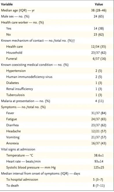
Eighty patients who had symptoms meeting the definition of suspected EVD were admitted to the two treatment facilities in Conakry. Among these patients, 37 (46%) were confirmed to have EVD, 36 (97%) by means of RT-PCR and 1 who had negative results on RT-PCR assay but had positive results for IgG antibodies on ELISA (Table 1). The latter patient had a clinical syndrome com-patible with EVD and was a close contact of an-other patient with confirmed disease.
The median age of the confirmed cases was 38 years (range, 19 to 61), and 24 (65%) were men. The most common mechanism of contact was through household clusters, which account-ed for 23 cases (62%). Fourteen patients (38%)
Table 1. Characteristics, Symptoms, Vital Signs, and Time Course of Clinical Progression of 37 Patients with Confirmed Ebola Virus Disease (EVD).*
Variable Value
Median age (IQR) — yr 38 (28–46)
Male sex — no. (%) 24 (65)
Health care worker — no. (%)
Yes 14 (38)
No 23 (62)
Known mechanism of contact — no./total no. (%)†
Health care 12/34 (35)
Household 23/37 (62)
Funeral 6/37 (16)
Known coexisting medical condition — no. (%)
Hypertension 2 (5)
Human immunodeficiency virus 2 (5)
Diabetes 1 (3)
Renal insufficiency 1 (3)
Tuberculosis 1 (3)
Malaria at presentation — no. (%) 4 (11)
Symptoms — no./total no. (%)
Fever 31/37 (84)
Fatigue 24/37 (65)
Diarrhea 23/37 (62)
Headache 12/21 (57)
Vomiting 21/37 (57)
Anorexia 16/37 (43)
Vital signs at admission
Temperature — °C 38.6±1
Heart rate — beats/min 93±14
Systolic blood pressure — mm Hg 125±25
Median interval from onset of symptoms (IQR) — days
To hospital admission 5 (3–7)
To death 8 (7–11)
were health care workers, in whom nosocomial transmission was implicated in 12 of 34 patients (35%). Participation in funeral ceremonies of confirmed cases was an additional risk factor for 6 of these patients (16%).
Clinical features at presentation were nonspe-cific and included fever in 31 patients (84%; mean temperature, 38.6°C), fatigue in 24 patients (65%), gastrointestinal symptoms (in 23 patients with diarrhea [62%] and 21 with vomiting [57%]), headache in 12 of 21 patients who were evaluated (57%), and anorexia in 16 patients (43%) (Table 1). At admission, patients had mild tachycardia (mean [±SD], 93±14 beats per min-ute) with a mean systolic blood pressure of 125±25 mm Hg. Hiccups occurred in 28% of patients during the period of hospitalization. The median time from symptom onset to pre-sentation was 5 days (interquartile range, 3 to 7), and the median time from symptom onset to death was 8 days (interquartile range, 7 to 11).
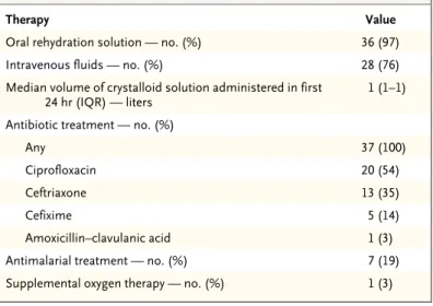
Oral rehydration solution was given to 36 pa-tients (97%), and 28 papa-tients (76%) received ad-ditional intravenous fluid resuscitation (Table 2). A median of 1 liter of intravenous crystalloids was administered during the first 24 hours after admission. Antibiotics were administered empiri-cally in 37 patients (100%) with gastrointestinal symptoms, and artemisinin-based combination therapy was administered in 7 patients (19%), of whom 4 had confirmed Plasmodium falciparum infection on rapid diagnostic testing. One pa-tient (3%) received supplemental oxygen therapy for hypoxemia.
For the first 3 weeks of the outbreak, no rou-tine clinical laboratory testing was available. For approximately 1 week, we used an i-STAT System point-of-care device with CHEM8+ and CG4+ cartridges (Abbott Point of Care) to perform lim-ited diagnostic testing in 3 patients with clinical symptoms in the treatment center. In 1 patient, we found severe prerenal kidney dysfunction (creatinine, 13.9 mg per deciliter [1229 μmol per liter]; and blood urea nitrogen, >140 mg per deci-liter [>50.0 mmol per deci-liter]) and accompanying metabolic acidosis (pH, 7.21; and lactate, 7.4 mmol per liter), results that improved after the admin-istration of approximately 5 liters of intravenous crystalloid fluids per day for 3 days (creatinine, 2.4 mg per deciliter [212 μmol per liter]; blood urea nitrogen, 40 mg per deciliter [14.3 mmol per liter]; pH, 7.46; and lactate, 1.1 mmol per
liter). Similar findings were noted in a second patient (creatinine, 4.9 mg per deciliter [433 μmol per liter]; and blood urea nitrogen, 73 mg per deciliter [26.1 mmol per liter]), findings that were probably caused by profound diarrhea (potassium, 2.4 mmol per liter; and bicarbonate, 15 mmol per liter), which also resolved after the administration of approximately 4 liters of intra-venous crystalloid fluids per day for 3 days along with potassium. Among 3 patients in whom anemia was suspected, including 1 patient with clinical evidence of lower gastrointestinal bleed-ing, hematocrit levels were not profoundly low (mean, 31.2±3.4).
Among the 16 patients (43%) who died, the median duration of hospital stay was 5 days, as compared with 9 days (interquartile range, 6 to 11) among survivors. The most common clinical complication was hemorrhage, which was report-ed in 19 patients (51%), most frequently gastro-intestinal bleeding (in 9 patients), of whom 8 pa-tients had melena and 1 patient each had hematemesis and hematochezia (Table 3).
In univariate analyses, the viral load appeared to be higher among patients who died than in survivors (Table 4), but this finding was influ-enced by the deaths of all 4 patients who had a viral load of more than 100,000 copies per mil-liliter on admission, and viral load was not signifi-cantly associated with death on nonparametric testing. Mortality was not significantly higher among the few patients who had coexisting con-ditions than in those without such concon-ditions (57.1% vs. 40.0%, P = 0.44). However, an older
Table 2. Therapies Received by 37 Patients Hospitalized for EVD.
Therapy Value
Oral rehydration solution — no. (%) 36 (97)
Intravenous fluids — no. (%) 28 (76)
Median volume of crystalloid solution administered in first
24 hr (IQR) — liters 1 (1–1)
Antibiotic treatment — no. (%)
Any 37 (100)
Ciprofloxacin 20 (54)
Ceftriaxone 13 (35)
Cefixime 5 (14)
Amoxicillin–clavulanic acid 1 (3)
Antimalarial treatment — no. (%) 7 (19)
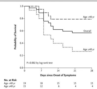
age was associated with an increased risk of death, with a median age of 29 years in survivors as compared with 45 years in those who died (P = 0.005) (Fig. 1). In a multivariable Poisson regression analysis that was adjusted for age, viral load, and time from symptom onset to presentation, patients who were 40 years of age or older had a relative risk of death of 3.49 (95% confidence interval [CI], 1.42 to 8.59), as com-pared with those under the age of 40 years
(P = 0.007). There were no significant differences between survivors and nonsurvivors in the num-ber of days between symptom onset and admis-sion (relative risk in survivors, 0.94; 95% CI, 0.86 to 1.04; P = 0.22) and viral load on admission (relative risk, 0.98; 95% CI, 0.91 to 1.07; P = 0.72).
Discussion
Patients, on average, presented 5 days after symptom onset, and the most common manifes-tations of EVD during hospitalization were fever, vomiting, diarrhea, and related volume depletion requiring the administration of intravenous flu-ids and electrolyte therapy. Overall mortality among patients presenting for treatment was 43%, and only the age of the patient was a sig-nificant predictor of outcome. In contrast to pre-vious Ebola virus outbreaks in which an older age was also associated with a worse outcome, the mean age of nonsurvivors in our study was low.13,14 The association between an older age
and a worse outcome among patients with viral infections is often attributed to an increased number of coexisting conditions. However, in our study, the relative absence of known coexist-ing conditions suggests that an older age may have an independent association with mortality. We also found that patients who presented for care with the highest viral loads were the least likely to survive, as has been shown for other strains of Ebola virus.15 After adjustment for
dif-Table 3. Clinical Complications and Outcomes for 37 Patients with EVD.
Variable Value
Hospital mortality — no. (%) 16 (43)
Median length of stay in hospital (IQR) — days 8 (6–11) Known complications in hospital — no. (%)
Hemorrhage
Any 19 (51)
Gastrointestinal 9 (24)
Subconjunctival 4 (11)
Intravenous catheter site 4 (11)
Nasorespiratory tract 2 (5)
Renal failure* 2 (5)
Seizure 2 (5)
Oral candidiasis 1 (3)
Hypoxemia 1 (3)
* Renal failure was defined as a serum creatinine level of more than 4 mg per deciliter (350 μmol per liter).
Table 4. Characteristics of Survivors and Nonsurvivors.
Characteristic Survivors (N = 21) Nonsurvivors(N = 16) P Value
Median age (IQR) — yr 29 (26–37) 45 (40–47) 0.005
Male:female ratio 14:7 10:6 1.00
Viral load at admission
Mean ±SD — copies/ml 8207±17,189 68,361±111,340 0.02
Median (IQR) — copies/ml 1079 (148–5059) 1915 (141–12,998) 0.47
>100,000 copies/ml — no. (%) 0 4 (25) 0.02
Health care worker — no. (%) 6 (29) 8 (50) 0.31
Clinical features
Hemorrhage — no. (%)
Any visible 8 (38) 11 (69) 0.1
Gastrointestinal 4 (19) 6 (38) 1.0
Interval from symptom onset to presentation (IQR) —
ferences in age and time to presentation, this relationship was not significant. However, our study had limited power to detect all predictors of outcome because of the small number of pa-tients.
The case fatality rate that we observed in this cohort in the capital city of Conakry was lower than the rate reported in most studies of previ-ous EVD outbreaks1 (although not in all
stud-ies7) and was lower than the rate in most other
regions in Guinea at that point in the epidem-ic.7,13 Clinical care at the main isolation facility
near Donka Hospital was jointly provided by the Ministry of Heath, Médecins sans Frontières, and the WHO during the study period. Adher-ence to new guidelines promoting increased medical interventions, particularly related to the use of oral and intravenous fluids and electrolyte replacement, appropriate antibiotics, and targeted clinical laboratory testing,9 may have
contribut-ed to the rcontribut-educcontribut-ed case fatality rate, as comparcontribut-ed with past outbreaks. However, assessing associa-tions between treatments and outcomes in small observational studies is challenging. In our study, there were approximately three clinical rounds per day, with two or three doctors and two or three nurses for each round, and depend-ing on the type of personal protective equipment that was used, rounds were limited to either 1 hour or 3 hours because of the intense heat and hu-midity inside some types of personal protective equipment. Although we attempted to deliver oral and intravenous fluids to correct dehydra-tion and metabolic abnormalities, care was still suboptimal. With more clinical personnel in each treatment center, better supportive care could be delivered more consistently, and we think that mortality could be driven lower.
The predominant clinical syndrome of EVD involves substantial volume loss due to vomiting and diarrhea. This requires aggressive oral and intravenous volume repletion and close follow-up to avoid further complications and hypoper-fusion-associated organ dysfunction. Point-of-care diagnostic testing provided additional insights in a small number of patients, suggesting inad-equate tissue perfusion — lactic acidosis, base deficits, prerenal kidney dysfunction, and low venous oxygen saturations. However, we could not perform such testing early or frequently enough to properly define the patterns. The sub-stantial volume loss and profound electrolyte
derangement from copious diarrhea represent opportunities to intervene clinically to improve outcomes. Notably, hypoxemia was rarely seen in these patients, despite attempts at aggressive volume repletion. However, this finding may still represent inadequate volume administration, and hypoxemia caused by pulmonary vascular leak may be more common in other care settings. Consideration may also be given to the empirical use of antimalarial therapy, especially if rapid diagnostic testing is not immediately available. Patients with severe gastrointestinal symptoms were also routinely treated with a finite empiri-cal course of antibiotics with activity against gram-negative, gram-positive, and anaerobic or-ganisms. However, the effect of this intervention remains unknown.
As has been seen in other disease outbreaks, but never before with EVD, large urban settings present special challenges to emergency health care facilities, and nosocomial transmission among health care staff members and patients represents an important potential outbreak am-plification and new lines of transmission.16,17
This finding highlights the importance of rapid support for infection control, not only in dedi-cated isolation facilities but also within existing treatment centers that will typically receive
un-Probability of Survival
1.0
0.8
0.6
0.4
0.2
0.0
0 7 14 21 28
Days since Onset of Symptoms P=0.002 by log-rank test
No. at Risk Age <40 yr
Age ≥40 yr 1915 1812 156 154 154
Age <40 yr
Age ≥40 yr Overall
differentiated patients with fever and nonspe-cific symptoms. Infection-control practices to protect patients and health care workers can have unanticipated negative consequences, in-cluding fewer clinical assessments, which may be compounded by limited clinician time at the bedside because of heat exposure in personal protective equipment.18,19
Limitations of our study include reliance on estimates from a discrete but relatively small cohort of patients. However, the observed mor-tality in Conakry (43%) has remained relatively stable (40%) between April and October 2014.20
Despite an active public health system for trac-ing contacts, case findtrac-ing, and referral to an acute care facility, we inevitably are unable to identify all patients with suspected and con-firmed EVD, since some will choose not to pre-sent to health care facilities and some will die before seeking medical attention. This potential selection bias will underestimate the number of cases and have uncertain effects on the case fa-tality rate for this epidemic. The inclusion of only patients who could be transported to our health care facilities may lead to a survivorship and immortal time bias, since patients with EVD needed to have survived long enough to get to a facility in order to be described and to receive certain treatments. Furthermore, selection of patients for certain treatments is subject to bias according to indication, which can lead to an overestimation of harm for certain therapies.21
Therefore, in this observational study, we are unable to validly explore relationships between
treatments received and clinical outcomes, which underscores the importance of enhanced strategies for supportive care and specific thera-pies in future clinical trials.
Our recording of simple clinical data was also limited by an inability to take any material, in-cluding paper, outside the treatment center, by limited electricity to power onsite electronic data capture, and by unreliable Internet access. Inside the treatment facility, with too few clinical staff members, there is often a traoff between de-livering and recording care. A further limitation is the paucity of basic data regarding blood chem-istry and hematology that would better charac-terize metabolic abnormalities and help to direct care for future patients. Point-of-care testing inside treatment centers is challenging because of a lack of time to perform testing due to high temperatures and dehydration of health care providers. Routine deployment of basic chemis-try and hematology analyzers in addition to RT-PCR assays for EBOV in international mobile laboratories would alleviate this limitation and may guide further improvements in patient care.
In conclusion, we found that among patients admitted to the hospital with confirmed EVD in Conakry, Guinea, the most common clinical syndrome was one of gastrointestinal illness, intravascular volume depletion, and related com-plications, which highlight the importance of enhanced levels of clinical assessment and diag-nostic testing, along with fluid management.
Disclosure forms provided by the authors are available with the full text of this article at NEJM.org.
Appendix
The authors’ affiliations are as follows: Donka Hospital (E.I.B., M.-C.L., S.M.S., B.M.), Projet de Fièvre Hémorragique Guinée, Univer-sité Gamal Abdel Nasser (B.S., N.M.), and Institut National de Santé Conakry (L.K.) — all in Conakry, Guinea; University of Liverpool, Liverpool (T.F.), and Emergency Department University Hospital of South Manchester, Manchester (S.M.) — both in the United King-dom; Hospital Mulago, Masaka, Uganda (S.T.J.); the Department of Medicine, University of Washington, Seattle (S.T.J.); Preparedness and Mass Gatherings, Global Preparedness, Surveillance and Response Operations, Global Capacities Alert and Response, Health Se-curity and Environment (D.M.B.-M.), and the Department of Pandemic and Epidemic Diseases (N.S., C.V.), World Health Organization, Geneva; Institut Pasteur de Dakar, Dakar, Senegal (A.A.S., Ousmane Faye, Oumar Faye); the Division of Pulmonary and Critical Care Medicine, University of North Carolina School of Medicine, Chapel Hill (W.A.F.); Centre de Recherche du Centre Hospitalier Universi-taire de Sherbrooke, Université de Sherbrooke, Sherbrooke, QC (F.L.), and the Trauma Emergency and Critical Care Program (R.P.) and Departments of Medicine and Critical Care Medicine (R.A.F.), Sunnybrook Health Sciences Centre and University of Toronto, Toronto — both in Canada; Virology and Emerging Infections Department, U.S. Naval Medical Research Unit No. 6, Lima, Peru (D.G.B.); the Department of Tropical Medicine, Tulane School of Public Health and Tropical Medicine, New Orleans (D.G.B., F.A.J., M.F.M.B.); Médecins sans Frontières, Brussels (T.J., A.S.); Naval Medical Research Center–Frederick, Frederick, MD (J.V.L.); and Service de Réani-mation Polyvalente, Centre Hospitalier de Dax, Dax (T.M.), and the Intensive Care Unit, Private Hospital Polyclinique Bordeaux Nord Aquitaine, Bordeaux (C.C.) — both in France.
References
1. World Health Organization. Ebola virus disease fact sheet (http://www.who .int/mediacentre/factsheets/fs103/en).
2. Beeching NJ, Fletcher TE, Hill DR, Thomson GL. Travellers and viral hae-morrhagic fevers: what are the risks? Int J
Antimicrob Agents 2010;36:Suppl 1:S26-S35.
Pre-vention. Outbreaks chronology: Ebola vi-rus disease (http://www.cdc.gov/vhf/ebola/ outbreaks/history/chronology.html).
4. World Health Organization. Guinea: statistics (http://www.who.int/countries/ gin/en).
5. World Health Organization. Ebola vi-rus disease in Guinea (http://www.afro .who.int/en/clusters-a-programmes/dpc/ epidemic-a-pandemic-alert-and-response/ outbreak-news/4063-ebola-hemorrhagic -fever-in-guinea.html).
6. World Health Organization. Industry leaders and key partners discuss trials and production of Ebola vaccine (http:// who.int/csr/disease/ebola/en/).
7. World Health Organization. Ebola response roadmap update (http://apps .who.int/iris/bitstream/10665/136645/1/ roadmapupdate17Oct14_eng.pdf?ua=1).
8. Weidmann M, Mühlberger E, Hufert FT. Rapid detection protocol for filovi-ruses. J Clin Virol 2004;30:94-9.
9. Sterk E, Médecins Sans Frontières. Filovirus haemorrhagic fever guideline. 2008 (http://www.medbox.org/preview/ 53f1e3e2-a078-464d-ba8e-257e1fcc7b89/ doc.pdf).
10. World Health Organization. Prise en charge clinique des cas de fièvre hémor-ragique virale. March 2014 (http://www .unicef.org/cbsc/files/VHF_pocket_book_ Guinea-2014-French.pdf).
11. Zou G. A modified Poisson regression approach to prospective studies with bi-nary data. Am J Epidemiol 2004;159:702-6.
12. Vittinghoff E, McCulloch CE. Relax-ing the rule of ten events per variable in logistic and Cox regression. Am J Epide-miol 2007;165:710-8.
13. MacNeil A, Farnon EC, Wamala J, et al. Proportion of deaths and clinical fea-tures in Bundibugyo Ebola virus infec-tion, Uganda. Emerg Infect Dis 2010;16: 1969-72.
14. WHO Ebola Response Team. Ebola virus disease in West Africa — the first 9 months of the epidemic and forward projections. N Engl J Med 2014;371:1481-96.
15. Towner JS, Rollin PE, Bausch DG, et al. Rapid diagnosis of Ebola hemorrhagic fever by reverse transcription-PCR in an outbreak setting and assessment of pa-tient viral load as a predictor of outcome. J Virol 2004;78:4330-41.
16. Borchert M, Mutyaba I, Van Kerkhove MD, et al. Ebola haemorrhagic fever break in Masindi District, Uganda: out-break description and lessons learned. BMC Infect Dis 2011;11:357.
17. Fowler RA, Lapinsky SE, Hallett D, et al. Critically ill patients with severe acute respiratory syndrome. JAMA 2003;290: 367-73.
18. Stelfox HT, Bates DW, Redelmeier DA. Safety of patients isolated for infection control. JAMA 2003;290:1899-905.
19. Brearley MB, Heaney MF, Norton IN. Physiological responses of medical team members to a simulated emergency in tropical field conditions. Prehosp Disas-ter Med 2013;28:139-44.
20. World Health Organization. Ebola vi-rus disease outbreak — West Africa (http:// www.who.int/csr/don/2014_09_04_ebola/ en).
21. Delgado-Rodríguez M, Llorca J. Bias. J Epidemiol Community Health 2004;58: 635-41.
Copyright © 2014 Massachusetts Medical Society.
receiveimmediatenotificationwhenanarticle
ispublishedonlinefirst