Volume 2010, Article ID 238792,5pages doi:10.1155/2010/238792
Research Article
The Influence of SiO
2
Shell on Fluorescent Properties of
LaF
3
:Nd
3+
/SiO
2
Core/Shell Nanoparticles
Cui Kai,
1, 2Gao Chao,
1Peng Bo,
1, 3and Wei Wei
31State Key Laboratory of Transient Optics and Photonics, Xi’an Institute of Optics and Precision Mechanics,
Chinese Academy of Science (CAS), Xi’an Shaanxi 710119, China
2Graduate School of the Chinese Academy of Sciences, Beijing 100039, China
3Institute of Advanced Materials, Nanjing University of Posts and Telecommunications, Nanjing 210003, China
Correspondence should be addressed to Peng Bo,[email protected]
Received 1 July 2010; Accepted 1 October 2010 Academic Editor: Hongchen Chen Gu
Copyright © 2010 Cui Kai et al. This is an open access article distributed under the Creative Commons Attribution License, which permits unrestricted use, distribution, and reproduction in any medium, provided the original work is properly cited.
Distinct effects of the SiO2 shell on fluorescence properties of LaF3:Nd3+/SiO2 core/shell nanoparticles were demonstrated by
annealling the nanoparticles at different temperatures. Emission spectra, excitation spectra, and decay curves of the nanoparticles
were measured. A significant improvement of fluorescence intensity was observed for LaF3:Nd3+/SiO2core/shell nanoparticles
annealed at 900◦C. The phenomenon is ascribed to the change of environment of LaF
3:Nd3+core which is imposed by SiO2shell.
And the change is confirmed by the excitation spectra. It provides a useful way to improve fluorescent intensity of the SiO2-coated
LaF3:Nd3+nanoparticles. The lifetime for nanoparticles annealed at 900◦C shows a slight decrease contrast with nanoparticles
annealed at 400 and 600◦C. This is caused by the higher phonon energy of SiO
2than that of LaF3.
1. Introduction
In the past decade, the synthesis of lanthanide-doped nanoparticles has attracted a great deal of attention, since the materials are considered as potentially useful phosphors in lamps, display devices [1], components in optical telecom-munication [2], new optoelectronic devices [3],and probes in biomedical imaging and detection [4]. LaF3 possessing
low phonon energy, adequate thermal and environmental stability, is regarded as excellent host matrixes for per-forming luminescence [5,6]. Nanoparticles of LaF3 doped
with lanthanide ions have been studied for years for their luminescence properties [7, 8]. However, the water and organic molecules absorbed on nanoparticles noticeably hampered their optical efficiency, when the nanoparticles are dispersed into aqueous and organic environment. The O-H and C-H groups have a high vibration frequency and can efficiently quench the luminescence of lanthanide ions [9, 10]. This is in particular true for the lanthanide ions emitted in the near-infrared region, like Nd3+, Yb3+,
and Er3+ because the energy gap between excited state and
ground state is small [11]. Fortunately, these problems can
be overcome when an appropriate shell is grown around the lanthanide ions doped LaF3 core, and silica is usually used
as a coating layer due to its high chemical stability, optical transparency, and biocompatibility [12]. LaF3nanoparticles
with different thickness of SiO2 shell were synthesized
and the LaF3:Nd3+/SiO2 core/shell nanomaterials used for
biological NIR probes has been reported [13,14]. However, except the protection effect of SiO2 layer, the influence of
SiO2shell on fluorescent properties of the Lanthanide-doped
LaF3core has seldom been discussed.
In this work, To investigate the interactions between SiO2 shell and lanthanide ions doped LaF3 core, a series
of Nd3+-doped LaF
3 nanoparticles capped with SiO2 shell
were synthesized and annealed at different temperatures. When the anneal temperature is 900◦C, spectroscopic evidence for the change of LaF3 environment created by
SiO2 shell was observed. And the change of environment
leads to a significant improvement of the fluorescent intensity of LaF3:Nd3+/SiO2 core/shell nanoparticles. This
provides a simple and useful way to improve the fluores-cent properties of lanthanide-doped LaF3/SiO2 core/shell
20 40 60 80 2θ(deg)
Figure 1: XRD patterns of LaF3:Nd3+/SiO
2core/shell nanoparticles
annealed at different temperatures.
2. Experimental
The SiO2-coated LaF3:Nd3+ nanoparticles were
synthe-sized as follows. NH4F (0.44 g, 12 mmol) was dissolved in
methanol (20 mL), and then the solution was heated to 65◦C. Another solution of La(NO)3·6H2O (1.694 g, 3.89 mmol),
Nd(NO)3·6H2O (0.052 g, 0.11 mmol) in methanol (10 mL)
was added dropwise to the NH4F solution. The
result-ing solution was stirred at 65◦C for 2 h. And the LaF3:Nd3+ nanoparticles were collected by centrifugation.
St¨ober method was adopted for the SiO2 coating process.
LaF3:Nd3+nanopartilces (0.5 g) were dispersed in ethanol
(100 mL). Then tetraethyl orthosilicate (TEOS) (0.2 mL, 1 mmol) was added dropwise to the solution. After mixing for 1 min, NH4OH (25%) (3 mL) were added in the mixture
under stirring. The mixture was stirred for 3 h to get the as-grown sample. For better crystallinity and enhanced luminescence, the as-grown sample was annealed in the air for 4 h to get the final product.
The Inductively coupled plasma (ICP) analyses were carried out on a Hitachi P-4010 inductively coupled plasma emission spectrometer. X-ray diffraction (XRD) patterns were measured on a Rigaku D/max-2400 X-ray powder diffractometer. The size and morphology of nanoparticles were determined at 300 kV by a JEOL JEM-3010 transmis-sion electron microscope (TEM) and XL30 field-emistransmis-sion scanning electron microscope (SEM). Photoluminescence emission spectra were recorded on a Zolix Omini-k 300 spectrophotometer pumped by a laser diode at 800 nm. The Fourier transform infrared (FTIR) spectra were made with a Shimadzu FT-IR 8900 spectrometer. The excitation spectra were recorded on an Edinburgh Instruments FLS920 spectrofluorimeter.
3. Results and Discussion
The concentrations of Nd, La, and Si in LaF3:Nd3+/SiO2
core/shell nanoparticles were determined to be 1.79, 57.1, and 7.93% by ICP. The XRD patterns of samples annealed
3 2
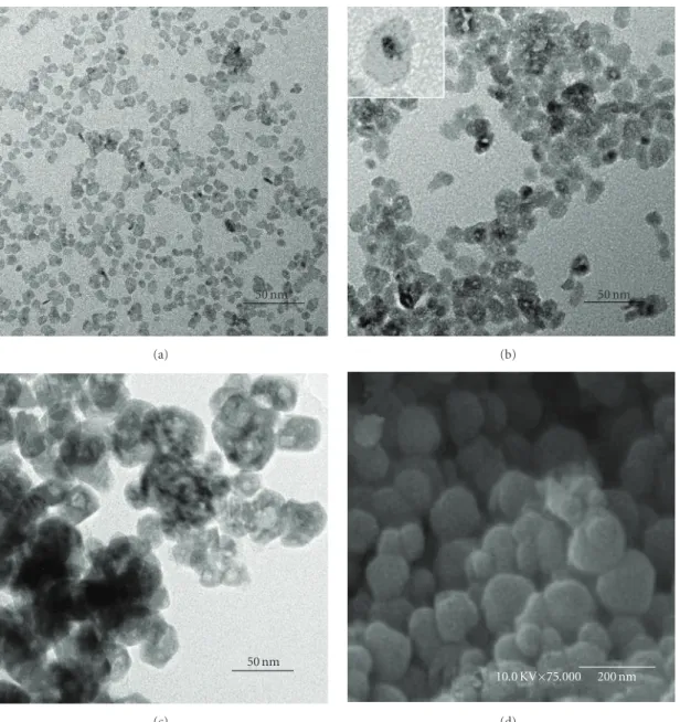
4 h, the morphology of the sample aggregates with a size from 30–60 nm, and the SEM image (Figure 2(d)) showed that the annealed LaF3:Nd3+/SiO2nanoparticles consists of spherical
particles with a size between 50–150 nm.
The FTIR spectra of LaF3:Nd3+/SiO2core/shell
nanopar-ticles are presented inFigure 3. Strong vibrational absorption bands at 3400–3600 and 1350–1600 cm−1were observed in as-prepared sample, which correspond to O-H mode. So the physically adsorbed solvent and O-H groups on the as-prepared nanoparticles are still not removed. Whereas for the nanoparticles annealed at 400◦C, the former absorption peaks show a great decrease. When the anneal tempera-ture raised to 900◦C, the absorption peaks of O-H mode completely disappeared. Thus, the nonradiative vibrational excitation of Nd3+ in the nanoparticles created by O-H and
C-H groups can be excluded.
Figure 4shows the room temperature emission spectra of LaF3:Nd3+/SiO2 core/shell and LaF3:Nd3+nanoparticles
under excitation at 808 nm. The emission lines centered at 880, 1060, and 1330 nm correspond to the transitions from 4F
3/2to 4I9/2, 4I11/2, and 4I13/2, respectively [15]. For
LaF3:Nd3+/SiO2 core/shell nanoparticles as-prepared and
annealed at 400 and 600◦C, the emission pattern is similar with that of LaF3:Nd3+ nanoparticles in both the peak
positions and shapes, which means that the SiO2 shell
have minimal effect on LaF3:Nd3+ core. However, when
the annealed temperature is 900◦C, emission spectra of the LaF3:Nd3+/SiO2 core/shell nanoparticles show a very
unusual manner. (1) Their fluorescence intensity show a great increase compared with that of the LaF3:Nd3+/SiO2
core/shell nanoparticles annealed at 600◦C. (2) The strongest emission line for 4F
3/2 → 4I11/2 transition peaking
at 1073 nm are redshifted by about 17 nm contrasted with that of nanoparticles annealed at 600◦C (1056 nm). (3) The 4F
3/2 → 4I9/2 and 4F3/2 → 4I13/2 emission of
LaF3:Nd3+/SiO2core/shell nanoparticles annealed at 900◦C
have remarkable Stark splittings which have not been found in other samples. In contrast, emission spectrum of 900◦C annealed LaF3:Nd3+ nanoparticles which have no SiO2shell
has also been recorded, and is shown inFigure 4(b). The flu-orescence intensity is only about 1/5 compared with that of LaF3:Nd3+/SiO2core/shell nanoparticles annealed at 900◦C,
the4F
3/2 → 4I11/2emission is peaking at about 1058 nm, and
no emission lines have Stark splittings. The results mean, for LaF3:Nd3+/SiO2 core/shell nanoparticles, that the influence
50 nm
(a)
50 nm
(b)
50 nm
(c)
10.0 KV×75.000 200 nm
(d)
Figure 2: TEM images of (a) LaF3:Nd3+, (b) LaF
3:Nd3+/SiO2, (c) LaF3:Nd3+/SiO2 annealed at 900◦C,SEM image of (d) LaF3:Nd3+/SiO2
annealed at 900◦C.
of SiO2shell has become clearly when the anneal temperature
is 900◦C, and the remarkable improvement of fluorescence intensity is caused by the SiO2shell.
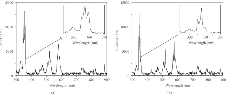
To get more information on the functions of SiO2
shell, the excitation spectra by monitoring the 4F
3/2 →
4I
11/2emission in LaF3:Nd3+/SiO2core/shell and LaF3:Nd3+
nanoparticles annealed at 900◦C are compared inFigure 5. The spectra displays well-resolved lines, centered at 328, 352, 430, 472, 519, 578.9, 737, 801, and 881 nm corresponding to the direct excitation of Nd3+ from 4I
9/2 to the higher
excited states:2D
5/2, 2P1/2, 4G11/2, 2K15/2 + 2D3/2 + 2G9/2,
2K
13/2+4G7/2+4G9/2,2G7/2+2G5/2,2H11/2,4F9/2,4S3/2+4F7/2,
4F
5/2+2H9/2, and4F3/2. Interestingly, remarkable differences
are observed in excitation spectra of the two samples. As shown in the inset of Figure 5, the shape of peaks for
4I
9/2 → 2P1/2 transition are different between the two
nanoparticles, and the lines for 4I
9/2 → 4S3/2 + 4F7/2,
and4F
5/2 + 2H9/2 transitions of LaF3:Nd3+/SiO2 core/shell
nanoparticles are much broader compared with that of LaF3:Nd3+ nanoparticles. The changes of excitation spectra
mean that the environment of LaF3:Nd3+ is different for
the two nanoparticles. The Stark splittings of4F
3/2 → 4I9/2
and4F
3/2 → 4I13/2 emission for LaF3:Nd3+/SiO2 core/shell
nanoparticles annealed at 900◦C also confirmed the change. In previous reports, the phenomena were often caused by the host matrixes or surfactants [15]. In this work, for SiO2
shell and LaF3 core have not the same lattice structures,
the shell could bring a noncentrosymmetric environment of the LaF3:Nd3+core [16]. And the new noncentrosymmetric
environment causes a significant improvement of fluores-cent intensity of LaF3:Nd3+/SiO2 core/shell nanoparticles
4000 3500 3000 2500 2000 1500 1000 500 Wave number (cm−1)
Figure 3: FTIR spectra of LaF3:Nd3+/SiO
2core/shell nanoparticles
annealed at different temperatures.
Table 1: Lifetimes of nanoparticles annealed at different
tempera-tures.
Nanoparticles Annealed temperature Lifetime
(◦C) (μs)
LaF3:Nd3+/SiO2core/shell
400 159
600 156
900 148
LaF3:Nd3+ 900 197
To have a further investigation of influence of the SiO2 shell on LaF3:Nd3+ core, fluorescent decay curves of
LaF3:Nd3+/SiO2 core/shell nanoparticles were measured by
monitoring the4F
3/2 → 4I11/2emission at about 1060 nm.
As shown in Table 1. For as-prepared nanoparticles, the lifetime is very difficult to be detected due to the presence of a large amount of O-H groups. When the nanoparticles were annealed at 400◦C, the lifetime is 159μs. Surprisingly, with a further rising of the anneal temperature, the lifetimes show a slight decrease. When annealed at 600 and 900◦C, the lifetimes are 156μs and 148μs, respectively. However, for 900◦C annealed LaF3:Nd3+nanoparticles which have no
SiO2 shell, the lifetime is 196μs. It can be deduced that the
lifetimes decrease in LaF3:Nd3+/SiO2core/shell nanoparticles
is not caused by the LaF3:Nd3+ core itself. In general, there
are two factors that could affect radiative lifetime of Nd3+
in nanoparticles. (1) The existence of O-H groups and impurity on nanoparticles surfaces which could effectively reduce the lifetime of Nd3+; (2) vibrational energies of host
matrix. For our samples, the difference is just the anneal temperature. For LaF3:Nd3+/SiO2 core/shell nanoparticles
annealed at 900◦C, the effect of O-H groups can be obviated (Figure 3) and the LaF3host has a good crystallinity
(Figure 1).
The LaF3:Nd3+/SiO2 core/shell nanoparticles were
syn-thesized in two steps. In as-prepared nanoparticles, SiO2shell
0
900 1000 1100 1200 1300 1400
(a)
0 1000 2000
In
te
nsit
y
(a.u.)
900 1000 1100 1200 1300 1400
Wavelength (nm) 900◦C
600◦C
(b)
Figure 4: Emission spectra of nanoparticles annealed at different
temperatures: (a) LaF3:Nd3+/SiO2, (b) LaF3:Nd3+.
is just coated on LaF3:Nd3+ core and interaction between
the two parts is very weak. When the anneal temperature is 400◦C, most solvent remained on nanoparticles is removed. Thus its lifetime is of 159μs. When the anneal temperature was raised to 900◦C, the interaction between SiO2shell and
LaF3:Nd3+ core increases. Since the phonon energy of SiO2
(1100 cm−1) is higher than that of LaF3 (350 cm−1) and
the nanoparticles have a larger surface-to-core ratio [16], lifetime of Nd3+ near or on the surfaces of nanoparticles is
slightly shorter than that of Nd3+within the core structures
and the measured lifetime of LaF3:Nd3+/SiO2 core/shell
nanoparticles is decreased.
4. Conclusions
To summarize, we have shown that, for the SiO2-coated
LaF3:Nd3+ nanoparticles, the SiO2 shell has an interesting
influence on their LaF3core. When the anneal temperature
is 900◦C, the change of environment of LaF3:Nd3+ core
caused by SiO2 shell has become obvious. As a result, Stark
splittings of 4F
3/2 → 4I9/2 and 4F3/2 → 4I13/2 transition
peaks and the redshift of 4F
3/2 → 4I11/2 transition peak
were observed in their emission spectrum. The fluorescent intensity of LaF3:Nd3+/SiO2 core/shell nanoparticles also
has a great improvement, which can be very beneficial for applications.
0 5000 10000 15000
In
te
nsit
y
(a.u.)
300 400 500 600 700 800 900
Wavelength (nm)
330 360 390
Wavelength (nm)
(a)
0 5000 10000 15000
In
te
nsit
y
(a.u.)
300 400 500 600 700 800 900
Wavelength (nm)
330 360 390
Wavelength (nm)
(b)
Figure 5: Excitation spectra of nanoparticles annealed at 900◦C: (a) LaF3:Nd3+/SiO2, (b) LaF3:Nd3+.
Acknowledgments
This work was financially supported by the National Natural Science Foundation of China (no. 10876009) and one Hundred Talents Programs of the Chinese Academy of Sciences.
References
[1] H. Song, B. Chen, B. Sun, J. Zhang, and S. Lu, “Ultravi-olet light-induced spectral change in cubic nanocrystalline
Y2O3:Eu3+,” Chemical Physics Letters, vol. 372, no. 3-4, pp.
368–372, 2003.
[2] M. Nishi, S. Tanabe, M. Inoue, M. Takahashi, K. Fujita, and K. Hirao, “Optical-telecommunication-band fluorescence
properties of Er3+-doped YAG nanocrystals synthesized by
glycothermal method,” Optical Materials, vol. 27, no. 4, pp. 655–662, 2005.
[3] Y. Pan, Q. Su, H. Xu et al., “Synthesis and red luminescence of
Pr3+-doped CaTiO
3nanophosphor from polymer precursor,”
Journal of Solid State Chemistry, vol. 174, no. 1, pp. 69–73, 2003.
[4] F. Wang, W. B. Tan, Y. Zhang, X. Fan, and M. Wang, “Lumines-cent nanomaterials for biological labelling,” Nanotechnology, vol. 17, no. 1, pp. R1–R13, 2006.
[5] X.-J. Wang, S. H. Huang, R. Reeves et al., “Studies of the
spec-troscopic properties of Pr3+ doped LaF
3 nanocrystals/glass,”
Journal of Luminescence, vol. 94-95, pp. 229–233, 2001. [6] S. Tanabe, H. Hayashi, T. Hanada, and N. Onodera,
“Fluores-cence properties of Er3+ions in glass ceramics containing LaF
3
nanocrystals,” Optical Materials, vol. 19, no. 3, pp. 343–349, 2002.
[7] J. Wang, J. Hu, D. Tang, X. Liu, and Z. Zhen, “Oleic acid
(OA)-modified LaF3:Er,Yb nanocrystals and their polymer hybrid
materials for potential optical-amplification applications,” Journal of Materials Chemistry, vol. 17, no. 16, pp. 1597–1601, 2007.
[8] D. Pi, F. Wang, X. Fan, M. Wang, and Y. Zhang, “Luminescence
behavior of EU3+doped LaF3 nanoparticles,” Spectrochimica
Acta A, vol. 61, no. 11-12, pp. 2455–2459, 2005.
[9] W Dew, Harrocks Jr., and D. R. Sudnick, “Lanthanide ion luminescence probes of the structure of biological macro-molecules,” Accounts of Chemical Research, vol. 14, no. 12, pp. 384–392, 1981.
[10] S. T. Frey, C. A. Chang, J. F. Carvalho et al., “Charac-terization of lanthanide complexes with a series of amide-based macrocycles, potential MRI contrast agents, using
EU3+luminescence spectroscopy and molecular mechanics,”
Inorganic Chemistry, vol. 33, no. 13, pp. 2882–2889, 1994. [11] J. W. Stouwdam, G. A. Hebbink, J. Huskens, and F. C.
J. M. Van Veggel, “Lanthanide-doped nanoparticles with excellent luminescent properties in organic media,” Chemistry of Materials, vol. 15, no. 24, pp. 4604–4616, 2003.
[12] L. M. Liz-Marz´an and P. Mulvaney, “The assembly of coated nanocrystals,” Journal of Physical Chemistry B, vol. 107, no. 30, pp. 7312–7326, 2003.
[13] Y. Wang, W. Qin, J. Zhang, C. Cao, S. L¨u, and X. Ren,
“Photoluminescence of colloidal YVO4:Eu/SiO2 core/shell
nanocrystals,” Optics Communications, vol. 282, no. 6, pp. 1148–1153, 2009.
[14] X.-F. Yu, L.-D. Chen, M. Li et al., “Highly efficient fluorescence
of NdF3/SiO2core/shell nanoparticles and the applications for
in vivo NIR detection,” Advanced Materials, vol. 20, no. 21, pp. 4118–4123, 2008.
[15] J. W. Stouwdam and F. C. J. M. Van Veggel, “Near-infrared
emission of redispersible Er3+, Nd3+, and Ho3+doped LaF
3
nanoparticles,” Nano Letters, vol. 2, no. 7, pp. 733–737, 2002. [16] Q. L¨u, A. Li, F. Guo, L. Sun, and L. Zhao, “The two-photon
excitation of SiO2-coated Y2O3:Eu3+nanoparticles by a
near-infrared femtosecond laser,” Nanotechnology, vol. 19, no. 20, Article ID 205704, 2008.
[17] H.-X. Mai, Y.-W. Zhang, R. Si et al., “High-quality sodium rare-earth fluoride nanocrystals: controlled synthesis and optical properties,” Journal of the American Chemical Society, vol. 128, no. 19, pp. 6426–6436, 2006.
Submit your manuscripts at
http://www.hindawi.com
Scientifica
Hindawi Publishing Corporation
http://www.hindawi.com Volume 2014
Hindawi Publishing Corporation
http://www.hindawi.com Volume 2014
Ceramics
Journal ofNanoparticles
Journal of Hindawi Publishing Corporationhttp://www.hindawi.com Volume 2014
Hindawi Publishing Corporation
http://www.hindawi.com Volume 2014
International Journal of
Biomaterials
Hindawi Publishing Corporation
http://www.hindawi.com Volume 2014
Nanoscience
Journal ofTextiles
Hindawi Publishing Corporation
http://www.hindawi.com Volume 2014 Journal of
Crystallography
Journal of Hindawi Publishing Corporationhttp://www.hindawi.com Volume 2014
The Scientific
World Journal
Hindawi Publishing Corporation
http://www.hindawi.com Volume 2014
Hindawi Publishing Corporation
http://www.hindawi.com Volume 2014
Coatings
Journal ofAdvances in
Materials Science and Engineering
Hindawi Publishing Corporation
http://www.hindawi.com Volume 2014
Materials
Journal ofHindawi Publishing Corporation
http://www.hindawi.com Volume 2014
N
a
no
ma
te
ria
ls
Hindawi Publishing Corporation
http://www.hindawi.com Volume 2014
Journal of