© 2020 by the Serbian Biological Society How to cite this article: Đorđević M, Grdović N, Mihailović M, Arambašić Jovanović 117 J, Uskoković A, Rajić J, Đorđević M, Tolić A, Mišić D, Šiler B, Poznanović G, Vidaković M, Dinić S. Centaurium erythraea extract reduces redox imbalance and improves insulin expression and secretion in pancreatic β-cells exposed to oxidative and nitrosative stress. Arch Biol Sci. 2020;72(1):117-28.
Centaurium erythraea
extract reduces redox imbalance and improves insulin expression
and secretion in pancreatic β-cells exposed to oxidative and nitrosative stress
Miloš Đorđević1, Nevena Grdović1, Mirjana Mihailović1, Jelena Arambašić Jovanović1, Aleksandra Uskoković1,
Jovana Rajić1, Marija Đorđević1, Anja Tolić1, Danijela Mišić2, Branislav Šiler2, Goran Poznanović1, Melita
Vidaković1 and Svetlana Dinić1,*
1Department of Molecular Biology, Institute for Biological Research “Siniša Stanković”, National Institute of Republic of Serbia,
University of Belgrade, Bulevar despota Stefana 142, 11060, Belgrade, Serbia
2Department of Plant Physiology, Institute for Biological Research “Siniša Stanković”, National Institute of Republic of Serbia,
University of Belgrade, Bulevar despota Stefana 142, 11060, Belgrade, Serbia
*Corresponding author: [email protected]
Received: January 27, 2020; Revised: January 31, 2020; Accepted: January 31, 2020; Published online: February 10, 2020
Abstract: Oxidative stress is one of the major mechanisms that underlies the damage of pancreatic β-cells and defects in insulin secretion in diabetes. As herbal preparations can alleviate oxidative stress through their redox-active secondary metabolites, in this study we investigated the cytoprotective effects of Centaurium erythraea extract (CEe) against H2O2- and SNP-induced oxidative/nitrosative stress in Rin-5F β-cells. The antioxidant activity of CEe and its effect on cell survival and insulin expression/secretion were evaluated. The CEe increased cell viability and ameliorated the disturbance of redox homeostasis in H2O2- and SNP-treated cells by decreasing DNA damage, lipid peroxidation and protein S-glutathionylation. The CEe restored GSH homeostasis in H2O2-treated β-cells and attenuated the SNP-induced disturbance of the GSH/ GSSG ratio. The H2O2- and SNP-induced disruption of CAT, GPx, GR, MnSOD and CuZnSOD activities was adjusted by the CEe towards control values, as well as mRNA and protein levels of GPx, MnSOD and CAT. The CEe increased insulin expression/secretion particularly in H2O2-treated β-cells, which was in accordance with the more pronounced antioxidant effect of the CEe observed in H2O2-treated β-cells as compared to SNP-treated cells. These findings support the beneficial effect of the CEe in preventing or slowing down β-cell damage and dysfunction caused by oxidative/nitrosative stress dur-ing diabetes development.
Keywords: oxidative stress; nitrosative stress; β-cells; Centaurium erythraea;antioxidant; cytoprotective
Abbreviations and acronyms: CEe – Centaurium erythraea extract; SNP – sodium nitroprusside; GSH – glutathione; GSSG – glutathione disulfide; GSSP – S-glutathionylated proteins; CAT – catalase; GPx – glutathione peroxidase; GR – glutathione reductase; MnSOD – manganese superoxide dismutase; CuZnSOD – copper-zinc superoxide dismutase
INTRODUCTION
Diabetes is a complex metabolic disorder with insulin producing pancreatic β-cell loss and dysfunction at its core. Studies indicate that oxidative stress-activat-ed rstress-activat-edox-signaling pathways damage β-cells, cause defects in insulin expression and secretion, leading to eventual cell death [1]. In type 1 diabetes, β-cell death is driven by pro-inflammatory cytokines and free radical species such as the superoxide anion (O2-)
and nitric oxide radical (NO•) released from immune
cells infiltrating pancreatic islets [2]. Elevated glucose
concentrations in type 2 diabetes lead to β-cell dys-function through increased glucose metabolism asso-ciated with mitochondrial production of reactive oxy-gen species (ROS) [2]. In the physiological state, ROS such as O2-, the hydroxyl radical (•OH) and hydrogen
peroxide (H2O2), and reactive nitrogen species (RNS), including NO• and the peroxynitrite anion (ONOO-),
NO• is a physiological regulator of insulin secretion
at lower concentrations [5]. High concentrations of ROS and RNS, resulting either from their overproduc-tion or from the disrupoverproduc-tion of antioxidant protecoverproduc-tion, produce oxidative stress associated with damage and dysfunction of proteins, lipids and DNA [6]. To main-tain redox homeostasis, the cell must ensure proper functioning of the endogenous antioxidant system. MnSOD and CuZnSOD catalyze the dismutation of O2- to H
2O2, which is neutralized to H2O by CAT or
by GPx that utilizes GSH as a reducing factor [7-9]. During catalytic activity of GPx, GSH is oxidized to GSSG, which is reduced to GSH by GR [10]. As a non-enzymatic antioxidant, GSH plays an important role in cellular redox buffering [11].
Because of the relatively low expression and activ-ity of antioxidant enzymes and redox buffers in com-parison to other cell types, β-cells are more susceptible to oxidative stress [6,12]. As redox-homeostasis in β-cells can easily shift to a state of harmful oxidative stress, exogenously assisted lowering of excess ROS/ RNS levels and/or the improvement of endogenous antioxidant defenses can contribute to the preserva-tion of structural and funcpreserva-tional properties of β-cells in diabetes.
A growing body of evidence indicates that herbal preparations can alleviate oxidative stress through additive and synergistic effects of their redox active polyphenolic compounds. Centaurium erythraea Rafn (CE), also known as a common centaury, is a plant from the Gentianaceae family, which is used in Serbian traditional medicine for treating different diseases, including diabetes [13]. We previously presented the phytochemical characterization of the extract pre-pared from CE (CEe), with secoiridoid glycosides and phenolics (including xanthones and flavonoids) as its major components [14]. We demonstrated a signifi-cant in vitro antioxidant potential of CEe, including strong H2O2- and NO-scavenging activities, as well as the ability to improve the structural and functional properties of pancreatic islets in streptozotocin (STZ)-induced diabetic rats [14, 15].
The aim of the present study was to examine the antioxidant effects of CEe in β-cells treated with H2O2 and with SNP, a prooxidant with a different mecha-nism of action: while hydrogen peroxide directly leads
to oxidative damage and is a source of highly reactive
•OH [16], SNP is a complex compound that in
aque-ous solution releases NO• and is most commonly used
to investigate the cytotoxicity induced by nitrosative stress [17,18]. The antioxidant action of the CEe was estimated by analyzing DNA damage, lipid peroxida-tion, protein S-glutathionylation (GSSP), the GSH/ GSSG ratio, as well as the expression and activities of CAT, GPx, GR, MnSOD and CuZnSOD antioxidant enzymes. The protective effect of the CEe was also evaluated by analyzing β-cell survival as well as insulin expression and secretion.
MATERIALS AND METHODS CE methanol extract
Procedures for the collection of plant material, prep-aration and phytochemical characterization of the methanol CEe used in the present study were de-scribed previously [14].
Cell culture
deter-mination of secreted insulin, the medium in which the cells were grown and treated was harvested and centri-fuged at 500 x g for 10 min; 10 µL of the obtained su-pernatant was used to determine the concentration of released insulin by enzyme immunoassay (Rat/Mouse Insulin ELISA Kit, EMD Millipore, MO, USA).
MTT viability assay
The cell viability test is based on the measurement of the mitochondrial activity detected by the forma-zan formation in living cells after reduction of the tetrazolium ring of 3-(4,5-dimethylthiazol-2-yl)-2,5-diphenyl tetrazolium bromide (MTT, Sigma). Rin-5F cells cultured in 96-well plates were treated with the indicated concentrations of H2O2 (for 3 h), SNP (for 24 h) and CEe (for 3 h and 24 h as indicated); imme-diately after the treatments, 200 µL of MTT (0.5 mg/ mL RPMI medium) was added to each well, followed by incubation for 2 h in the dark. The insoluble forma-zan was dissolved in dimethyl sulfoxide (DMSO) and quantified by measuring the absorbance at 570 nm. Cell viability was expressed in comparison to control cells assumed to be 100%.
Comet assay
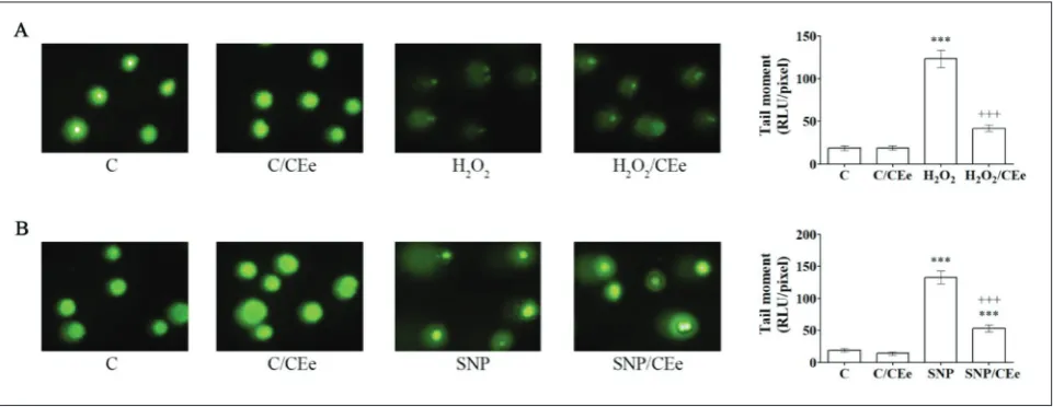
DNA damage was estimated using the Comet as-say performed as previously described [15]. The tail moment, which takes into account both the relative amount of DNA in the tail and the migration of the DNA, was used as an indicator of the displacement of the genetic material between the nucleus (‘comet head’) and the resulting ‘comet tail’. Quantification of images was performed by TriTekCometScore Freeware version 1.5.
Lipid peroxidation assay
Cells grown in 6-well plates were resuspended in 200 µL of phosphate-buffered saline (PBS); 10 µL of the cell suspension was used for the Comet assay, while the rest was centrifuged at 300 x g for 10 min. The cell pellet was dissolved in 1.15% KCl solution, sonicated (5 s/30 kHz/+4oC three times), and the obtained
ho-mogenate was stored at -80oC until use. The level of
lipid peroxidation was estimated by measuring the level of malondialdehyde (MDA) in the
thiobarbitu-ric acid-reactive substance (TBARS) assay [19]. The cell homogenate (0.1 mL) was mixed with 0.2 mL of 8.1% sodium dodecyl sulfate (SDS), 1.5 mL of 20% acetic acid (pH 3.5), 1.5 mL of 0.8% thiobarbituric acid (TBA) and 0.7 mL of water and heated at 95°C for 60 min. After cooling to room temperature, 1 mL of water and 5 mL of n-butanol-pyridine (15:1, v/v) were added to samples that were mixed and centrifuged at 3000 × g for 10 min. The absorbance of the upper colored layer was measured at 532 nm. The concentra-tion of MDA was calculated from a calibraconcentra-tion curve prepared using MDA standards (concentration range of 25 to 500 nM/mL) and expressed as nM MDA/100 mg of proteins. Protein concentrations were deter-mined according to Lowry et al. [20].
Determination of GSH, GSSG and GSSP
Measurement of GSH, GSSG and GSSP was per-formed as described [21]. Rin-5F cells were resus-pended in PBS (500 µL) and centrifuged at 300 x g
Antioxidant enzyme activity assays
Homogenates were prepared by resuspension of cells in sucrose buffer (0.25 M sucrose, 1 mM EDTA and 0.05 M Tris-HCl, pH 7.4); cell suspension was soni-cated at 20 kHz/30 s on ice, centrifuged at 14000 ×
g/4°C for 1 min, and the supernatants were used for the determination of enzyme activities and protein concentrations. CAT activity was measured by the rate of H2O2 decomposition as described by Beutler [22]. Total SOD activity was determined according to the epinephrine method [23]. MnSOD activity was per-formed after pre-incubation with 8 mM KCN, and CuZnSOD activity was calculated from the difference between total SOD and MnSOD activities. The ac-tivity of GPx was determined following oxidation of NADPH as a substrate with tert-butyl hydroperoxide [24]. The activity of GR was measured as described by Glatzle et al. [25]. Enzyme activity was expressed as U activity/mg of protein.
Sodium dodecyl sulfate (SDS) polyacrylamide gel electrophoresis (PAGE) and Western blot analysis
Homogenate proteins (20 μg) were separated by SDS-PAGE and transferred onto polyvinylidene difluoride membranes (Amersham Hybond P 0.45 PVDF, GE Healthcare Life Sciences, UK). After blocking with 5% non-fat dry milk (Blotto, non-fat dry milk, Santa Cruz Biotechnology, USA) in blotto base buffer (0.2% Tween 20, 20 mM Tris-HCl pH 7.6, 150 mM NaCl) for 1 h at room temperature, samples were subjected to analysis using the following antibodies: anti-CAT, anti-GR and anti-GPX 1 (all from Abcam, USA) and anti-MnSOD (FL-222), anti-CuZnSOD (C-17)and anti-GAPDH (FL-335) (all from Santa Cruz Biotech-nology, USA). The membranes were probed with adequate HRP-conjugated secondary antibodies (all from Santa Cruz Biotechnology, USA). Staining was performed by chemiluminescence according to the manufacturer’s instructions (Western Blotting Lumi-nol Reagent, Santa Cruz BiotechLumi-nology, USA). Im-munoreactive bands were quantified using TotalLab (Phoretix, USA) electrophoresis software (v1.1).
RNA isolation and real-time quantitative PCR analysis (RT-qPCR)
Total RNA was isolated from Rin-5F cells using the GeneJET RNA Purification Kit (Thermo Fisher Scien-tific, USA) following the manufacturer’s instructions. Cells were seeded in 6-well plates and treated with H2O2 (75 µM) for 3 h or with SNP (1.25 mM) for 24 h in parallel with the CEe (0.25 mg/mL). Synthesis of cDNA was performed using total RNA (1 μg) treated with
DN-ase I and reverse transcribed with oligo(dT) primers using the RevertAid First Strand cDNA Synthesis Kit (Thermo Fisher Scientific, USA). The levels of mRNA were quantified by Maxima SYBR Green/ROX qPCR Master Mix (2X) (Thermo Fischer Scientific, USA) and QuantStudio 3 Real-Time PCR system (Applied Bio-systems, Carlsbad, CA, USA). The cycles for RT-qPCR included an initial denaturation step (95°C/10 min), followed by 40 cycles of a two-step PCR program at 95°C for 15 s and at 60°C for 1 min. In all RT-qPCR reactions negative controls without the template were used. The expression levels of the target genes were calculated in relation to the averaged expression of rat GAPDH gene. RT-qPCR reactions were carried out in triplicate. The fragments were amplified using the following primers (Invitrogen, USA): for the rat CAT gene: Fw 5'-GCGAATGGAGAGGCAGTGTAC-3' and Rev 5'- GAGTGACGTTGTCTTCATTAGCACTG -3' (652 bp); for the rat GPx gene: Fw 5'-AGTTCGGA-CATCAGGAGAATGG-3' and Rev 5'-TAAAGAGC-GGGTGAGCCTTC-3' (141 bp); for the rat GR gene: Fw 5'-CACTTCCCGGTAGGAAACCC-3' and Rev 5'-GATCGCAACTGGGGTGAGAA-3' (227 bp); for the rat MnSOD gene: Fw 5'-CAGATCATG-CAGCTGCACCA-3' and Rev 5'-AGTCCAGGCT-GAAGAGCA-3' (133 bp); for the CuZnSOD gene: Fw 5'-GCAGAAGGCAAGCGGTGAAC-3' and Rev 5'-CGGCCAATGATGGAATGCTC-3' (282 bp); for the rat insulin 1 (Ins1) gene: Fw 5'-ATGGCCCT-GTGGATGCGCTT-3' and Rev 5'-ACAATGCCAC-GCTTCTGCCG-3' (275 bp); for the rat GAPDH gene: Fw 5'-CAAGGTCATCCATGACAACTTTG-3' and Rev 5'-GTCCACCACCCTGTTGCTGTAG-3' (496 bp).
Statistical analysis
differ-ences between groups in comparison to the corresponding control group were an-alyzed using one-way analysis of variance (one-way ANOVA), followed by Bonfer-roni’s Multiple Comparison Test. The data were expressed as the mean±SEM. (standard error of mean). The difference was considered statistically significant at
p<0.05. The statistical significance (p) for all obtained results is shown in the tables and figures.
RESULTS
CEe increases cell-viability and insulin expression/secretion in H2O2- and SNP-treated β-cells
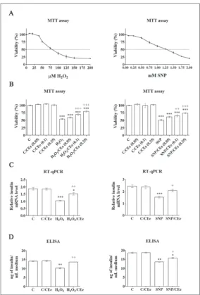
Treatment of Rin-5F cells with a series of concentrations of either H2O2 or SNP revealed their IC50 doses as follows, 75 μM for H2O2 and 1.25 mM for SNP (Fig. 1A), which were then used in all further analyses. Combined treatment of Rin-5F cells with the IC50 dose of H2O2 and in-creasing concentrations of CEe resulted in an increase in the percentage of viable cells in comparison to the H2O2 treatment (Fig. 1B). A slight increase (5%) was ob-served when 0.05 mg/mL of the extract was applied, while concentrations of the CEe of 0.1 and 0.25 mg/mL increased the number of viable cells by 15% and 26%, respectively. Co-treatment of cells with IC50 SNP and increasing concentra-tions of the CEe revealed that the greatest improvement in cell viability (24%) was when 0.25 mg/mL of the CEe was applied (Fig. 1B). Therefore, the concentration of 0.25 mg/mL of the CEe was chosen for subsequent investigation in combination with IC50 doses of H2O2 and SNP.
The effect of the CEe on the func-tional properties of β-cells was examined by measuring the mRNA level of the in-sulin gene (Ins1), and by determining the level of insulin released into the growing
Fig. 1. The CEe increases cell-viability and insulin expression/secretion in H2O2- and SNP-treated β-cells. A – Viability test performed on Rin-5F cells treated with increasing concentrations of H2O2 (3 h) and SNP (24 h) revealed IC50 doses for H2O2 (75 µM) and SNP (1.25 mM). B – Viability test of Rin-5F cells co-treated with H2O2 (75 µM) and increasing concentrations of CEe for 3 h or cells co-treated with SNP (1.25 mM) and increasing concentrations of CEe for 24 h. C – Quan-titative RT-PCR analysis of insulin mRNA in Rin-5F cells co-treated with H2O2 (75 µM) and CEe (0.25 mg/mL) or co-treated with SNP (1.25 mM) and CEe (0.25 mg/mL). D – The level of insulin secreted in medium after co-treatment of Rin-5F cells with H2O2 (75 µM) and CEe (0.25 mg/mL) or co-treatment with SNP (1.25 mM) and CEe (0.25 mg/mL). The values are means±SEM from at least three separate experiments. C – control cells; C/CEe – control cells treated with CEe (0.25 mg/mL) for 3 h for H2O2 treatments or for 24 h for SNP treatment; H2O2 – cells treated with H2O2 (75 µM) for 3 h; H2O2/CEe – cells co-treated with H2O2 (75 µM) and CEe (0.25 mg/mL) for 3 h; SNP – cells treated with SNP(1.25 mM) for 24 h; SNP/CEe – cells co-treated with SNP (1.25 mM) and CEe (0.25 mg/mL) for 24 h. * p<0.05, **p<0.01, ***p<0.001 as compared to control cells (C); ++p<0.01, +++p<0.001 when H
medium (Fig. 1C, D). Following H2O2 treatment, the relative level of insulin mRNA was reduced by 45% relative to the mRNA level observed in control cells. Simultaneous H2O2/CEe treatment increased the level of insulin mRNA relative to H2O2-treated cells by 46%. Accordingly, reduction of insulin secretion by 28% after H2O2 treatment returned to the control level after H2O2/CEe co-treatment. Both insulin expression and secretion were also impaired in SNP-treated Rin-5F cells as the level of insulin mRNA was reduced by 38% (Fig. 1C), while insulin release was decreased by 27% (Fig. 1D) when compared to the control. SNP/ CEe co-treatment increased the level of insulin mRNA by 36% and increased the level of released insulin by 16% in comparison to SNP-treated cells. Treatment of control cells with the CEe had no influence on gene expression and the level of insulin secretion.
CEe improves the β-cell response to H2O2- and SNP-mediated redox challenge
To assess the impact of the CEe on the degree of oxi-dative damage in β-cells exposed to H2O2 and SNP treatments, oxidative stress parameters such as DNA damage, lipid peroxidation, GSSP formation, the GSH/ GSSG ratio and antioxidant enzyme activities were
analyzed. As can be seen in Fig. 2, in H2O2- and SNP-treated cells the tail moment was increased 6.6- and 7-fold, respectively, relative to the control cells. Both H2O2/CEe and SNP/CEe co-treatments reduced the quantity of DNA in the comet tail (by 2.9- and 2.5-fold, respectively). In comparison to the control, the levels of MDA and GSSP in H2O2-treated cells were in-creased (2.3- and 3.2-fold, respectively), while the GSH/ GSSG ratio was reduced 39% (Table 1). In H2O2/CEe co-treated cells, the GSH/GSSG ratio was at the level of the control while the levels of MDA and GSSP were reduced (34% and 55%, respectively) when compared to H2O2-treated cells. Treatment of Rin-5F cells with H2O2 affected the activities of the antioxidant enzymes by inducing CAT, MnSOD and CuZnSOD (by 32%, 51% and 48%, respectively), and by lowering the activi-ties of GPx and GR (by 30% and 23%, respectively). In H2O2/CEe co-treated cells, the activities of CAT and CuZnSOD were lowered (both for 17%), whereas the activities of GPx and GR were increased (40% and 20%, respectively) in comparison to the H2O2 treatment.
SNP treatment of β-cells induced lipid peroxida-tion (a 4.9-fold increase in MDA), increased the level of GSSP (3.6-fold) and decreased the GSH/GSSG ratio (21%) (Table 2). Compared to control cells, increases
Fig. 2. CEe-mediated protection of DNA in H2O2- and SNP-treated Rin-5F cells.The level of DNA damage was analyzed by the alka-line comet assay. Nuclear displacement of DNA (tail moment) was quantified by TriTekCometScoreTM Freeware software (v. 1.5) and graphically presented as the mean±SEM. Representative images of comets from three independent experiments are shown. C – control cells; C/CEe – control cells treated with CEe (0.25 mg/mL) for 3 h for the H2O2 treatment or for 24 h for the SNP treatment; H2O2 – cells treated with H2O2 (75 µM) for 3h; H2O2/CEe – cells co-treated with H2O2 (75 µM) and CEe (0.25 mg/mL) for 3h; SNP – cells treated with SNP(1.25 mM) for 24 h; SNP/CEe – cells co-treated with SNP (1.25 mM) and CEe (0.25 mg/mL) for 24 h. *** p<0.001 as compared to control cells (C); +++p<0.001 when H
in MnSOD and CuZnSOD activities (2.1- and 1.9-fold, respectively) and decreases in CAT, GPx and GR activities (by 26%, 30% and 51%, respectively) were observed after SNP treatment (Table 2). In relation to the SNP treatment, co-treatment with SNP/CEe was characterized by lower levels of MDA and GSSP (by 27% and 21%, respectively), as well as by decreases in MnSOD and CuZnSOD activities (by 29% and 20%, respectively) and increases in CAT, GPx and GR activ-ities (by 19%, 32% and 43%, respectively). Compared
to the SNP treatment, the GSH/GSSG ratio was increased in SNP/CEe co-treated cells although the observed increase was not sta-tistically significant.
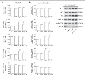
The CEe modulates the expression of antioxidant enzymes in H2O2- and SNP-treated β-cells
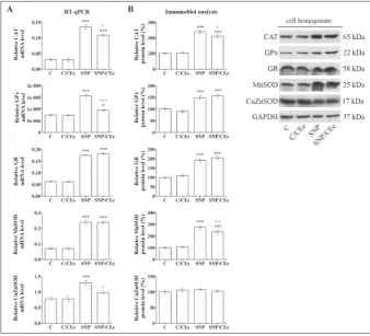
To examine whether the observed changes in the activities of antioxidant enzymes in H2O2- and SNP-treated β-cells are associated with alterations in enzyme expression, fur-ther investigation included analysis of their relative mRNA and protein levels. After the H2O2 treatment, the levels of GPx and Mn-SOD mRNA were increased (by 83% and 76%, respectively), whereas following H2O2/ CEe co-treatment their mRNA levels were lowered (23% and 25%, respectively) (Fig. 3). Immunoblot analysis of cell homogenates revealed an increase in the levels of GPx, GR and MnSOD proteins (by 54%, 62% and 73%, respectively) after the H2O2 treat-ment, and their reduction after the H2O2/ CEe co-treatment (by 13%, 10% and 14%, respectively). SNP treatment of Rin-5F cells was accompanied by an increase in the lev-els of mRNAs for CAT, GPx, GR, MnSOD and CuZnSOD (by 4.5-, 2.1-, 2.8-, 3.4- and 1.6-fold, respectively) (Fig. 4). SNP/CEe co-treatment lowered the levels of CAT, GPx and CuZnSOD mRNAs (by 20%, 39% and 24%, respectively). Immunoblot analysis of SNP-treated cells revealed increases in the protein levels of CAT and MnSOD enzymes (2.4-fold and 2.7-fold, respectively), which decreased (12% and 14%, respectively) after the SNP/CEe co-treatment. The protein levels of GPx and GR that were increased (49% and 90%, respective-ly) after the SNP treatment remained elevated after the SNP/CEe co-treatment.
DISCUSSION
Studies on the animal model of diabetes have revealed the antidiabetic and antioxidant effects of extracts
pre-Table 1. Parameters of oxidative stress in H2O2-treated Rin-5F cells. Presented arelipid peroxidation (MDA) level, ratio of reduced and oxidized glutathione (GSH/GSSG), S-glutathionylated proteins (GSSP), activities of antioxidant enzymes – catalase (CAT), glutathione peroxidase (GPx), glutathione reduc-tase (GR), manganese and copper-zinc superoxide dismureduc-tase (MnSOD and CuZnSOD).
C C/CEe H2O2 H2O2/CEe
MDA a 23.73±1.19 29.58±1.48 54.07±2.70 *** 35.45±1.77 *; +++
GSH/GSSG 23.15±1.16 24.01±1.2 14.16±0.71 ** 24.97±1.25 +++
GSSP b 48.69±2.43 30.8±1.54 153.46±7.67 *** 68.79±3.44 +++
CAT c 3.42±0.15 3.58±0.09 4.54±0.3 ** 3.77±0.11 +
GPx c 2.97±0.17 2.9±0.19 2.07±0.12 * 2.9±0.22 +
GR c 40.52±0.92 38.62±1.79 31.11±1.69 *** 37.25±0.26 +
MnSOD c 1.18±0.05 1.26±0.07 1.78±0.1 *** 1.51±0.05 *
CuZnSOD c 3.21±0.18 3.3±0.19 4.75±0.17 *** 3.93±0.17 +
The values are means±SEM from three experiments performed in triplicate. C – control cells; C/CEe – control cells treated with CEe (0.25 mg/mL) for 3 h; H2O2
– cells treated with H2O2 (75 µM) for 3 h; H2O2/CEe – cells co-treated with H2O2 (75
µM) and CEe (0.25 mg/mL) for 3 h. * p<0.05, ** p<0.01, *** p<0.001 as compared to C; +p<0.05, +++ p<0.001 when H
2O2/CEe is compared to H2O2. anM MDA/100 mg of
proteins; bµM GSH/mg of proteins; cU/mg of proteins.
Table 2. Oxidative stress parameters in SNP-treated Rin-5F cells. Presented arelipid peroxidation (MDA) level, ratio of reduced and oxidized glutathione (GSH/GSSG), S-glutathionylated proteins (GSSP), activities of antioxidant enzymes – catalase (CAT), glutathione peroxidase (GPx), glutathione reduc-tase (GR), manganese and copper-zinc superoxide dismureduc-tase (MnSOD and CuZnSOD).
C C/CEe SNP SNP/CEe
MDA a 16.23±2.87 23.12±3.34 79.43±3.17 *** 58.16±1.14 ***; +++
GSH/GSSG 22.59±1.13 23.48±1.17 17.80±0.89 * 21.41±1.07
GSSP b 74.42±3.72 58.24±2.91 265.57±13.28 *** 209.43±10.47 ***; +
CAT c 3.74±0.08 3.96±0.07 2.77±0.20 *** 3.30±0.10 +
GPx c 1.88±0.14 1.90±0.06 1.32±0.11 ** 1.74±0.04 +
GR c 42.53±1.03 44.84±1.70 20.71±2.71 *** 29.62±2.17 **; +
MnSOD c 1.34±0.12 1.45±0.22 2.78±0.17 *** 1.96±0.21 +
CuZnSOD c 3.18±0.16 3.25±0.28 5.99±0.29 *** 4.78±0.26 **; +
The values are means±SEM from three experiments performed in triplicate. C – control cells; C/CEe – control cells treated with CEe (0.25 mg/mL) for 24 h; SNP – cells treated with SNP(1.25 mM) for 24 h; SNP/CEe – cells co-treated with SNP (1.25 mM) and CEe (0.25 mg/mL) for 24 h. * p<0.05, **p<0.01, ***p<0.001 as
pared from CE, supporting its traditional use in diabe-tes management and stimulating further investigation of its biological activities [14,15,26,27]. Medicinal plants are a valuable source for drug discovery, and a number of active components have been isolated for direct use as drugs, or as a lead compound or pharmacological agent [28,29]. Thus, galegine, prepared from the plant
Galega officinalis L., which has been used in traditional
medicine for the treatment of diabetes has provided
a template for the synthesis of the oral hypoglycemic agent metfor-min and opened up interest in the synthesis of other biguanidine-type antidiabetic drugs [28]. In the pres-ent study we investigated the cy-toprotective effects of the CEe on β-cells in H2O2- and SNP-induced oxidative and nitrosative stress. Our results revealed that the CEe ame-liorated disturbed redox homeo-stasis in β-cells, contributing to a decrease in DNA, lipid and protein damage and to an increase in cell viability and insulin expression/ secretion. Suppression of oxida-tive and nitrosaoxida-tive stress in β-cells points to the beneficial effect of the CEe in preventing or slowing down the processes of β-cell loss and dys-function in diabetes.
The increase in DNA damage, lipid peroxidation and protein S-glutathionylation, the decrease in the GSH/GSSG ratio and altera-tions in the activities of antioxi-dant enzymes in β-cells observed after treatments with H2O2 and SNP are in accordance with their prooxidant action. H2O2 exerts its effect through the oxidation of DNA, lipids and protein thiol (-SH) groups and inactivation of enzymes [30]. Also, H2O2 is a source of •OH that causes
dam-age to DNA, proteins and lipids [16,31]. H2O2 can be converted to hypochlorous acid (HOCl), which in reaction with the amino group (-NH2), followed by free radical formation, leads to protein degradation, enzyme inactivation, nucleic acid denaturation and lipid peroxidation [32]. On the other hand, SNP reacts with -SH groups of proteins and releases NO•, which in reaction with other radicals
such as O2- forms extremely reactive ONOO- [33,34].
The protonated form of peroxynitrite (ONOOH) leads to depletion of -SH groups, fragmentation of DNA, lipid and protein oxidation [16,31]. The
pre-Fig. 3. The CEe modulates the expression of the antioxidant enzymes in H2O2-treated Rin-5F cells. A – The levels of CAT, GPx, GR, MnSOD and CuZnSOD mRNAs were analyzed using quantitative RT-PCR. Preparation of RNA and complementary DNA (cDNA) was performed after treatment of Rin-5F cells with H2O2 (75 µM) for 3 h with or without CEe (0.25 mg/mL). The levels of mRNA for antioxidant enzymes were calculated against GAPDH mRNA and the plotted values are the means±SEM from three separate experiments performed in triplicate. B – CAT, GPx, GR, MnSOD and CuZnSOD protein levels were determined by immunoblot analysis of cell homogenates isolated from Rin-5F treated with H2O2 (75 µM) for 3 h with or without CEe (0.25 mg/mL). Blots were quantified using TotalLab (Phoretix) electrophoresis software. The relative protein levels of antioxidant enzymes were calculated against GAPDH protein level and are presented on the graphs as the means±SEM relative to the control arbitrarily taken as 100%. CAT – catalase; GPx – glutathione peroxidase; GR – glutathione reductase; MnSOD – manganese superoxide dismutase; CuZnSOD – copper-zinc superoxide dismutase; GAPDH – glyceraldehyde 3-phosphate dehydrogenase. C – control cells; C/CEe – control cells treated with CEe (0.25 mg/ mL) for 3 h; H2O2 – cells treated with H2O2 (75 µM) for 3 h; H2O2/CEe – cells co-treated with H2O2 (75 µM) and CEe (0.25 mg/mL) for 3 h. ** p< 0.01, *** p<0.001 as compared to C; +p<0.05, ++p<0.01, +++ p<0.001 when H
sented results indicate that both agents caused about the same level of DNA damage in β-cells, while SNP had a greater effect on the in-duction of lipid peroxidation and protein S-glutathionylation than H2O2, which in turn had a more pronounced influence on the de-crease of the GSH/GSSG ratio. Such differences in the extent of changes of oxidative stress param-eters could be ascribed to differ-ent mechanisms of H2O2 and SNP prooxidant actions.
The CEe alleviated H2O2- and SNP-induced DNA damage in β-cells that is crucial for their sur-vival and functioning, especially with regard to the fact that β-cells possess a weak repair machinery for oxidative DNA damage [35]. An additional contribution of the CEe to β-cell protection was achieved by the reduction of lipid peroxidation that causes oxidative damage of membrane proteins, in-activation of receptors, enzymes and transport proteins and conse-quently cell death [16,36]. It can be assumed that by lowering lipid peroxidation, CEe also reduced the production of various stable aldehydes known to act as sec-ondary messengers at sites distant from the site of their formation, leading to DNA and protein dam-age [36]. By increasing the GSH/ GSSG ratio, which contributes to the redox potential of the cell and
to redox homeostasis [11], CEe further suppressed the H2O2-and SNP-induced redox imbalance in β-cells. Oxidative stress leads to oxidation of GSH to GSSG and to S-glutathionylation (GSSP) through the forma-tion of mixed disulfide bonds between GSH or GSSG and redox-sensitive protein -SH groups [37]. Nitro-sative stress, through the reaction of NO• with GSH,
produces high levels of S-nitrosoglutathione (GSNO) that contribute to protein S-glutathionylation as well
[38]. Although S-glutathionylation was assumed to be a by-product of oxidative or nitrosative stress, under physiological conditions this modification is involved in redox regulation and signaling, while during oxida-tive stress it leads to functional alterations of proteins, including modulation of enzyme activities [37,39]. By lowering the level of protein S-glutathionylation in H2O2-and SNP-treated β-cells, CEe could lessen the negative effects of this modification on the structural
Fig. 4. The CEe affects the expression of antioxidant enzymes in SNP-treated Rin-5F cells. A – Quantitative RT-PCR analysis of CAT, GPx, GR, MnSOD and CuZnSOD mRNA levels. Prepara-tion of RNA and complementary DNA (cDNA) was performed after treatment of Rin-5F cells with SNP (1.25 mM) for 24 h with or without CEe (0.25 mg/mL). The levels of mRNA for antioxidant enzymes were calculated against GAPDH mRNA and the plotted values are the means±SEM from three separate experiments performed in triplicate. B – Immunoblot analysis of CAT, GPx, GR, MnSOD and CuZnSOD protein levels from cell homogenates isolated from Rin-5F treated with SNP (1.25 mM) for 24 h with or without CEe (0.25 mg/mL). Blots were quantified using TotalLab (Phoretix) electrophoresis software. The relative protein levels of antioxidant enzymes were calculated against GAPDH protein and are presented on the graphs as the means±SEM in relation to the control arbitrarily taken as 100%. CAT – catalase; GPx – glutathione peroxidase; GR – glutathione reductase; MnSOD – manganese superoxide dismutase; CuZnSOD – copper-zinc superoxide dismutase; GAPDH – glyceraldehyde 3-phosphate dehydrogenase. C – control cells; C/CEe – control cells treated with CEe (0.25 mg/mL) for 24 h; SNP – cells treated with SNP (1.25 mM) for 24 h; SNP/CEe – cells co-treated with SNP (1.25 mM) and CEe (0.25 mg/ mL) for 24h. * p<0.05, *** p<0.001 as compared to C; +p<0.05, ++p<0.01, +++ p<0.001 when SNP/
and functional properties of enzymes and regulatory proteins. CEe was more efficient in lowering lipid peroxidation and S-glutathionylation and in raising the GSH/GSSG ratio in H2O2-treated β-cells in com-parison to the SNP treatment, suggesting its higher antioxidant potential in ROS-induced oxidative stress.
Suppression of the detrimental effects of H2O2 and SNP by CEe could be because of the H2O2- and NO-scavenging activities [14]. The CEe used in this study was previously phytochemically characterized and its free radical scavenging activity was attributed to major phenolic compounds quantified, such as quercetin, isoquercitrin, naringenin, astragalin, luteolin, api-genin, caffeic, p-coumaric, ferulic and sinapic acids and xanthones [14]. The mechanism involved in the scavenging activity of polyphenolic compounds in-cludes the availability of their hydrogen atom and the possibility of stabilization of •OH and NO• through
hydrogen donation or expansion of electron delocal-ization [40]. Accordingly, sinapic acid was reported to possess an appreciable NO• scavenging activity [41],
while quercetin, apigenin and luteolin display DNA protective capacity against H2O2-generated free radi-cals [42]. Modulation of the activities of antioxidant enzymes in H2O2- and SNP-treated β-cells towards control values could also be due to the potential of CEe to neutralize free radicals [14]. By this mecha-nism of action CEe could reduce the need for fur-ther increases in enzyme activities, i.e. activation of CAT after the H2O2 treatment and activation of SOD enzymes after both treatments. On the other hand, CEe-mediated scavenging of free radicals could re-duce their inhibitory effect on the enzyme activities of CAT, GPx and GR in SNP-treated β-cells. Namely, it has been shown that NO• reduces GPx activity and
CAT activity by blocking the catalytic center of the enzyme [43,44]. Such inhibitory effects of free radi-cals in SNP-treated β-cells could drive the decrease in CAT, GPx and GR activities despite the detected increases in their gene and protein expression. Simi-larly, in H2O2-induced oxidative stress, GPx and GR activities were reduced although their protein levels were increased, suggesting ROS-mediated inhibition of their catalytic activity.
The observed increase in mRNA and protein lev-els of CAT, GPx and GR in SNP-induced nitrosative stress points to their regulation at the transcriptional
level. In H2O2-treated β-cells, upregulation of GPx mRNA accompanied by the increase in GPx protein level suggests it is regulated at the transcriptional level, while the increase in GR protein level that is accom-panied by a control level of GR mRNA, points to the greater stability of its mRNA or the involvement of post-translational regulation of protein stability. The rise in mRNA and protein levels of MnSOD after the H2O2 and SNP treatments suggests that the enzyme is regulated at the transcriptional level. By lowering the mRNA and protein levels of GPx and MnSOD after the H2O2 treatment, as well as of CAT after the SNP treatment, CEe affected their transcriptional regu-lation. Also, the decrease in the protein level of GR in H2O2-treated β-cells by CEe revealed its potential effect on post-translational regulatory mechanisms. These findings suggest that the antioxidant activity of CEe can influence the stability and/or the activity of redox-sensitive regulatory proteins involved in the control of gene and protein expression of antioxidant enzymes [15].
Elevated levels of H2O2 and NO• have detrimental
effects on β-cell proliferation and insulin expression and secretion [45-47]. The underlying mechanisms are complex and are based on the post-translational modifications of proteins involved in the regulation of insulin gene expression, such as pancreatic and duodenal homeobox 1 (PDX1) and V-Maf avian mus-culoaponeurotic fibrosarcoma oncogene homolog A (MafA) [48]. We cannot exclude the possibility that CEe improved insulin expression and secretion in H2O2- and SNP-treated β-cells not just by increasing their viability, but also through the protection of regu-latory proteins from oxidative damage. This assump-tion is further supported by the previously reported positive effect of CEe on PDX1 and MafA activities, as well as on the increase in insulin expression and secretion in STZ-treated β-cells [15]. It should be mentioned that CEe was more effective in enhancing the mRNA level and insulin secretion in H2O2-treated β-cells when compared to the SNP treatment.
Conclusion
The methanol extract prepared from Centaurium
ery-thraea (CEe) attenuates the H2O2- and SNP-induced
contrib-uting to increased cell-viability and insulin expression and secretion. Application of the CEe improved the GSH/GSSG ratio and reduced DNA damage, lipid peroxidation and protein S-glutathionylation and modulated the activities of the antioxidant enzymes towards control values. Attenuation of the H2O2- and SNP-mediated detrimental effects in β-cells could rely on the H2O2 and NO• scavenging properties of
the CEe. The antioxidant effect of the CEe was more pronounced in H2O2-treated β-cells, indicating its higher efficiency against ROS-mediated oxidative stress, which was followed by a more effective induc-tion of insulin expression/secreinduc-tion. These findings disclose the beneficial effect of the CEe in preventing or slowing down the processes of β-cell damage and dysfunction. They support the traditional use of this plant in diabetes management.
Funding: This work was supported by the Ministry of Education, Science and Technological Development of the Republic of Serbia, Grant No. 173020.
Author contributions: S.D. and M.V. planned and designed the experiments. M.Đ., N.G., M.M., J. R., M. Đ., A.T., J.A.J., A.U. con-tributed to data acquisition. D. M. and B. Š. prepared and charac-terized plant extract. M.Đ. and S.D. contributed to data analysis and data interpretation. G.P. contributed to manuscript writing. S.D. wrote the manuscript. All authors have read and approved the final manuscript.
Conflicts of interest disclosure: The authors declare no conflict of interest.
REFERENCES
1. Cernea S, Dobreanu M. Diabetes and beta cell function: from mechanisms to evaluation and clinical implications. Biochem Med (Zagreb). 2013;23(3):266-80.
2. Newsholme P, Cruzat VF, Keane KN, Carlessi R, de Bitten-court PI Jr. Molecular mechanisms of ROS production and oxidative stress in diabetes. Biochem J. 2016;473(24):4527-50. 3. Valko M, Leibfritz D, Moncol J, Cronin MT, Mazur M,
Telser J. Free radicals and antioxidants in normal physiologi-cal functions and human disease. Int J Biochem Cell Biol. 2007;39(1):44-84.
4. Pi J, Zhang Q, Fu J, Woods CG, Hou Y, Corkey BE, Collins BE, Andersen ME. ROS signaling, oxidative stress and Nrf2 in pancreatic beta-cell function. Toxicol Appl Pharmacol. 2010;244(1):77-83.
5. Smukler SR, Tang L, Wheeler MB, Salapatek AM. Exog-enous nitric oxide and endogExog-enous glucose-stimulated beta-cell nitric oxide augment insulin release. Diabetes. 2002;51(12):3450-60.
6. Ježek P, Jabůrek M, Plecitá-Hlavatá L. Contribution of Oxida-tive Stress and Impaired Biogenesis of Pancreatic β-Cells to Type 2 Diabetes. Antioxid Redox Signal. 2019;31(10):722-51. 7. Fridovich I. Superoxide anion radical (O2-.), super-oxide dismutases, and related matters. J Biol Chem. 1997;272(30):18515-7.
8. Johansson LH, Borg LA. A spectrophotometric method for determination of catalase activity in small tissue samples. Anal Biochem. 1988;174(1):331-6.
9. Ursini F, Maiorino M, Brigelius-Flohe R, Aumann KD, Roveri A, Schomburg D, Flohe L. Diversity of glutathione peroxi-dases. Methods Enzymol. 1995;252:38-53.
10. Cole L, Kramer PR. Chapter 1.3 - Sugars, Fatty Acids, and Energy Physiology. In: Cole L, Kramer PR, editors. Human Physiology, Biochemistry and Basic Medicine. Boston: Aca-demic Press; 2016. p. 17-30
11. Sun R, Eriksson S, Wang L. Oxidative stress induced S-glu-tathionylation and proteolytic degradation of mitochondrial thymidine kinase 2. J Biol Chem. 2012;287(29):24304-12. 12. Lenzen S. The mechanisms of alloxan- and
streptozotocin-induced diabetes. Diabetologia. 2008;51(2):216-26.
13. Jarić S, Macukanović-Jocić M, Djurdjević L, Mitrović M, Kostić O, Karadžić B, Pavlović P. An ethnobotanical survey of traditionally used plants on Suva planina mountain (south-eastern Serbia). J Ethnopharmacol. 2015;175:93-108. 14. Đorđević M, Mihailović M, Arambašić Jovanović J, Grdović
N, Uskoković A, Tolić A, Sinadinović M, Rajić J, Mišić D, Šiler B, Poznanović G, Vidaković M, Dinić S. Centaurium erythraea methanol extract protects red blood cells from oxidative damage in streptozotocin-induced diabetic rats. J Ethnopharmacol. 2017;202:172-83.
15. Đorđević M, Grdović N, Mihailović M, Arambašić Jovanović J, Uskoković A, Rajić J, Sinadinović M, Tolić A, Mišić D, Šiler B, Poznanović G, Vidaković M, Dinić S. Centaurium ery-thraea extract improves survival and functionality of pancre-atic beta-cells in diabetes through multiple routes of action. J Ethnopharmacol. 2019;242:112043.
16. Halliwell B, Gutteridge J. Free radicals in biology and medi-cine. 5th ed. Oxford: Oxford University Press; 2015. 17. Dominiak A, Wilkaniec A, Wroczynski P, Jesko H, Adamczyk
A. Protective Effects of Selol Against Sodium Nitroprusside-Induced Cell Death and Oxidative Stress in PC12 Cells. Neu-rochem Res. 2016;41(12):3215-26.
18. Hottinger DG, Beebe DS, Kozhimannil T, Prielipp RC, Belani KG. Sodium nitroprusside in 2014: A clinical concepts review. J Anaesthesiol Clin Pharmacol. 2014;30(4):462-71.
19. Ohkawa H, Ohishi N, Yagi K. Assay for lipid peroxides in animal tissues by thiobarbituric acid reaction. Anal Biochem. 1979;95(2):351-8.
20. Lowry OH, Rosebrough NJ, Farr AL, Randall RJ. Protein measurement with the Folin phenol reagent. J Biol Chem. 1951;193(1):265-75.
22. Beutler E. Catalase. In: Beutler E, editor. Red cell metabo-lism, a manual of biochemical methods. New York: Grune and Stratton, Inc; 1982. p. 105-6.
23. Misra HP, Fridovich I. The role of superoxide anion in the autoxidation of epinephrine and a simple assay for superoxide dismutase. J Biol Chem. 1972;247(10):3170-5.
24. Tamura M, Oshino N, Chance B. Some characteristics of hydrogen- and alkylhydroperoxides metabolizing systems in cardiac tissue. J Biochem. 1982;92(4):1019-31.
25. Glatzle D, Vuilleumier JP, Weber F, Decker K. Glutathione reductase test with whole blood, a convenient procedure for the assessment of the riboflavin status in humans. Experien-tia. 1974;30(6):665-7.
26. Sefi M, Fetoui H, Lachkar N, Tahraoui A, Lyoussi B, Bou-dawara T, Zeghal N. Centaurium erythrea (Gentianaceae) leaf extract alleviates streptozotocin-induced oxidative stress and beta-cell damage in rat pancreas. J Ethnopharmacol. 2011;135(2):243-50.
27. Stefkov G, Miova B, Dinevska-Kjovkarovska S, Stanoeva JP, Stefova M, Petrusevska G, Kulevanova S. Chemical charac-terization of Centaurium erythrea L. and its effects on car-bohydrate and lipid metabolism in experimental diabetes. J Ethnopharmacol. 2014;152(1):71-7.
28. Fabricant DS, Farnsworth NR. The Value of Plants Used in Traditional Medicine for Drug Discovery. Environ Health Perspect. 2001;109(Supl.1):69-75.
29. Tafesse TB., Hymete A, Mekonnen Y, Tadesse M. Antidia-betic activity and phytochemical screening of extracts of the leaves of Ajuga remota Benth on alloxan-induced diabetic mice. BMC Complement Altern Med. 2017;17:243.
30. Halliwell B. Lipid peroxidation, antioxidants and cardiovas-cular disease: how should we move forward? Cardiovasc Res. 2000;47(3):410-8.
31. Kohen R, Nyska A. Oxidation of biological systems: oxidative stress phenomena, antioxidants, redox reactions, and meth-ods for their quantification. Toxicol Pathol. 2002;30(6):620-50.
32. Panasenko OM, Gorudko IV, Sokolov AV. Hypochlorous acid as a precursor of free radicals in living systems. Biochemistry (Mosc). 2013;78(13):1466-89.
33. Cardaci S, Filomeni G, Rotilio G, Ciriolo MR. Reactive oxy-gen species mediate p53 activation and apoptosis induced by sodium nitroprusside in SH-SY5Y cells. Mol Pharmacol. 2008;74(5):1234-45.
34. Terwel D, Nieland LJ, Schutte B, Reutelingsperger CP, Ramaekers FC, Steinbusch HW. S-nitroso-N-acetylpenicil-lamine and nitroprusside induce apoptosis in a neuronal cell line by the production of different reactive molecules. Eur J Pharmacol. 2000;400(1):19-33.
35. Modak MA, Parab PB, Ghaskadbi SS. Pancreatic Islets Are Very Poor in Rectifying Oxidative DNA Damage. Pancreas. 2009;38(1):23-9.
36. Repetto M, Semprine J, Boveris A. Lipid Peroxidation: Chemical Mechanism, Biological Implications and Analyti-cal Determination. In: Catala A, editor. Lipid Peroxidation. Rijeka, Croatia: Intech Open; 2012. p. 2-30.
37. Cooper AJ, Pinto JT, Callery PS. Reversible and irreversible protein glutathionylation: biological and clinical aspects. Expert Opin Drug Metab Toxicol. 2011;7(7):891-910. 38. Grek CL, Zhang J, Manevich Y, Townsend DM, Tew KD.
Causes and consequences of cysteine S-glutathionylation. J Biol Chem. 2013;288(37):26497-504.
39. Boguszewska-Mańkowska D, Nykiel M, Zagdańska B. Protein Oxidation and Redox Regulation of Proteolysis. In: Gowder SJT, editor. Basic Principles and Clinical Significance of Oxi-dative Stress. Rijeka, Croatia: IntechOpen; 2015. p. 17-35. 40. Erkan N, Akgonen S, Ovat S, Goksel G, Ayranci E. Phenolic
compounds profile and antioxidant activity of Dorystoechas hastata L. Boiss et Heldr. Food Res Int. 2011;44(9):3013-20. 41. Nićiforović N, Abramovič H. Sinapic Acid and Its Derivatives:
Natural Sources and Bioactivity. Compr Rev Food Sci Food Saf. 2014;13(1):34-51.
42. Romanova D, Vachalkova A, Cipak L, Ovesna Z, Rauko P. Study of antioxidant effect of apigenin, luteolin and quercetin by DNA protective method. Neoplasma. 2001;48(2):104-7. 43. Asahi M, Fujii J, Suzuki K, Seo HG, Kuzuya T, Hori M, Tada
M, Fujii S, Taniguchi N. Inactivation of Glutathione Per-oxidase by Nitric Oxide. Implication for Cytotoxicity. J Biol Chem. 1995;270(36):21035-9.
44. Brown GC. Reversible binding and inhibition of catalase by nitric oxide. Eur J Biochem. 1995;232(1):188-91.
45. Corbett JA, Sweetland MA, Lancaster JR Jr, McDaniel ML. A 1-hour pulse with IL-1 beta induces formation of nitric oxide and inhibits insulin secretion by rat islets of Langerhans: evi-dence for a tyrosine kinase signaling mechanism. FASEB J. 1993;7(2):369-74.
46. Quintana-Lopez L, Blandino-Rosano M, Perez-Arana G, Cebada-Aleu A, Lechuga-Sancho A, Aguilar-Diosdado M, Segundo C. Nitric Oxide Is a Mediator of Antiproliferative Effects Induced by Proinflammatory Cytokines on Pancreatic Beta Cells. Mediators Inflamm. 2013;2013:10.
47. Pi J, Bai Y, Zhang Q, Wong V, Floering LM, Daniel K, Reece JM, Deeney JT, Andersen ME, Corkey BE, Collins S. Reac-tive Oxygen Species as a Signal in Glucose-Stimulated Insulin Secretion. Diabetes. 2007;56(7):1783-91.