AJPRHC
Research Article
ANTIBACTERIAL EFFICACY OF VARIETY PLANTS AGAINST THE RESISTANT STREPTOCOCCUSWHICH CAUSE CLINICAL MASTITIS IN COWS
2
L AL FADE D
MOHAMMA S
FRDOO ,
*
1
SHAZA ANWAR AL LAHAM
For Author affiliations see end of the text
This paper is available online at www.jprhc.in
ABSTRACT:
Streptococcus is considered to be one of the most dangerous causes of Clinical mastitis in cows. The aim of this study
was to investigate the effectiveness of extracts prepared from different parts of the flowing plants: Oleaeuropea
Linn(Oleaceae), Myrtuscommunis Linn (Liliaceae) ,thymus vulgaris Linn (Laminaceae), Rosemery Linn(Laminaceae),
Ficuscarica Linn (Moraceae), and Achilleafalcata Linn(Asteraceae) against resistant Streptococcus in 1371 Samples of
milk. This work was achieved in four stages: First of all, the presence of Streptococcus in 1371 Samples of milk, by using blood agar, Bile Esculin agar, and some bio-chemical tests were investigated .Secondly, the antibacterial activity of many antibiotics on these bacteria by using disc diffusion method were determined.Thirdly, the plants were extracted with water, absolute alcohol, and petroleum Ether by using soxhlet apparatus and rotary vacuum evaporator. Fourthly, the antibacterial activity of the extractions on resistant Streptococcus was determined by using disc diffusion method. This study has shown the presence of different antibacterial effectiveness of the extracts prepared from different parts of those plants. The extract of thymus vulgaris is more effective when compared to the extract of Oleaeur opaea against resistant Streptococcus.
KEY WORDS:
Oleaeuropea, Myrtuscommunis ,thymus vulgaris, Rosemery, Ficuscarica ,Achilleafalcata, mastitis, Streptococcus.
INTRODUCTION:
Therapeutic effects of medicinal plants have beenrecognized several years ago. Different studies have shown that medicinal plants can have a potential activity against drug resistant strains of bacteria, especially medicinal plants rich in various antimicrobial compounds such as tannins, flubatanins, trepenoides, saponin, steroids, alkaloids and flavonoids.
One of the important plants in this regard is the Ficuscarica(Fig), which recently is discussed due to high amounts of calcium as a major fruit for the survival of other plants and animals.Fruits, leaves and roots of Ficuscarica(fig) have been used in traditional medicine to treat various digestive disorders respiratory, inflammatory, cardiovascular diseases and cancer. Different studies indicated that figs have antimicrobial effects on various positive and gram-negative bacteria as well as drug resistant bacteria, yeasts and mold(1).
effectiveness(5).Thymus vulgarisis from the Laminaceae, the leaves are short-petioled, linear or oblong-round, acute, glandular-punctate with an involute margin and a tomentose under surface (4). There are many studies that pointed to its anti-bacterialeffect(6). As a member of the Laminaceae family, Rosemary Linn (Rosmarinus officinalis), grows wild in Mediterranean countries. The leaves are linear, coriaceous, entire-margined, light green and somewhat rugose above (4). Many studies have shown its anti-bacterial effect (7). Finally Achillea falcata Linn backs to the Asteraceae, is a herb perennial, ranging in height from 20 to 70 cm and sometimes higher. The leaves are light green, small, long, and gear.The flowers have yellow color, which grows in the Badia region of Syria(4). Some studies have pointed to its anti-bacterial effect(8).
Bovine mastitis is one of the most important bacterial diseases in dairy cattle throughout the world, and it is responsible for great economic losses to milk producers as well as to the milk processing industries. These losses include reduced milk production, discarded milk, replacement cost, extra labor, treatment, and veterinary services. Many factors can influence the development of mastitis; however, the inflammation of the mammary gland is usually a consequence of invasion and colonization in the secretory tissue by one or more microorganisms, especially Staphylococcus aureus,
Streptococcus agalactiae, Streptococcus dysgalactiae, Streptococcus uberis, and Escherichia coli(9).Those strains that
can cause infections, are becoming resistant to antibiotics. Prolonged usage of antibiotics has led to the emergence of drug resistance. That directed the researcher to medical plants. Therefore we tried in our research to investigate the possibility of owning those plants the capacity of confront these bacteria.We believe thatour researchisthe first report testing the activity of six Syrianplants against multidrug-resistant Streptococcus.
MATERIAL & METHODS:
Collection of plant materials :Oleaeuropea, Myrtus communis, Thymus vulgaris and Ficu scarica leaves ,and Achillea
falcata flowers were Collected in the early morning hours during the periodfromJunetoAugust fromDamascus rural
area, while the Rosmarinuis officinalis leaves were purchased from Damascus markets, which were identified by Prof. Dr.AnwarAl-Khatib from Damauscs university. The plant were washedwith cold water, distilled water, then dried with hot airat a temperaturenot exceeding60°C in shadow .Then were crushedproperly by metal mortaruntilafine homogeneous powder was obtained, kept inpaper bags with free humidity conditions (10).
Preparing plant extracts:
Plant parts were extracted separately by continuous extraction device(Soxhlet apparatus), adopted method described by Wang(11) for preparing plant extracts by organic solvents. 50 gof plant powder were placed byan electricmortar, inside the thimble-holder of Soxhlet apparatus, with 500 ml of each organic solvent (rate 1:10weight: volume). Threedifferentpolarsolventshavebeen selectedto extract the components of the plants, which are respectively: water, absoluteethanol, thenLight Petroleum. Extraction period was 4 hours, until the solvent that comes out of thimble became colorless. Then to concentrate the extracts, the ethanol and petroleum ether extracts, were driedusing rotary vacuum evaporatorat a temperaturenot exceeding40°C, while the aqueous extract was dried using lyophilizer (freeze dryer). The thicklayerof thebottom was stored insterilebottles at 4°C for further experiments.
All extracts were filter-sterilized using a 0.45μm membrane filters (whatman,UK)(10). Sampling method:
During a 1 year period (2010–2011), 1371 milk samples were collected from dairy cows with clinical mastitis (as veterinary diagnosis) sent daily to theCentral Laboratory of Veterinary in Damascus. The samples were preserved at 2-4°C and transported to the laboratory in sterile tubes, fitted with a strap closure, and card number includes name of the sample, place, and date of collection. These samples were investigated for the presence of Streptococcus.
Cultured Method of pathological sample:
Media cultures were Prepared according to the manufacturer’
s
instructions. Thensterilizedby autoclaving at
121˚Candunderthepressureof 15 pounds for 15 minutes.
sediment, then were spread on a Blood agar (Haimedia) plate, and incubated for 24 hours at 37°C. All the samples were planted within two hours from the time of sampling.
Selective cultures for bacterial growth:
-MacConky agar (Haimedia) which was used in order to distinguish Streptococcus faecalis, by using the sterilized platinumrod, then incubated for 40 to 48 hours at 35-37 °Cat an aerobic culture incubator.
-Bile Esculin Agar (Haimedia) was used for the growth and isolation of Streptococcus bacteria, and checked for Esculin and blood hydrolysis, by using the sterilized platinum rod, then incubated for 40 to 48 hours at 35-37 °Cat an aerobic culture incubator.
Identification Method of the bacteria:
The bacteria were identified culturally, morphologically and biochemically. Microscopic examination:
Microscopic examination was conducted after 24 hours of incubation on blood agar (HiMedia), India plates. The staining and cellular morphological features of organisms were ascertained by microscopic examination of Gram stained smears.
Biochemical tests:
All of the following tests were conducted:
oxidase, catalase ,Indole, urease hydrolysis, coagulated test. Fermentation reactions of the following sugars:
D-Mannitol,ramnose , Lactose,Sorbitol, Trihalose.
A bacterial growth inhibition test of antibiotics by the disk diffusion method: Pure cultures of udder pathogens were tested for antibacterial susceptibility by the disc diffusion method (Kirby-Bauer Disk Diffusion Susceptibility Test Protocol) using the 18 antimicrobial substances (Becton Dikinson, Microbiology Systems, MD, USA )on Mueller- Hinton agar medium. Testing was performed according to the recommendation of the Clinical and Laboratory Standards Institute (CLSI) document M100-S17 in 2009 (12, 13) .
5 mm diameter standard discs contain certain concentrations of the following antibiotics (Bioanalyse): amikacin (30 µg), ampicillin (10 µg), Cephalexin (30 µg), cephalothin(30 µg), Doxycyclin(30 µg), Cefadroxil (30 µg), ciprofloxacin (5 µg), clindamycin (2 µg), chloramphenicol (30 µg), erythromycin (15 µg, gentamicin (10 µg, Norfloxacin (10 µg, Oxytetracycline (30 µg), Pefloxacin (5 µg), Oxacillin (1 µg), Enrofloxacin (5 µg), tetracycline (30 µg), and Amoxicillin (25 µg). The resistance breakpoints were those defined by the National Committee for Clinical Laboratory Standards (NCCLS, 2000) for gram-positive bacteria(12, 13) .
4-5colonies ofbacteria were suspended (afterpure isolation andidentification) to the test tube in 2 ml of physiological solution, Mixed thoroughly until aturbidhomogeneous obtained. Sterile swab stick simmersed in suspension, and spread onto the surface of the Muller Hintonagar plates, then the agar plates were covered partly with lids to dry before proceeding to the next step. The antibiotic discs were placed and gently pressed, by sterilized forceps, onto the middle plates (Forceps was sterilized after each antibiotic), finally the agar plates were covered and incubated in aerobic incubator at 37° C for 24hours. The result was recorded on the result sheet and sent back to the documentation accompanying by the manufacturer. Negative controls were prepared using the same solvents as used to prepare the extracts.
The plates were observed for the presence of inhibition of bacterial growth that was indicated by a clear zone around the wells. The size of the zones of inhibition was measured and the antibacterial activity expressed in terms of the average diameter of the zone inhibition in millimeters.
A bacterial growth inhibition test of plant extracts by the disk diffusion method against Streptococcusthat showed resistance to all antibiotics:
Hinton agarand incubatedfor 17 hours at 37 ˚C. After incubation, all dishes were observed for zones of growth inhibition, and the diameter of these zones was measured in millimeters with a ruler. Results were expressed as the percentage of inhibition of bacterial growth, determined by comparing it with Control disks, and standard susceptibility After completing the work the Petri dishes were settled by using the autoclave.
RESULTS:
Identification of the bacteria:
Bacteria samples that gave us the following results were selected:
Gram staining :round, Gram-positive, nonmotile, nonsporing bacteria that form winding chains, compatible with kayser and Ebeid(14).
Culture on blood agar: small, whitish-gray colonies surrounded by large hemolysis zones.These results were depended according to Kayser(14). The results of Culture of Streptococcus type on selective media as shown in Table (1) according to Ibrahim(15)
Table (1): the results of Culture of Streptococcus type on culture medium
Bile Esculin Agar MaCconkey agar Streptococcus type - - Streptococcus agalactiae - - Streptococcus dysgalactiae + - Streptococus uberis + + Streptococcus faecalis
6-1-3- The results of biochemical tests:
The results were shown in Table 2, These results were depended according to(16). Table(2):the results of biochemical tests of Streptococcus type
Streptococcus agalactiae Streptococcus dysgalactiae Streptococusuberis Streptococcus faecalis Test + + + + Oxidase - - - - Catalase - - - - Indol β-hemolysis α-hemolysis α-hemolysis α-hemolysis Hemolysis + + + + Lactose - - + + Mannitol - - - - Raffinose - - + + Sorbitol + + + + Trehalose
Table (3): the percentage of affected samples out of the total number
Antimicrobial susceptibility results against Streptococcus types:
Testing was done according to manufacturer’s instructions and according to guidelines developed by the National Committee for Clinical Laboratory Standards (NCCLS) (13). The percentage of antimicrobial sensibility of Streptococcus isolates to the antibiotics are shown in Table (4).
Table(4):The percentage of antimicrobial sensibility to streptococciisolates
Resistant % Intermediate
sensitive% Susceptible%
Antibiotic
87.5 9.3
3.2 Oxytetracycline(T)
90.07 6.2
3.1 Amoxicillin(AX)
65.83 17.56
16.61 Oxacillin(OX)
56.04 27.53
16.43 Cefadroxil(CER)
66.9 22.56
10.54 Pefloxacin(PEF)
84.67 11.53
3.8 Amikacin(AK)
84.7 9.6
5.7 Tetracyclin(TE)
51.93 28.84
19.23 Ciprofloxacin(CIP)
72.24 18.57
9.19 Norfloxacin(NOR)
18.62 69.02
12.36 Gentamycin(CN)
55.76 22.12
22.12 Chloramphenicol(C)
80.82 15.38
3.8 Enrofloxacin(ENR)
76.3 14.4
9.3 Doxycyclin(DO)
70.64 19.62
9.74 Cephalexin(CL)
88.6 8.6
2.8 Cephalotin(KF)
Table(5): Showsthe number of samples infected and their percentage
The total number of samples infected=1371 Number of samples infected
Percentage of samples infected Infected samples by Streptococcus bacteria of the total
number of samples
192 14.01%
Infected samples by resistant Streptococcus to all antibiotic of the total number of samples
98 7.14%
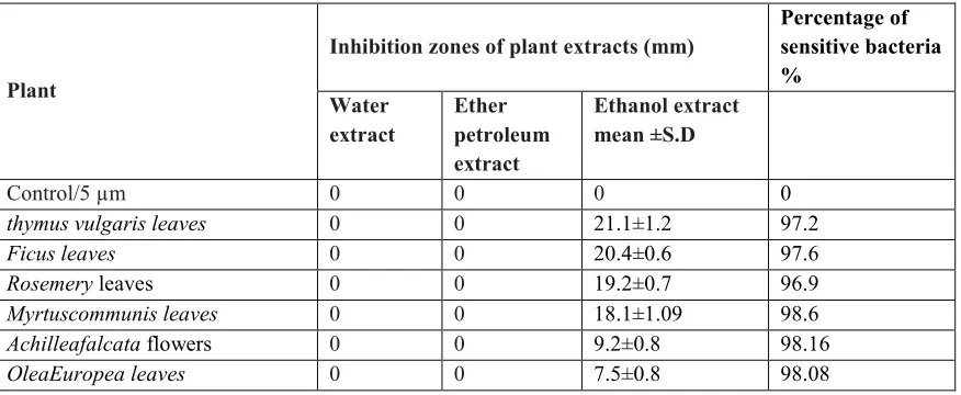
The results of Antibacterial Efficacy of plant extracts:
Table (6): Antibacterial activity of different extracts of studied plants
Plant
Inhibition zones of plant extracts (mm)
Percentage of sensitive bacteria %
Water extract
Ether petroleum extract
Ethanol extract mean ±S.D
Control/5 µm 0 0 0 0
thymus vulgaris leaves 0 0 21.1±1.2 97.2
Ficus leaves 0 0 20.4±0.6 97.6
Rosemery leaves 0 0 19.2±0.7 96.9
Myrtuscommunis leaves 0 0 18.1±1.09 98.6
Achilleafalcata flowers 0 0 9.2±0.8 98.16
OleaEuropea leaves 0 0 7.5±0.8 98.08
DISCUSSION:
Mastitis is the most important disease in dairy milk production worldwide. Staphylococcus and Streptococcus (S. aureus, Str. uberis, Str. dysgalactiaeand Str. Agalactiae), are the major contagious pathogens of bovine mastitis, frequently combined with E. coli(17). Out of 1371 milk samples were obtained from clinical cases of mastitis cows,192 (14.01%) infected samples by Streptococcusbacteria, and (7.4%) infectedsamples by resistant Streptococcus to all antibiotic ofthe totalnumber of samples. The same as Sumathi study has (16%) of Streptococcus spp(18)and Myllys found (11.3%) of Streptococci strains isolated from bovine mastitis in Finland(19). The bacterial species isolated were
Streptococcusagalactiae(22.11%), Streptococcus dysgalactiae(11.43%) in Atyabi study (20). Other research reveald that
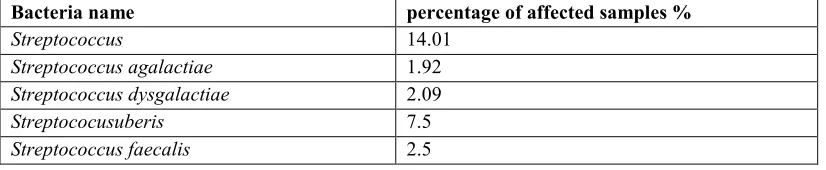
Str. uberis(53%),Str. dysgalactiae(29%), and Str. agalactiae(27%)(17), while in our study were Str. agalactiae(1.29%), Str. Dysgalactiae(2.09%), Str. Uberis(7.5% ),and Streptococcus faecalis (2.5%).
The in vitro antibiogram studies of the bacterial isolates from mastitis milk proved theChloramphenicol to be the drug of choice in this study (22.12%),and ciprofloxacin (19.23%),then Oxacillin (16.61%),Cefadroxil (16.43%), and Gentamycin(12.36%).Those results weren’t in agreement with sumathi study, who revealed the gentamicin to be most effective drug (90%) followed by enrofloxacin (88%),ciprofloxacin (85%), and chloramphenicol (75%)(18).
On the contrary, the present study showed that streptococci isolated from the infected udder milk samples were resistance to 14 studied antibiotics out of 15.A higher resistance was recorded against Amoxicillin (90.07%), Cephalotin (88.06%), Oxytetracycline (87.5%),and Amikacin (84.67%), Tetracyclin (84.7%).While Martín study revealed a high resistance against lincomycin (61.9%) and enrofloxacin (38.1%)(9). This difference may be due to the effects of antibiotics by antibiotic commonly used for treatment.That lead us that there is a need to develop alternative antimicrobial drugs for the treatmentof infectious diseases. So we have used six plants with different extracts. In the present study, the ethanol extracts of the plants exhibited disparate antibacterial activity (Table 6), while no effect for water, neitherpetroleum ether extracts was observed. The ethanolic extracts showed strong activity (inhibition zone 21-18 mm), and weak inhibition (zone 7-9 mm). According to this, the major effectiveness was achieved by the ethanolic extracts from thymus vulgaris,Ficuscarica,Rosemery, and Myrtus communis leaves, followed by Achillea falcata flowers, and the less effectiveness was Olea Europea leaves extaract.
As we concluded in our research, Thymus Vulgaris extract had a significant antimicrobial activity against
Streptococci(21).Essential oils derived from Thymus have been found to possess significant antifungal, insecticidal, and
that asynergistic effect is achieved (24).The structure of thymol is similar to that of carvacrol; both substances seem to make the membrane penetrable. Their structure shatteredthe external membrane of gram-negative bacteria, releasing lipopolysaccharides (LPS) and increasing the permeability of the cytoplasmic membrane to ATP (25).
Ficus carica posse's large amounts of polyphenolic, benzaldehyde and coumarin compounds that has anti-cancer properties. Different studies indicated that figs have antimicrobial effects on various positive and gram-negative bacteria, and Flavonoids acting as antioxidants (1).Ficus capensis revealed the presence of alkaloids, flavonoids, tanins, terpenes, resins, and sterols .The alkaloidsreveals its activity against pathogenic bacteria (26).The petroleum ether extract
of Ficus racemosa leaves had effectiveness against Escherichia coli, and Staphylococcus aureus(27), whereas that was
disagree with our results .Also water extract of methanol extracts of Ficus carica leaves had inhibited growth of Staphylococcus aureus, and Streptococcus pyogenes(28).
There is not much knowledge concerning anti-streptococcal activity of rosemary (Rosmarinuso fficinalis); the mechanism underlying this effect is not known.It was reported that both water and methanolrosemary extracts had different efficacy as antimicrobial agent, which linked to their different polyphenol compositions. Methanol rosemary extract containing carnosic acid, carnosol and rosmarinic acid was the most effective antimicrobial against Gram positive bacteria, Gram negative bacteria. By contrast, water extract containing only rosmarinic acid revealed a narrow efficacy. Therefore, the antimicrobial rosemary extractsactivity was associated with their specific phenolic composition (29).
Ourextract has the same impact, where just the ethanol extract has the antibacterialefficacy. Another reported that aqueous and methanolic extracts of rosemary inhibited S. sobrinus growth and its glucosyl transferase activity(30), this result comply with my extracts, but only ethanol extract has the antibacterial efficacy.
Myrtuscommunis leaves contain different polyphenolic classes as flavonols and galloyl derivatives, which areresponsible to their antimicrobial action. Active phenolic compounds might have several targets which could lead to the inhibition of bacteria. Most of the studies on the mechanism of phenolic compounds have focused on their effects on cellular membranes. They attacked not only cell walls and cell membranes, but affecting their permeability and the release of intracellular constituents, and interfering with membrane functions such as electron transport, enzyme activity or nutrient uptake (31). Moreover the inhibition activity may be due to presence of Tannin via producing hydrogen bonds with proteins, that converted its structure and lead to block the protein synthesis.Tannins considered as a phenolic compounds of plants which have anti oxidative effects (32). Mansouriet al evaluated the antibacterial activity of methanol crude extract of M. communis against Streptococcus pneumoniae, S. pyogenes, and S. agalactiae(33). The inhibition effect of Myrtus communis may refer to the polyphenolic and Tannin they contain.
Achillea, and Oleahad the same impact,9-7 mm inhibition zonesdiameters.
Information about the antimicrobial activity of Achillea extracts is limited. The study of the 13 Turkish Achillea species demonstrated that not all of the Achillea species possess antibacterial activity. Achillea falcata showed mild to low antibacterial activity(8), which matched our results.
As well as the essential oil of Achillea millefolium showed antimicrobial activity against Streptococcus pneumonia. However, water-insoluble parts of the methanolic extracts exhibited slight or no activity (34).The composition of the extract of Achillea clavennaeare alkanes, fatty acids, monoterpenes, guaianesesquiterpenes, and flavonoids (apigenin and centaureidin) (35).The observed activity of the plants studied here in might be due to the presence of sesquiterpene lactones and flavonoids, and possibly due to synergistic interactions between the components of these extracts.
Researchers have published numerous studies concluding that olive leafs and its active ingredient oleuropein act as a natural antibiotic agent. Oleuropein had strong antimicrobial activity against both negative and Gram-positive bacteria, as well as mycoplasma(36).unfortunately, the ethanol extract we had was less or no effectiveness than another researches and other stusdied plants; However, the exact mechanism of the antimicrobial activity of oleuropein is still not completely established(36).
membrane functions such as electron transport, enzyme activity or nutrient uptake. Thus, active phenolic compounds might have several targets which could lead to the inhibition of bacteria (31).
CONCLUSION: The ethanol extracts of the studied plants revealed different antibacterial activity against
Streptococcus, and had shown antibiotics resistance. While the water and ether petroleum extracts had no antibacterial
effectiveness.
REFERENCES:
1.Hosainzadegan H, Alizadeh M, Karimi F, Pakzad P. Study of antibacterial effects of ripped and raw fig alone and in combination. Journal of Medicinal Plants Research. 2012;6(14):2864-7.
2.Belaj A, Satovic Z, Rallo L, Trujillo I. Genetic diversity and relationships in olive (Olea europaea L.) germplasm collections as determined by randomly amplified polymorphic DNA. TAG Theoretical and Applied Genetics. 2002;105(4):638-44.
3.Pereira AP, Ferreira IC, Marcelino F, Valentão P, Andrade PB, Seabra R, et al .Phenolic compounds and antimicrobial activity of olive (Olea europaea L. Cv. Cobrançosa) leaves. Molecules. 2007;12(5):1153-62. 4.Gruenwald J, Brendler T, Jaenicke C. Physicians' Desk Reference(PDR) For Herbal Medicines: Medical
Economics Company; 2000.
5.Al-Anbori DK, Al-Nimer MS, Al-Weheb AM. Athraa M:Antibacterial activity of ethanolic extract of Myrtus communis. L leaves against salivary Mutans streptococci. Saudi Dental Journal. 2008;20(3):82-7.
6.Qaralleh HN, ABBOUD MM, KHLEIFAT KM, Tarawneh K ,Althunibat O. Antibacterial activity in vitro of Thymus capitatus from Jordan. Pak J Pharm Sci. 2009;22(3):247-51.
7.Bozin B, Mimica-Dukic N, Samojlik I, Jovin E. Antimicrobial and antioxidant properties of rosemary and sage (Rosmarinus officinalis L. and Salvia officinalis L., Lamiaceae) essential oils. Journal of agricultural and food chemistry. 2007;55(19):7879-85.
8.Karaalp C, Yurtman AN, Karabay Yavasoglu NU. Evaluation of antimicrobial properties of Achillea L. flower head extracts. Pharmaceuticalbiology. 2009;47(1):86-91.
9.Martín BS, Kruze J, Morales MA, Agüero H, Iragüen D, Espinoza S, et al. Antimicrobial Resistance in Bacteria Isolated From Dairy Herds in Chile. The International Journal of Applied Research in Veterinary Medicine. 2009;4(7)
10. Radulović N, Stankov-Jovanović V, Stojanović G, Šmelcerović A, Spiteller M, Asakawa Y. Screening of in vitro antimicrobial and antioxidant activity of nine Hypericum species from the Balkans. Food chemistry. 2007;103(1):15-21.
11. Wang L, Weller CL .Recent advances in extraction of nutraceuticals from plants. Trends in Food Science & Technology. 2006;17(6):300-12.
12. Hudzicki J. Kirby-Bauer Disk Diffusion Susceptibility Test Protocol. Kirby-Bauer Disk Diffusion Susceptibility Test Protocol. 2009.
13. Wilker MA, Cockerill FR, Craig WA, Dudley MN, Eliopoulos GM, Hecht DW. M100-S17 Performance Standards for Antimicrobial Susceptibility Testing;Seventeenth Informationl Supplement. 2007;27(1). 14. Kayser FH. Medical microbiology: Georg Thieme Verlag; 20 05.
15. Ibrahim S, alRifai H. The role of Staphylococcus and Streptococcus in the incidence of mastitis in sheep in the central region in Syria. the Baath Journal. 2009;31(4).
16. Ibrahim S, alRifai H. The role of Staphylococcus and Streptococcus in the incidence of mastitis in sheep in the central region in Syria. Journal of the Baath. 2009;31(4).
17. Cheng DR, Zhu SY, Yin ZH, Ding WW, Mu ZX, Su ZR, et al. Prevalence of bacterial infection responsible for bovine mastitis. African Journal of MicrobiologyResearch. 2010;4(11):1110-6.
18. Sumathi B, Veeregowda B, Amitha RG. Prevalence and antibiogram profile of bacterial Isolates from clinical bovine mastitis. Veterinary World. 2008;1(8):237-8.
19. Myllys V. Staphylococci in heifer mastitis before and afterparturition. Journal of Dairy Research. 1995;62(01):51-60.
20. Atyabi N, Vodjgani M, Gharagozloo F, Bahonar A. Prevalence of bacterial mastitis in cattle from the farms around Tehran. Iranian J Vet Res. 2006;7:76-9.
22. González M, Marioli J. Antibacterial activity of water extracts and essential oils of various aromatic plants against Paenibacillus larvae, the causative agent of American Foulbrood. Journal of invertebrate pathology. 2010;104(3):209-13.
23. Ivanovic J, Misic D, Zizovic I, Ristic M. In vitro control of multiplication of some food-associated bacteria by thyme, rosemary and sage isolates. Food Control. 2011.
24. Rota MC, Herrera A, Martínez RM, Sotomayor JA, Jordán MJ. Antimicrobial activity and chemical composition of Thymus vulgaris, Thymus zygis and Thymus hyemalis essential oils. Food Control. 2008;19(7):681 -. 7
25. Silva N, Fernandes Júnior A. Biological properties of medicinal plants: a review of their antimicrobial activity. J Venom Anim Toxins incl Trop Dis. 2010;16(3):402-13.
26. Oyeleke S, Dauda B, Boye O. Antibacterial activity of Ficus capensis. African Journal of Biotechnology. 2010;7(10).
27. Mandal SC, Saha B, Pal M. Studies on antibacterial activity of Ficus racemosa Linn. leaf extract. Phytotherapy Research. 2000;14(4):278-80.
28. Ali NH, Faizi S, Kazmi SU. Antibacterial activity in spices and local medicinal plants against clinical isolates of Karachi, Pakistan. Pharm Biol. 2011 Aug;49(8):833-9.
29. Moreno S, Scheyer T, Romano CS, Vojnov AA. Antioxidant and antimicrobial activities of rosemary extracts linked to their polyphenol composition. FreeRadical Research. 2006;40(2):223-31.
30. Tsai PJ, Tsai TH, Ho SC. In vitro inhibitory effects of rosemary extracts on growth and glucosyltransferase activity of Streptococcus sobrinus. Food chemistry. 2007;105(1):311-6.
31. Amensour M, Bouhdid S, Fernández-López J, Idaomar M, Senhaji NS, Abrini J. Antibacterial activity of extracts of Myrtus communis against food-borne pathogenic and spoilage bacteria. International Journal of Food Properties. 2010;13(6):1215-24.
32. Khder AK. Effect of Allium sativum and Myrtus communis on the elimination of antibiotic resistance and swarming of Proteus mirabilis. Jordan Journal of Biological Sciences (JJBS). 2008:3124.
33. Mansouri S, Foroumadi A, Ghaneie T, Najar AG. Antibacterial activity of the crude extracts and fractionated constituents of Myrtus communis. Pharmaceutical biology. 2001;39(5):399-401.
34. Candan F, Unlu M, Tepe B, Daferera D, Polissiou M, Sökmen A, et al. Antioxidant and antimicrobial activity of the essential oil and methanol extracts of Achillea millefolium subsp millefolium Afan.(Asteraceae). Journal of Ethnopharmacology. 2003;87(2):215-20.
35. Stojanović G, Radulović N, Hashimoto T, Palić R. In vitro antimicrobial activity of extracts of four Achillea species: The composition of Achillea clavennae L.(Asteraceae) extract. Journal of ethnopharmacology. 2005;101(1):185-90.
36. Omar SH. Oleuropein in olive and its pharmacological effects. Scientia pharmaceutica. 2010;78(2):133. 37. H, Alizadeh M, Karimi F, Pakzad P. Study of antibacterial effects of ripped and raw fig alone and in
combination. Journal of Medicinal Plants Research. 2012;6(14):2864-7.
AUTHORS AFFILIATION AND ADDRESS FOR CORRESPONDENCE