ANTITUMOR AND ANTIOXIDANT ACTIVITY OF
E
SCHERICHIA
C
OLI
IS
ACCOMPANIED BY CHANGES IN THE L-ARGININE
DICHOTOMOUS PATHWAYS IN PERITONEAL AND BLOOD
LEUKOCYTES FOLLOWING EHRLICH ASCITES
CARCINOMA
N. Kh. Alchujyan
[a]*, H. A. Movsesyan
[a], A. A. Aghababova
[a], A. G. Guevorkyan
[b], N. H.
Movsesyan
[a], H. F. Khachatryan
[a], V. H. Barseghyan
[a], H. L. Hairapetyan
[a], L. S.
Sahakyan
[c],
L. G. Avanesyan
[d], L. H. Melkonyan
[a], K. A. Barseghyan
[a]and G. A.
Kevorkian
[a]Keywords: arginase, cytoplasm, Ehrlich ascites carcinoma, Escherichia coli, leukocyte, lipid peroxidation, mitochondria, nitric oxide synthase.
The effectiveness and mechanisms of antitumor activity of non-pathogenic Escherichia coli EM0 strain using mouse model of Ehrlich ascites carcinoma (EAC) is studied. 48 h after inoculating EAC cells, a single noninvasive treatment of the 2-month-old male white mice’s eyes and mouths with live E. coli isolate increased the life span by 75% (P<0.001). Furthermore, a significant decrease in the volume of ascites fluid (66.5 %, P<0.01) was determined, accompanied by down-regulation of EAC-activated lipid peroxidation processes and changes in the L-arginine metabolic profile in leukocytes as early as within 9 days of post-treatment compared to non-treated EAC-bearing mice. We found EAC-induced stimulation of arginase and nitric oxide synthase (NOS) activity correlated with the levels of their common substrate, L-arginine and products (arginase-derived L-ornithine and NOS-derived NO and L-citrulline) in the cytoplasm and mitochondria of peritoneal and blood leukocytes. The biochemical pattern was differentially modulated by E.coli treatment depending on whether leukocytes were localized in the ascitic fluid or the peripheral blood. E. coli concentrated in the ascitic fluid directly affected the surrounding cells including peritoneal leukocyte in which it decreased the activity of two arginase isoforms and a total NOS in the cytoplasm. Negligible number of E coli at sites remote of tumor suggests its indirect effects particularly via stimulation of LPS-mediated non-specific immune response associated with activation of arginase and NOS in the cytoplasm of blood leukocytes. The data obtained should be taken into account in the further study aimed to use non-pathogenic E. coli strainsin the adjuvant therapy of ascites tumors. * Corresponding Authors
E-mail: [email protected]
[a] H. Buniatian Institute of Biochemistry NAS RA, 5/1 P. Sevak St., 0014, Yerevan, Republic of Armenia
[b] Yerevan State Medical University after M. Heratsi (YSMU) [c] R. O. Eolyan Hematology Center, MH RA (HC)
[d] Kh. Abovyan Armenian State Pedagogical University (ASPU)
INTRODUCTION
Although conventional anticancer treatment methodologies are effectively used, but for about half of cancer patients these are ineffective.1 Outcomes of cancer treatments depend on plenty of factors, many of which are still unknown. Leukocytes infiltrate and interact with tumor and can cause either destruction of the tumor or promotion of malignancy.2 Understanding the metabolic interplay between tumor and immune system will guide the development of optimal metabolic interventions on cancer.3 Recent findings in tumor bearing mice and most human cancers indicate that accumulation of myeloid-derived suppressor cells (MDSC), heterogeneous cell population consisted of granulocyte, and mononuclear cells play a significant role in tumor-associated inflammation and immunosuppression.4 Myeloid-derived cell immuno-suppressive activity is provided by production of reactive
oxygen species, and over-expression of cytoplasmic arginase (ARG1) and inducible NO synthase (iNOS).5The critical interplay between arginase and NOS sharing a common substrate, L-arginine is involved in the cellular immune response and outcome of several pathologic conditions by modulating either the amount of NO produced or metabolic intermediates of the arginase pathway comprising L-ornithine, the precursor of polyamines essential for cell proliferation and urea which participates in detoxification of protein degradation.6Up-regulation ARG1 and iNOS expression is implicated in the suppression of T cell proliferation and IFN-γ production by MDSCs.7 Inversely, inhibition of suppressive effects of CD11b+/Gr-1+ MDSCs is associated with a down-regulation of the mentioned enzymes, accompanied by enhanced T cell infiltration of the tumor and reduction of tumor growth.8 Modulation of expression and activity of both arginase and NOS by various pathogenic microbes has been reported.9,10 At the same time certain bacteria could be used for treatment of neoplasia.11 They accumulate in the tumor microenvironment and induce specific and non-specific antitumor immune response, inhibiting carcinogenesis and thus offering a potential tool for cancer therapy.12
lipopolysaccharide (LPS) (the major component of the cell wall of gram-negative bacteria) cause a production of tumor necrosis factor by activated macrophages, which suppresses proliferation of tumor cells prolonging a survival time of mice in Ehrlich ascites carcinoma (EAC).13,14Phagelysates of E. coli potentiate antitumor effects of drugs (combination of doxorubicin, cyclophosphan, and fluorouracil) elevating the intrinsic resistance to a cytotoxicity of EAC cells, and inhibiting a selective growth advantage of tumor cells over normal cells by 80-90% without apparent side effects.15 Nevertheless, the impact of E. coli on the tumor-induced molecular changes in the immune cells is not fully elucidated. Notably, E. coli could be eliminated by iNOS-derived nitric oxide (NO), the central component of innate immunity contributed to antibacterial activity in the host.16 Peritoneal macrophages has shown express high levels of the iNOS, and released substantial amounts of NO in ascitic fluid of tumor-bearing animals.17 As a consequence, the occurred E. coli deficit may contribute to alterations in microbiota that should affect various metabolic pathways including those of L-arginine.9 This could be prevented by administration of E. coli, which plays a selective role in regional gut immunity via transient modulation of the host intestinal microflora and the capacity to interact with the immune system directly or mediated by the autochthonous microflora.18 We propose that E. coli could affect reactive oxygen species production and arginine metabolizing enzymes considered as therapeutic targets in cancer treatment. This study was undertaken to evaluate the effectiveness and mechanisms of antitumor activity of E. coli using well-established mouse model of EAC.
EXPERIMENTAL
Materials
Dulbecco’s modified Eagle medium (DMEM) was from Serva (Heidelberg, Germany). Bovine serum albumin was from Carl Roth (GmbH, Karlsruhe). NG -monomethyl-L-arginine hydrochloride was obtained from Calbiochem (La Jolla, CA). All other reagents were purchased from Sigma-Aldrich (St. Louis, MO, USA).
Animals and treatments
All procedures involving animals were in accordance with the International Laboratory Animal Care and the European Communities Council Directive (86/809/EEC) and approved by the respective local committee on biomedical ethics (H. Buniatyan institute of biochemistry, Yerevan, RA). 2-month-old male white mice weighing 20.3 ± 0.2 g from our breeding colony were used. All animals were maintained on a 12 h light/dark cycle at normal room temperature and housed in groups of 6/cage with free access to food and tap water.
Experimental design
The animals were divided into groups (n = 18 mice/group): control group - native mice; the first experimental group - mice inoculated intraperitoneally (ip) with EAC cells (0.2 ml of 1.5 × 10⁷ cells); the second
experimental group - mice treated with E. coli (109CFU/ml) 48 h after EAC inoculation. A single non-invasive treatment of eyes and mouth of mice with live bacteria was performed. On the 11th day after tumor implantation, all the experimental animals were sacrificed by decapitation and the ascitic fluid was collected for measuring the volume and leukocyte separation.
Bacteria. The E. coli strain EM0 isolated from the fecal flora of a healthy human volunteer was kindly supplied by Dr. I. Hakobyan (“Armenicum”, Clinical Center, Armenia).
Commensal E coli isolate were propagated in Luria-Bertani broth (Difco Lab., Detroit, MI, USA) for 18-24 h at 37 °C under aerobic conditions, and suspended after centrifugation and adjusted to the appropriate concentration in sterile 20 mM HEPES buffer (pH 7.4).
Determination of survival time. Two groups of male white mice (18/group) were used. EAC cells were inoculated to each group of mice and treatment with E. coli was performed 48 h after tumor cells inoculation. Host survival was recorded and expressed as mean survival time (MST, in days) and percent of increase in life span (ILS) was calculated by the following formula: MST = (Day of first death + Day of last death) / 2; ILS = [(MST of EAC/E. coli group/MST of EAC group) –1] × 100.19
Isolation of resident peritoneal cells. After decapitation, healthy control mice were fixed and 20 mM HEPES buffer (pH 7.4) was injected by sterile syringe into the abdominal cavity, to collect peritoneal cells. Cell suspension was centrifuged at 1000 rpm for 10 min, the precipitated cells were suspended in DMEM (liquid medium (1X) with Na2CO3, without glutamine), cultured at 37 °C for 24 h, and centrifuged at 1000 rpm for 10 min. Supernatant was discarded, cells in the pellet were washed twice with 20 mM HEPES buffer (pH 7.4) and used to isolate resident peritoneal leukocytes.
Ehrlich ascites carcinoma was maintained in 3-month-old white mice in ascitic form under a week passage. Ascitic fluid from the mice was collected by ip puncture using a sterile syringe and the volume was measured to evaluate tumor growth. The ascitic fluid was centrifuged at 1000 rpm for 10 min, and the precipitated cells were washed twice with 20 mM HEPES buffer (pH 7.4) and used to isolate peritoneal leukocytes of EAC-bearing mice.
Isolation of peritoneal and blood leukocytes. Peripheral blood was drawn into 3.8% sodium citrate anticoagulant, mixed with 6 % dextran (prepared on 0.9 % NaCl) and incubated at 37 ºC for 60 min to remove erythrocytes from the fresh blood by gravity sedimentation. The decanted layer was centrifuged at 4 °C (6000 rpm, 10 min) and blood plasma was obtained in supernatant.
Leukocyte subsets were identified in fixed smears stained by the Romanovsky-Giemsa method.20 All cell preparations consisted of about 90 to 95 % viable cells as determined with the trypan blue exclusion test.
Preparation of cytoplasmic and mitochondrial fractions was performed by differential centrifugation.21 Leukocytes suspended in 20 мМ HEPES buffer containing 0.25 M sucrose (pH 7.4) were homogenized (1500 rpm, 3 min), centrifuged at 4 ºС (1200 rpm, 10 min) to remove nuclear fraction and cell debris. Pellet was discarded and the supernatant was further centrifuged at 4 ºС (11000 rpm, 20 min) to yield the crude mitochondrial preparation which was washed, suspended and homogenized in 20 mM Hepes buffer containing of 0.1 M NaCl, 10 % glycerol, 0.1 % Triton X-100-PC, and cocktail of protease inhibitors (Roche, Basel, Switzerland). The supernatant was used as the cytoplasm.
Measurement of L-arginine. Samples were deproteinized with 0.5 N NaOH and 10 % ZnSO4. Following a centrifugation (15000 rpm, 3 min), the protein-free supernatants were sampled and analyzed for L-arginine according to Akamatsy & Watanabe with our modification.24 Briefly: a sample of supernatant diluted with distilled water (1:1, v/v) was added in a ratio 3:1 (v/v) to a reagent (mixture of equal amounts of 0.4 % 8-oxychinoline in 96 % ethanol, 5 % sulfosalicylic acid in 0.01 M glycine buffer, 2.5 % NaOH), stirred with 1 % solution of sodium hypobromite and the absorbance was measured at 525 nm wavelength against reagent blank containing all the reagents minus the sample.
Arginase assay. The samples were added to the reaction mixture: 20 mM HEPES buffer containing 2 mM dithiothreitol (pH 7.4), 1.5 mM MnCl2·4H2O, 4.75 mM L-arginine·HCl and incubated at 37 °C for 60 min, followed by the addition of 10 % TCA to stop the reaction.22Parallel control experiments were conducted in the presence of 60 mM L-valine, a non-selective inhibitor of the arginase isoforms. Following a centrifugation (15000 rpm, 3 min) the supernatants were sampled and analyzed for L-ornithine content. The arginase activity expressed as produced in an hour L-ornithine per mg of total protein.
Measurement of L-ornithine. Samples were mixed with 4.5 % ninhydrin (1:1,v/v), heated (90 oC, 20 min), cooled to the room temperature and the absorbance was measured at 505 nm wavelength against reagent blank containing all the reagents minus the sample.22
Nitric oxide synthase assay. A total NOS activity was assessed by measuring stable intermediate of NO, nitrite (NO2) accumulated during a long-term incubation of samples (37 °C for 22 h) in 20 mM HEPES buffer (pH 7.4), containing 2 mM dithiothreitol and 3 mM MgCl2·6H2O, in the presence of NOS substrate, 5.5 mM L-arginine·HCl, and cofactors: 0.2 mM NADPH, 6 µM FAD, 5.5 µM FMN, 20 µM ((6R)-5,6,7,8-tetrahydro-L-biopterin dihydro- chloride) and 1.7 mM CaCl2. Parallel control experiments were conducted in the presence of 5 mM NG –monomethyl-L-arginine hydrochloride, non-selective inhibitor of all the NOS isoforms. Reaction was initiated by addition of samples to the incubation medium and terminated by subsequent addition of 0.5 N NaOH and 10 % ZnSO4. Following a centrifugation (15000 rpm, 3 min) the
supernatants were sampled and analyzed for nitrite content. The NOS activity expressed as produced in 22 h nitrite per mg of total protein.
Measurement of nitrite. Samples were deproteinized with 0.5 N NaOH and 10 % ZnSO4. Following a centrifugation (15000 rpm, 3 min), the protein-free supernatants were sampled and analyzed for nitrite using colorimetric technique based on diazotization.23 Samples were mixed in equal parts with Griess-Ilosvay reagent (1:1 mixture of 0.17 % sulfanilic acid and 0.05 % α-naphthylamine in 12.5 % acetic acid), and the absorbance was measured at 546 nm wavelength against reagent blank containing all the reagents minus the sample.
Measurement of L-citrulline. Samples were deproteinized with 10 % TCA, centrifuged (15000 rpm, 3min), then protein-free supernatants were mixed with 9.6 % H2SO4 and reagent (5 mÌ diacetylmonoxime, 0.9 mM thiosemicarbazide, and 0.025 mÌ FeCl3) at a ratio of 1:1:1
(v/v), heated in a boiling water bath for 10 min, cooled to the room temperature and the absorbance was measured at 490 nm wavelength against reagent blank containing all the reagents minus the sample.25
Indices of oxidative stress referring to lipid peroxidation processes were established by measuring malondialdehyde (MDA) using thiobarbituric acid.26 Samples were deproteinized with 10 % TCA and the precipitates were removed by centrifugation at 15000 rpm for 3 min, supernatants obtained were mixed with 0.72 % TBA and 0.6 N HCl in proportion 1: 0.8 : 0.2 (v/v), heated for 15 min in boiling water bath that resulted in the formation of pink-colored secondary product of MDA and the absorbance was measured at 535 nm wavelength against reagent blank containing all the reagents minus the sample.
Protein was determined using crystalline bovine serum albumin as standard.27
Statistical analysis. All data were analyzed using a one-way analysis of variance (ANOVA) followed by post hoc Holm-Sidak test (SigmaStat 3.5 for Windows). Relationships between biochemical parameters studied were determined calculating the Pearson linear correlation coefficient (r). Data are expressed as the mean ± S.E.M. Differences are considered significant at P < 0.05.
RESULTS AND DISCUSSION
mononuclears in EAC-bearing mice. Notably, there are the findings of other authors that activated granulocytes in advanced cancer patients and healthy donor with N-formyl-L-methionyl-L-leucyl-L-phenylalanine (both may inhibit cytokine production by T cells) exhibit low density and co-purified with low density peripheral blood mononuclears.30 At the same time, conventional mast cells have a greater density than granulocytes up to 1.2g • ml-1, while the mast cellsfrom tumor are recovered with malignant EAC cells in the lowest density fractions (up to 1.06 g • ml-1), because of high content of lipid and mucopolysaccharide laden vacuoles.31 It should be noted that mast cells were not studied in the present work.
Antitumor and antioxidant effects of E. coli following Ehrlich ascites carcinoma
E coli strain EM0 isolated from the feces of a healthy human volunteer was used for treatment of EAC-bearing mice. It is described as a non-pathogenic strain that had no adverse effect in germfree mice after colonization, despite the fact that EM0 may express hemolysin and cytotoxic necrotizing factor in vitro.32 Two days after EAC transplantation, a single non-invasive treatment of eyes and mouth of two-month-old white male mice with live E. coli
isolate prolonged a life span by 75 %. On 11th day of EAC, E. coli caused a decrease of approximately thrice in the volume of ascitic fluid that accompanied by appropriate changes in the body weight of animals. Previously, we have demonstrated that after administration, E. coli is mainly concentrated in the ascitic fluid of EAC-bearing mice, and a much less its number was detected at sites remote from tumor, particularly in blood.33 Concomitantly, E. coli induced inflammatory response which interfered with induction and maintenance of the oncogenic phenotype of cancer cells appearing as a diminished anaplasia, in contrast to non-treated animals.
Both macrophages and neutrophils are activated in tumor-bearing patients and experimental animals and produce the elevated levels of reactive oxygen species (ROS) and down-regulate the ROS scavengers and antioxidant enzymes thus contributing to the oxidative stress associated with immunosuppression and carcinogenesis.34,35Carcinoma cells also produce ROS and other free radicals at elevated rates in vitro, and, many tumors have developed resistance то oxidative stress in vivo.36 High activity of antioxidant enzymes may contribute to resistance of tumor cells to overproduction of ROS. Notably, a significant up-regulation of superoxide dismutase (SOD) is observed on the stationary phase of EAC cells.37 At the same time, impaired functioning of the antioxidant system is demonstrated in the leukocytes isolated from area of tumor growth and blood.38 Neutrophils from the blood of patients with larynx carcinoma exhibit low level of SOD prior to surgery, especially in the patients with advanced tumor stages.34
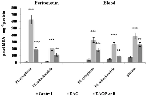
Our data also suggests the prevalence of oxidative stress in the leukocyte over that of tumor cells on the 11th day of EAC development, viz. the level of a marker of oxidative stress, MDA (a measure of lipid peroxidation) was two-fold higher in the homogenates of peritoneal leukocytes (PL) than in EAC cells (165.3 ± 26.4 vs. 82.0 ± 11.2 pmol MDA • mg-1 protein respectively). Lipid peroxidation processes were significantly stimulated in PL and blood in EAC, and
inhibited following E. coli treatment (Fig. 1). The MDA levels were increased to 41-, and 11-fold in the cytoplasm and mitochondria of PL, and to 7-, and 5-fold in those of blood leukocyte (BL), as well as to 4.6- fold in blood plasma of EAC-bearing mice compared respectively to the basal levels.
Figure 1. Lipid peroxidation in the peritoneum and blood following Ehrlich ascites carcinoma (EAC) and E. coli treatment. Data are expressed as M ± SEM, n=18. The confidence probability (p) of parameters evaluated for EAC-bearing mice compared to the control, and p for E. coli-treated mice compared to untreated EAC-bearing mice. Peritoneal leukocytes (PL): cytoplasm - F=59.3,
p<0.001; mitochondria - F=16.1, p<0.001. Blood leukocytes (BL): cytoplasm - F=30.2, p<0.001; mitochondria - F=40.9, p<0.001; plasma - F=24.4, p<0.001. # p >0.05, * p <0.05, ** p <0.01, *** p<0.001.
After E. coli treatment, the MDA amounts decreased by 3.2 and 1.8 times in the cytoplasm and mitochondria of PL, and by 1.8 and 2.8 times in those of BL compared respectively to EAC-bearing mice. Simultaneously, the MDA level in plasma dropped lower than control values. Hence, E. coli may counteract the oxidative stress, particularly EAC-induced redox imbalance in leukocytes preventing their oxidative damage and impairment in antitumor activity. The inhibition of ROS production by immune cells could cancel their suppressive effects in mice and patients with cancer.28,39 Antioxidant activity of E. coli appears to be due to its polyamines, which are effective scavengers of ROS, and could also trigger a transcription of protective proteins under conditions of strong oxidative stress.40 Capability of E. coli to rapidly adapt to the oxidative environment was demonstrated on the E. coli
strain MG1655 which metabolite profile was normalized in 40-60 min after exposure to a sub-lethal concentration of hypochlorite.41
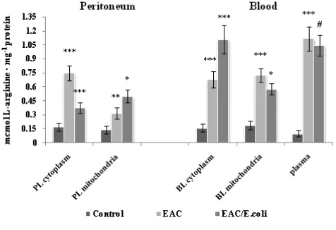
Figure 2. L-arginine level in the peritoneum and blood following Ehrlich ascites carcinoma (EAC) and E. coli treatment. Data are expressed as M ± SEM, n=18. The confidence probability (p) of parameters evaluated for EAC-bearing mice compared to control, and p for E. coli-treated mice compared to untreated EAC-bearing mice. Peritoneal leukocytes (PL): cytoplasm - F=22.4, p<0.001; mitochondria - F=9.6, p<0.001. Blood leukocytes (BL): cytoplasm - F=21.6, p<0.001; mitochondria - F=21.5, p<0.01; plasma - 32.2,
p<0.001. # p >0.05, * p <0.05, ** p <0.01, *** p<0.001.
An elevation of the arginine content might be a consequence of EAC-induced exacerbation of free radical oxidation that apparently may intensify intracellular protein degradation. In turn, the tRNA mediated, posttranslational, N-terminal arginylation of proteins (occurred in all eukaryotic cells) may lead to rapid ubiquitination of arginylated proteins degraded by cytosolic proteases by the N-end rule pathway.42 High amounts of L-arginine may affect the ion balance causing resistant hyperkalemia, a typical and the most life-threatening of tumor lysis syndrome.43,44Hence, negative consequences at this stage of EAC appear to be caused not by arginine deficiency, but rather of its increase.
Despite of a decrease in the arginase activity (vide infra), the L-arginine level dropped twice in the cytoplasm of PL following E. coli treatment. Simultaneously, L-arginine content was elevated by 1.6 times in the mitochondria of PL, possibly, due to E. coli-mediated normalization of the mitochondrial arginase activity (vide infra) and/or penetration of amino-acid from the cytoplasm. E. coli -induced decrease in oxidative stress might interfere with protein breakdown served as a source of L-arginine and diminish the arginine level. Notably, the cytoplasmic L-arginine high pool in contrast to other creatine analogues and related compounds can inhibit creatine transporters localized on inner and outer mitochondrial membrane fractions and interfere with the creatine transport from cytoplasm into mitochondria.45 Thus, E. coli mediated modulation of the L-arginine level in the cytoplasm of PL could facilitate their energy production.
L-arginine may be converted to agmatine by the cytosolic arginine decarboxylase of E. coli in which it exists in constitutive and inducible isoforms.46 Agmatine in turn may serve as a free radical scavenger by protecting against the
oxidation of sulfhydryl groups and decreasing hydrogen peroxide content, preserving from mitochondrial damage and apoptosis.47 At the same time, agmatine released into ascitic fluid may exert cytostatic activity in proliferating cancer cells, because of its preferable uptake by transformed cell with short cycling times lines (H-ras- and Src-transformed murine NIH/3T3 cells, Ras/3T3 and Src/3T3, respectively) accompanied by a suppression of cell growth via agmatine-mediated induction of the antizyme synthesis, which inhibits ornithine decarboxylase activity attenuating polyamine formation.48,49Further, E. coli lacks endogenous arginase and NO-synthase, enzymes that can compete with arginine decarboxylase.50,51
E. coli caused a 1.6-fold increase in the L-arginine level in the cytoplasm of BL and a slight decrease in the mitochondria of BL compared to non-treated EAC-bearing mice, whereas no changes were detected in blood plasma. As noted, after treatment a much less number of E. coli was observed in blood, compared to ascitic fluid, and signaling mechanisms appear to be involved, particularly E. coli’s LPS among other pro-inflammatory stimuli can upregulate the L-arginine cationic amino acid transporter 2 (CAT2).52 This may contribute to E. coli-induced elevation of the arginine level in BL. Presumably, L-arginine would be utilized by such arginine-metabolizing enzymes as ARG1 and NOS that are also activated in the cytoplasm of BL following E. coli administration (vide infra). Early concomitant induction of iNOS, ARG2, argininosuccinate synthase and ornithine decarboxylase, and CAT2 and late induction of ARG1 are demonstrated in LPS-activated peritoneal macrophages.53
Arginase activity in peritoneal and blood leukocytes following EAC and E. coli treatment.
Figure 3. Arginase activity in the peritoneum and blood following Ehrlich ascites carcinoma (EAC) and E. coli treatment. Data are expressed as M ± SEM, n=18. The confidence probability (p) of parameters evaluated for EAC-bearing mice compared to control, and p for E. coli-treated mice compared to untreated EAC-bearing mice. Peritoneal leukocytes (PL): cytoplasm - F=37.7, p<0.001; mitochondria - F=13.5, p<0.001. Blood leukocytes (BL): cytoplasm - F=59.6, p<0.001; mitochondria - F=35.4, p<0.001; plasma - F=7.1, p=0.002. # p >0.05, * p <0.05, ** p <0.01, *** p<0.001.
E. coli-treatment decreased the ARG1 activity by 1.44 times and normalized ARG2 in PL of EAC-bearing mice, while in BL a 3-fold increase in the ARG1 activity was detected and a 2.5-fold decrease in that of ARG2 respectively compared to non-treated mice. Inhibition of arginase isoforms in PL and ARG2 in BL indicated to ameliorating effect of bacterial treatment interfering with imminosuppresive effects of arginases. Besides, ARG2 plays a critical role in macrophage proinflammatory responses promoting mitochondrial ROS production.58 At the same time in blood E. coli/LPS-mediated non-specific immune response is, presumably, associated with the activation of both ARG1 and iNOS (vide infra). Interestingly, iNOS/NO could exclusively affect ARG1 activity (not ARG2) via S-nitrosylation of 2 cysteine residues (C168 and C303), and through S-nitrosylation of C303 stabilized ARG1 trimer and reduced its Km value 6-fold, both in vitro and ex vivo.59 In contrast to ARG1, ARG2 is not significantly modulated by Th1 or Th2 cytokines.60 Although, recent data suggests that IFNã interferes with arginase activity in the three murine renal cell carcinoma cell lines, perhaps by inhibiting transcription of the ARG2 gene there through inhibiting proliferation of cells.61 Whether IFN-γis implicated in E. coli impact on the ARG2 activity in leukocytes must be elucidated. It can be speculated that E. coli via stimulation of the ARG1 activity and subsequent urea overproduction in the cytoplasm of BL is metabolically aimed to down-regulate the iNOS activity. Urea inhibits the iNOS/NO in a dose-dependent manner facilitating proliferation of LPS-stimulated mouse macrophages (RAW 264.7).62 Interestingly, the same distribution of the intracellular arginase activities was observed both in PL and BL after E. coli treatment (ARG1 : ARG2 = 3.6 : 1).
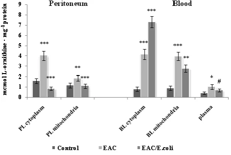
One of the major products of arginase is L-ornithine, a precursor in the production of polyamines, whose elevated levels are associated with neoplastic growth.63 Ornithine
levels are significantly dropped in blood plasma and other tissues in the double knockout ARG1 and ARG2-deficient mice, indicating that arginase is critical to the maintenance of ornithine homeostasis.64 Fig. 4 shows the alterations in the L-ornithine content in the peritoneum and blood following EAC and E. coli treatment. Positive correlation between arginase activity and L-ornithine level was determined in the PL cytoplasm (r=0.63, p=0.0053) and mitochondria (r=0.99, p<0.0001), and in the BL cytoplasm (r = 0.97, p <0.0001) and mitochondria (r = 0.96, p <0.0001), as well as in plasma (r = 0.55, p = 0.02). Although, L-ornithine may inhibit the two isoforms of arginase in vivo by a feedback mechanism, no inhibitory effect was observed in the studied leukocytes over L-ornithine concentration range determined in vitro.
Figure 4. L-ornithine level in the peritoneum and blood following Ehrlich ascites carcinoma (EAC) and E. coli treatment. Data are expressed as M ± SEM, n=18. The confidence probability (p) of parameters evaluated for EAC-bearing mice compared to control; and p for E. coli-treated mice compared to untreated EAC-bearing mice. Peritoneal leukocytes (PL): cytoplasm - F=28.6, p<0.001; mitochondria - F=8.27, p<0.001. Blood leukocytes (BL): cytoplasm - F=42.6, p<0.001; mitochondria - F=24.3, p<0.001; plasma - F=2.7, p=0.078. # p >0.05, * p <0.05, ** p <0.01, *** p<0.001.
tumor cell growth and not only through depletion of L-arginine, but also by providing them with polyamines, because the progression of a malignancy correlates with an elevated expression of both or specific isoform of arginase (depending on the type of tumor), while an inhibition of tumor growth is accompanied by a decrease in the expression and activity levels of arginase.61,66
Nitric oxide synthase activity in peritoneal and blood leucocytes following EAC and E. coli treatment.
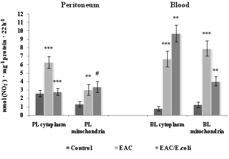
There are three main isoforms of NOS, namely, constitutive (cNOS), including neuronal and endothelial NOSs and iNOS, which are highly induced by LPS, lipoteichoic acid, and proinflammatory cytokines.69 A total NOS activity was assessed in the leukocyte cytolasmic and mitochondrial compartments, in which all the NOS isoforms are presented (Chen, K., 2010). S.S. Greenberg et al., 1998; T. Wallerath et al., 1997).70.71.72 As shown in Fig. 5, on the 11th day of EAC transplantation a total NOS activity increased by 2.4 and 2.3 times in the cytoplasm and mitochondria of PL, and 8.5 and 6.3 times in those of BL respectively compared to control. It should be noted, that enhanced mitochondrial NO production and increased free radical generation, disrupt the electron transport system and mitochondrial permeability transition.73 NO may potentially de-energise mitochondria via inhibition of creatine kinase, aconitase, cytochrome c oxidase.69,74 Interestingly, in some cases involving proteins with N-terminal Cys, arginylation can happen only after nitric oxide-dependent Cys oxidation and such oxidation-dependent arginylation are likely to target proteins for degradation.75E. coli decreased the NOS activity in cytoplasm up to basal values, whereas no changes in the NO production were detected in mitochondria of PL.
Figure 5. Nitric oxide synthase activity in the peritoneum and blood following Ehrlich ascites carcinoma (EAC) and E. coli
treatment. Data are expressed as M ± SEM, n=18. The confidence probability (p) of parameters evaluated for EAC-bearing mice compared to control; and p for E. coli-treated mice compared to untreated EAC-bearing mice. Peritoneal leukocytes (PL): cytoplasm - F=16.4, p<0.001; mitochondria - F=12.6, p<0.001. Blood leukocytes (BL): cytoplasm - F=32.5, p<0.001; mitochondria - F=28.3, p<0.001. Note: NOS activity was not determined in plasma. # p >0.05, * p <0.05, ** p <0.01, *** p<0.001.
EAC-enhanced arginine level in the leukocytes appear to up-regulate a total NOS activity, particularly the iNOS, which in contrast with the cNOS is a high-output form strongly dependent on the presence of intracellular L-arginine that is a rate-limiting factor in NO synthesis.76 As noted, peritoneal macrophages in ascites tumor-bearing animals express high levels of iNOS accompanied by overproduction of NO.17 The iNOS can produce NO for prolonged periods and contribute to excessive NO, which is the only biomolecule produced in high enough concentrations to out-compete SOD for superoxide and forming peroxynitite (ONOO-, PN), a potent oxidizing and nitrating agent that modifies tyrosine in proteins and creates nitrotyrosines leaving a footprint detectable in vivo.77 NO released from PL may interact with superoxide generated by extracellular NADPH oxidase, the activity of which was revealed in ascitic fluid and was about 28 % higher than that in serum of healthy mice, and 8-10 times higher than in the EAC cells on terminal stage of EAC.78 MnSOD and CuZnSOD are inactivated when exposed to simultaneous fluxes of superoxide and NO, and PN may inactivate the MnSOD via the direct reaction with the Mn center and a metal-catalyzed nitration of Tyr-34 in MnSOD.79 PN could be involved in the inactivation of T-cell receptor complex via nitration and subsequent inhibition of the protein tyrosine phosphorylation in purified lymphocytes and priming them to undergo apoptotic cell death after phytohemagglutinin or CD3-mediated activation.80 The presence of high levels of nitrotyrosine in T lymphocytes infiltrating human prostate carcinoma, suggests production of PN.67
Interestingly, population of circulating CD11b+IL-4 receptor α+ (CD11b+IL-4Rα++), inflammatory-type monocytes elicited by growing tumors and producing IL-13 and IFN-γ contributing to stimulation of both ARG1 and iNOS that in turn are involved in the suppression of antigen-activated CD8+ T lymphocytes.81 Notably, сNOS-derived NO could block the nuclear factor kappaB (NF-kB) activation and proinflammatory mediators release, vice versa iNOS/NO could stimulate these processes.82 This becomes even more important, since inhibition of NF-kB signaling in ARG1-expressing tumor-associated macrophages transformed them into tumor-cytotoxic effector cells.83 In addition, iNOS-derived NO may inhibit cNOS by a feedback mechanism through the formation of stable inhibitory ferrous nitrosyl complexes, whereas in case of the iNOS it appears weak.84
Thus, contrary to PL, E. coli may up-regulate both ARG1 and cytoplasmic NOS activity in BL of EAC-bearing mice that might be mediated by LPS. It is documented that LPS as well as various inflammatory stimuli may increase the NO production and the expression and activity of iNOS and arginase in rodents.86,87
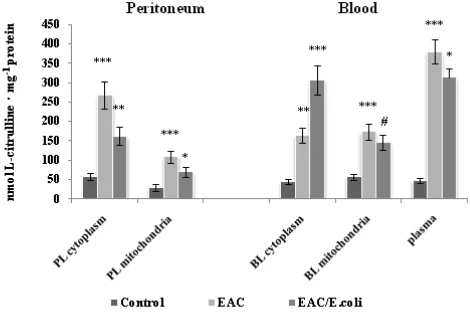
Subcellular changes in the NOS activity were associated with appropriate alterations in the levels of its products, stable metabolite of NO, nitrite and L-citrulline in the peritoneum and blood following EAC and E. coli treatment (Fig. 6 and 7, respectively).
Figure 6. L-citrulline level in the peritoneum and blood following Ehrlich ascites carcinoma (EAC) and E. coli treatment. Data are expressed as M ± SEM, n=18. The confidence probability (p) of parameters evaluated for EAC-bearing mice compared to control, and p for E. coli-treated mice compared to untreated EAC-bearing mice. Peritoneal leukocytes (PL): cytoplasm - F=18.2, p<0.001; mitochondria - F=9.1, p<0.001; Blood leukocytes (BL): cytoplasm - F=27.8, p<0.001, mitochondria - F=12.7, p<0.001, plasma -
F=58.8, p<0.001. # p >0.05, * p <0.05, ** p <0.01, *** p<0.001.
Figure 7. Nitrite level in the peritoneum and blood following Ehrlich ascites carcinoma (EAC) and E. coli treatment. Data are expressed as M ± SEM, n=18. The confidence probability (p) of parameters evaluated for EAC-bearing mice compared to control, and p for E. coli-treated mice compared to untreated EAC-bearing mice. Peritoneal leukocytes (PL): cytoplasm - F=13.6, p<0.001; mitochondria - F=15.3, p<0.001; Blood leukocytes (BL): cytoplasm - F=26.4, p<0.001; mitochondria - F=24.5, p<0.001; plasma - F=37.4, p<0.001. # p >0.05, * p <0.05, ** p <0.01, *** p<0.001.
Positive correlations between total NOS activity and L-citrulline levels were determined in the cytoplasm (r=0.89,
p<0.0001) and mitochondria (r = 0.78, p=0.0002) of PL, and in the cytoplasm (r = 0.97, p <0.0001) and mitochondria (r = 0.92, p<0.0001) of BL, as well as between total NOS activity and nitrite levels in the cytoplasm (r=0.88,
p<0.0001) and mitochondria (r=0.99, p<0.0001) of PL, and in the cytoplasm (r = 0.89, p<0.0001) and mitochondria (r = 0.78, p=0.0002) of BL.
It has been recently demonstrated that only modulating both enzymes arginase and NOS in vivo may reduce tyrosine nitration and restore responsiveness of tumor infiltrating T lymphocytes to human prostatic adenocarcinoma that was confirmed also on a transgenic mouse prostate model.67 We believe that the beneficial effect of the non-pathogenic E. coli strain is in part due to its antioxidant activity and associated impact on the mentioned enzymes in the PL. Our findings highlight also the importance of the subcellular location of arginine and arginine metabolizing enzymes as well as their substrate and products during EAC and bacterial treatment and should be further investigated.
CONCLUSION
In summary, we suggest that in the stationary to terminal phases of EAC an overproduction of ROS and an enhancement of L-arginine levels occurred and accompanied by stimulation of the ARG1 and ARG2 and total NOS activity in the cytoplasm and mitochondria of both peritoneal and blood leukocytes. The most important novel finding of this study is that antitumor activity of non-pathogenic E. coli EM0 strain appear to be associated with its inhibition of lipid peroxidation and differential modulation of L-arginine metabolic pattern depending on whether leukocytes are localized in the ascitic fluid or in the peripheral blood. After administration, E. coli was generally concentrated in the ascitic fluid and directly affected the surrounding cells including peritoneal leukocytes, in which the EAC-induced high activity of ARG1 decreased and a total cytoplasmic NOS and ARG2 were normalized. Negligible number of E coli were observed at sites remote of tumor which suggests its indirect effects, presumably via stimulation of LPS-mediated non-specific immune response associated with activation of both ARG1 and NOS in the cytoplasm of blood leukocytes in which they might contribute to inhibition of ARG2 and mitochondrial NOS, implicated in the reciprocal regulation of host antitumor response, and interfered with E. coli dissemination, as well. The data obtained should be taken into account in the further study aimed to use non-pathogenic E. coli strains in the therapy of ascites tumors. Overall, a precise understanding of indigenous bacteria impact on the arginine pathways in cancer may contribute to the development of more adequate therapy.
ACKNOWLEDGMENTS
Center "Armenikum") who provided us non-pathogenic E. coli EM0 strain, and for her help in establishing the methodological procedures necessary for this study, Ms. Ani Hakobyan (Master of English language and literature) for editing the manuscript.
REFERENCES
1Jemal, A., Siegel, R., Xu, J., Ward, E., CA Cancer J. Clin.,2010, 60(5), 277.
2Lewis, C. E. and Pollard, J. W., Cancer Res., 2006, 66(2), 605. 3Wang, T., Liu, G. and Wang, R., Front. Immunol., 2014, 5, 358. 4Raber, P., Ochoa, A. C. and Rodríguez, P. C., Immunol. Invest.
2012, 41(6-7), 614.
5Greten, T. F., Manns, M. P. and Korangy, F., Int. Immunopharmacol., 2011, 11(7), 802.
6Munder, M., British J. Pharmacol., 2009, 158(3), 638.
7Lim, H. X., Hong, H. J., Cho, D. and Kim, T. S., J. Immunol., 2014, 193(11), 5453.
8Serafini, P., Meckel, K., Kelso, M., Noonan, K., Califano, J. and
Koch, W., J. Exp. Med., 2006, 203(12), 2691.
9Das, P., Lahiri, Am., Lahiri, Ay. and Chakravortty, D., PLOSpathogens, 2010, 6(6),1-7.
10Chen, M., Bao, W., Aizman, R., Huang, P., Aspevall, O.,
Gustafsson, L.E., Ceccatelli, S. and Celsi, G., J Infect Dis., 2004, 190(1), 127.
11Hoption Cànn, S. A., van Netten, J. P. and van Netten,
C., Postgrad. Med. J., 2003, 79(938), 672.
12Patyar, S., Joshi, R., Byrav, P., Prakash, A., Medhi, B. and Das, B.
K., J. Biomed. Sci.,2010, 17(1), 21.
13Wong, C. K., Fung, K. P., Lee, C. Y. and Choy, Y. M., Cancer Lett., 1992, 63(1), 7.
14Fiore, N., Green, S., Williamson, B., Carswell, E., Old, L. J. and
Hlinka, J., Proc. Am. Assoc. Cancer Res., 1975, 16(1), 125.
15Gambashidze, K., Khorava, P., Azaladze, T., Kalandarishvili, K.,
Jaiani, E., Lasareishvil, B., Azaladze, A. and Tediashvili, M.,
Exp. Oncol., 2012, 34 (2), 107.
16Chakravortty, D. and Hensel, M., Microb. Infect., 2003, 5(7), 621. 17Nishikawa, M., Sato, E., Kashiba, M., Kuroki, T., Utsumi, K. and
Inoue, M., Hepatology, 1998, 28(6), 1474.
18Lodinová-Zádníková, R., Cukrowska, B. and
Tlaskalova-Hogenova, H., Inter. Arch. Allerg. Immunol., 2003, 131(3), 209.
19Mazumder, U. K., Gupta, M., Maiti, S. and Mukherjee, M., Indian J. Exp. Biol., 1997, 35(5), 473.
20Putintceva, O. V., Artukhov, V. G. and Koltakov, I. A., Immunology. PartII. Voronezh. 2008, 45.
21Dizhe, G. P., Eshchenko, N. D., Dizhe, A. A. and Krasouskaya, I.
E. Introduction to the techniques of biochemical experiments.
SPb. 2003, 86.
22Iyamu, E. W., Asakura, T. and Woods, G. W., Anal. Biochem., 2008, 383(2), 332.
23Schmidt, H.H.H.W. and Kelm, M. (Feelisch M. and Stamler J. S.,
eds.) Methods in Nitric Oxide Research. Wiley, Chichester. 1996, 491.
24Akamatsy S., Watanabe T.J., J. Biochem.,1961, 77(3), 484. 25Moore, R. B. and Kauffman, N. J., Anal. Biochem., 1970, 33(2),
263.
26Buge, J. A. and Aust, S. D., Method enzymol., 1978, 52, 302.
27Lowry, O. H., Rosebrough, N. J., Farr, A. L. and Randall, R. J., J. Biol. Chem., 1951, 193(1), 265.
28Diaz-Montero, C. M., Finke, J. and Montero, A. J., Semin. Oncol., 2014, 41(2),174.
29Gratchev, A., Kzhyshkowska, J., Kothe, K., Muller-Molinet, I.,
Kannookadan, S., Utikal, J. and Goerdt, S., Immunobiology, 2006, 211(6-8), 473.
30Schmielau, J., Finn, O.J., Cancer Res., 2001, 61(12), 4756. 31Hamburger, A. W., Dunn, F. E. and White, C. P., Br. J.
Cancer, 1985, 51(2), 253.
32Hudault, S., Guignot, J. and Servin, A.L., Gut, 2001, 49(1), 47. 33Aghababova, A. A., Movsesyan, N. H., Hakopyan, A. M. and
Avagyan, H. Kh., Reports of NAS of Armenia, 2013, 113(3), 303.
34Szuster-Ciesielska, A., Hryciuk-Umer, E., Stepulak, A., Kupisz,
K. and Kandefer-Szerszen, M., Acta Oncol., 2004, 43(3), 252-258.
35Corzo, C. A., Cotter, M. J., Cheng, P., Cheng, F., Kumartsev, S.,
Sotomayor, E., Padhya, T., McCaffrey, T. V., McCaffrey, J. C. and Gabrilovich D. I., J. Immunol., 2009, 182(9), 5693.
36Storz, P., Front. Biosci., 2005, 10, 1881-1896.
37Smirnova, L. P, Kondakov, I. V., Biomed. Chem., 2004, 50(6),
566-575.
38Potselueva, M. M, Naumov, A. A., Sukhomlin, T. K., Zinatullina,
G. G. and Shatalin, Iu. V., [Article in Russian]. Tsitologia, 2013, 55(5), 307.
39Kusmartsev, S., Nefedova, Y., Yoder, D. and Gabrilovich, D. I., J. Immunol., 2004, 172(2), 989.
40Tkachenko, A. G. and Fedotova, M. V., Biochemistry (Mosc), 2007, 72(1), 109.
41Drazic, A., Kutzner, E., Winter, J. and Eisenreich, W., PLoS One, 2015, 10(5), e0125823.
42Yu, M., Chakraborty, G., Grabow, M. and Ingoglia, N. A., Neurochem. Res., 1994, 19(1), 105.
43Krichevskaya, A. A., Lukash, A. I., Shugalei, V. S. and
Bondarenko, T. I., Aminoacids, its derivartives and regulation of metabolic pathways. Rostov, 1983, 112.
44Semenova, A. I., Practical oncology, 2006, 7(2), 101.
45Dolder, M., Walzel, B., Speer, O., Schlattner, U., Wallimann, T., J. Biol. Chem., 2003, 278(20), 17760.
46Wu, W.H. and Morris, D.R., J. Biol. Chem., 1973, 248(5),1687. 47Arndt, M. A., Battaglia, V., Parisi, E., Lortie, M. J., Isome, M.,
Baskerville, C. and Pizzo, D. P., Am. J. Physiol. Cell Physiol., 2009, 296(6), C1411.
48Isome, M., Lortie, M.J., Murakami, Y., Parisi, E., Matsufuji, S.,
Satriano, J., Am. J. Physiol. Cell Physiol., 2007, 293(2), C705.
49Satriano, J., Matsufuji, S., Murakami, Y., Lortie, M. J., Schwartz,
D., Kelly, C. J., Hayashi, S. and Blantz, R. C., J. Biol. Chem., 1998, 273(25), 15313.
50Philippovich, S.Y., Biochemistry (Mosc), 2010, 75(10), 1367. 51Gabrilovich, D. I., Ostrand-Rosenberg, S. and Bronte, V.,
Coordinated regulation of myeloid cells by tumors. Nat. Rev. Immunol.,2012, 12(4), 253.
52Thompson, R. W., Pesce, J. T., Ramalingam, T., Wilson, M. S.
and White, S., PLoS Pathog., 2008, 4 (3), 1.
53Salimuddin, Nagasaki, A., Gotoh, T., Isobe, H. and Mori, M., Am J Physiol.1999, 277(1 Pt 1), E110.
54Khallou-Laschet, J., Varthaman, A., Fornasa, G., Compain, C.,
55Munder, M., Mollinedo, F., Calafat, J., Canchado, J.,
Gil-Lamaignere, C., Fuentes, J.M, Luckner, C., Doschko, G., Soler, G., Eichmann, K., Muller, F.M., Ho, A.D., Goerner, M. and Modolell, M., Blood, 2005, 105(6), 2549.
56Matthiesen, S., Lindemann, D., Warnken, M., Juergens, U. R.
and Racké, K., Eur. J. Pharmacol., 2008, 579(1-3), 403.
57Gallardo-Soler, A., Gómez-Nieto, C., Campo, M. L., Marathe, C.,
Tontonoz, P., Castrillo, A. and Corraliza, I., Mol. Endocrinol., 2008, 22 (6), 1394.
58Ming, X-F., Rajapakse, A.G., Yepuri, G., Xiong, Y., Carvas, J.M.,
Ruffieux, J., Scerri, I., Wu, Z., Popp, K., J. Am. Heart Assoc., 2012, 1, e000992 doi: 10.1161/JAHA.112.000992.
59Santhanam, L., Lim, H. K., Lim, H. K., Miriel, V., Brown, T.,
Patel, M., Balanson, S., Ryoo, S., Anderson, M., Irani, K., Khanday, F., Di Costanzo, L., Nyhan, D., Hare, J. M., Christianson, D. W., Rivers, R., Shoukas, A. and Berkowitz, D. E., Circ. Res., 2007, 101(7), 692.
60Rodriguez, P. C., Zea, A. H., DeSalvo, J., Culotta, K. S., Zabaleta,
J., Quiceno, D. G., Ochoa, J. B. and Ochoa A.C., J. Immunol., 2003, 171(3), 1232.
61Tate D. J. Jr., Patterson J. R., Velasco-Gonzalez C., Carrol E. N.
and Trinh J., Int. J. Biol. Sci., 2012, 8(8), 1109.
62Moeslinger, T., Friedl, R., Volf, I., Brunner, M., Baran, H.,
Koller, E. and Spieckermann, P. G. Kidney Int., 1999, 56(2), 581.
63Casero R. A. and Pegg A. E., Polyamine catabolism and disease. Biochem J., 2009, 421(3), 323.
64Deignan, J. L., Livesay, J. C., Yoo, P. K., Goodman, S. I., Pegg,
A. E., O’Brien, W. E., Iyer, R. K., Cederbaum, S. D. and Grody, W. W., Mol. Genet. Metab., 2006, 89(1-2), 87.
65Popovic, P.J., Zeh III, H.J., Ochoa, J.B., J. Nutr., 2007, 137 (6 Suppl 2), 1681S.
66De Boniface, J., Mao, Y., Schmidt-Mende, J., Kiessling, R. and
Poschke, I., Oncoimmunol., 2012, 1(8), 1305.
67Bronte, V., Kasic, T., Gri, G., Gallana, K., Borsellino,
G., Marigo, I., Battistini, L., Iafrate, M., Prayer-Galetti, T., Pagano, F. and Viola, A., J. Exp.l Med., 2005, 201(8), 1257.
68Chen, F., Lucas, R. and Fulton, D., Front. Immunol., 2013, 4, 184. 69Alderton, W. K., Cooper, C. E. and Knowles, R. G., Biochem. J.,
2001, 357(Pt 3), 593.
70Wallerath, T., Gath, I., Aulitzky, W. E., Pollock, J. S., Kleinert, H.
and Forstermann, U., Thromb. Haemost., 1997, 77(1), 163.
71Greenberg, S. S., Ouyang, J., Zhao, X. and Giles, T. D., Nitric Oxide, 1998, 2(3), 203.
72Chen, K., Northington, F. J. and Martin, L. J., Brain Struct. Funct., 2010, 214 (2-3), 219.
73Jacobson, J., Duchen, M. R., Hothersall, J., Clark, J. B. and
Heales, S. J. J. Neurochem., 2005, 95(2), 388.
74Delwing, D., Cornelio, A. R., Wajner, M., Wannmacher C. M.
and Wyse A. T., Metab. Brain Dis., 2007, 22(1), 13.
75Hu, R. G., Sheng, J., Qi, X., Xu, Z., Takahashi, T. T. and
Varshavsky, A., Nature, 2005, 437(7061), 981.
76Lowenstein, C. J. and Padalko, E., J. Cell Sci., 2004,117(Pt 14),
2865.
77Beckman, J. S. and Koppenol, W. H., Am. J. Physiol., 1996, 271(5 PT 1), C1424.
78Alexanyan, Ì. Ê., Simonyan, R. M., Simonyan, G. M., Babayan,
M. A., Alexanyan, S. S. and Simonyan, Ì.À., Med. Sci. Arm., 2011, 51(4), 47.
79Demicheli, V., Quijano, C., Alvarez, B. and Radi, R., Free Radic. Biol. Med., 2007, 42(9), 1359.
80Brito, C., Naviliat, M., Tiscornia, A. C., Vuillier, F., Gualco, G.,
Dighiero, G., Radi, R. and Cayota, A. M., J. Immunol., 1999,
162(6), 3356.
81Gallina, G., Dolcetti, L., Serafini, P., De Santo, C., Marigo, I.,
Colombo, M. P., Basso, G., Brombacher, F. and Borrello, I.,
J. Clin. Invest., 2006, 116(10), 2777.
82Stefano, G. B., Coumon, Y., Bilfinger, T. V., Welters, I. D. and
Cadet, P., Prog. Neurobiol., 2000, 60(6), 513.
83Hagemann, T., Lawrence, T., McNeish, I., Charles, K. A., Kulbe,
H., Thompson. R. G., Robinson, S. C. and Balkwill, F. R., J. Exp. Med., 2008, 205(6), 1261-1268.
84Abu-Soud, H. M., Wang, J., Rousseeau, D. L., Fukuto, J. M.,
Ignarro, L. J. and Stuehr, D. J., J. Biol. Chem., 1995, 270(39), 22997.
85Galea, E., Regunathan, S., Eliopoulos, V., Feinstein, D. L. and
Reis, D. J., Biochem. J., 1996, 316(Pt 1), 247.
86Corraliza, I. M., Soler, G., Eichmann, K. and Modolell, M., Biochem. Biophys. Res. Commun., 1995, 206(2), 667.
87Hrabak, A., Bajor, T. and Csuka, I. Inflamm. Res., 2006, 55(1),
23.