Department of Radiology, Papageorgiou General Hospital, Thessaloniki, Greece
2Diagnostic Centre Platon, Thessaloniki, Greece
part a
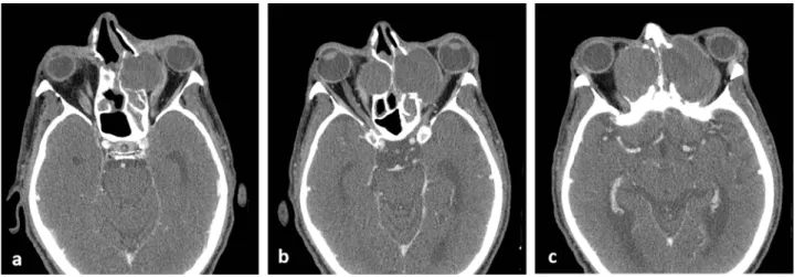
A 73-year-old male patient presented to the emergency de-partment for acute worsening of visual acuity and exoph-thalmos of the left eye. The patient reported that a piece of wood had fallen close to his left eye about a week prior toadmission. Ophthalmologic physical examination showed proptosis and papilloedema in the left eye. Visual acuity was 3/10 in the left eye and 7/10 in the right eye. A com-puted tomography (CT) scan was performed (Figs. 1-3).
SUBMISSION: 15/5/2020 | ACCEPTANCE: 14/7/2020
Corresponding
Author,
Guarantor
Eliza Stavride
Department of Radiology, Papageorgiou General Hospital, Ring Road, N. Eykarpia, 56403, Thessaloniki, Greece, Email: [email protected]
Fig. 1. Axial contrast-enhanced CT images of the sinuses and the orbit.
Fig. 2. Coronal CT image reconstruction of the sinuses and the orbit.
Fig. 3. a, b. Axial CT images of the frontal sinuses (bone window a, soft tissue window b) and c. sagittal CT image reconstruction of the right frontal sinus.
tion cysts which only partially occupy the cavity [2]. There is only a slight male gender predilection ac-cording to various studies and the highest incidence occurs in the third and fourth decades of life [1, 3]. Mucocoeles expand in a path of less resistance, in-ducing erosion and remodelling of the surrounding bone. New bone formation or thickening can also occur [2-4]. The most commonly affected sinus is the frontal, followed by the ethmoid (70-90% of cas-es), probably because of their complex and variable drainage. Maxillary (10%) and sphenoid sinuses (1%) are not usually involved [1, 3]. Mucocoeles can be primary or secondary. The most common causes of primary lesions are chronic inflammation (infection, allergy, mucociliary dysfunction), mucus drainage blockage, secretory duct obstruction, mucus gland cystic dilatation and cystic degeneration of polyps. Secondary mucocoeles are caused by trauma or sur-gery [1, 2, 5]. There is a strong association between the presence of nasal polyps and the mucocoele for-mation, probably due to the role of inflammation [6]. If a mucocoele is infected, it becomes a mucopyocoe-le [4].
The manifestation of paranasal sinus mucocoele is determined by the location and the vital structures involved and can be classified as rhinologic, opthal-mologic and neurologic symptoms and signs, with the ophthalmologic symptoms being the most com-mon [1]. When there is intraorbital extension, the mass effect upon the orbit leads to exophthalmos, di-plopia, paresis of extraocular muscles, epiphora and periorbital swelling, with or without pain. On the other hand, when there is direct optic nerve com-pression, the clinical manifestations include visual compromise or even unilateral blindness, impaired
primary site cannot be determined. The expanding mucocoele causes extensive osteolysis of the bone walls and destruction of anatomical structures and can obstruct the adjacent sinuses’ drainage ostia [2]. Serious complications of mucocoeles include epidur-al abscess, meningitis, subdurepidur-al empyema, brain ab-scess and cranial nerve palsies [4].
The diagnosis of sinus mucocoele is confirmed by CT or magnetic resonance imaging (MRI). CT can bet-ter delineate bone details and is preferable for head and neck evaluation. MRI has proven to be superior in terms of tissue contrast [4, 8]. On CT, a mucocoele appears homogeneous and isodense to the cerebral parenchyma, with sharp margins [2, 3, 9]. The ad-jacent osseous structures are remodelled with areas of thickening and erosion. There may be osteolysis of the osseous wall, without signs of infiltration of adjacent anatomical structures. However, in areas of lower resistance, such as the anterior cranial fossa, there may be herniation [2, 4]. After contrast media administration, there is a thin rim of enhancement, which corresponds to the mucoperiosteal lining of the cavity. If the rim is thick or the mass is hyper-dense in the pre-contrast images, a mucopyocoele must be suspected [1, 2, 9]. A distinguishing feature between mucocoeles and neoplasms is the absence of internal enhancement, exclusively seen in muco-coeles [2, 3, 9]. MRI is used to identify the sinona-sal tumours which may be the aetiological factor in some cases of large mucocoeles [2]. Mucocoeles show variable signal intensity on both T1- and T2-weight-ed images, depending on the fluid and protein con-tent, viscosity and degree of dehydration [1, 4]. Wa-ter-rich content results in low signal intensity on T1W and high signal intensity on T2W MR images.
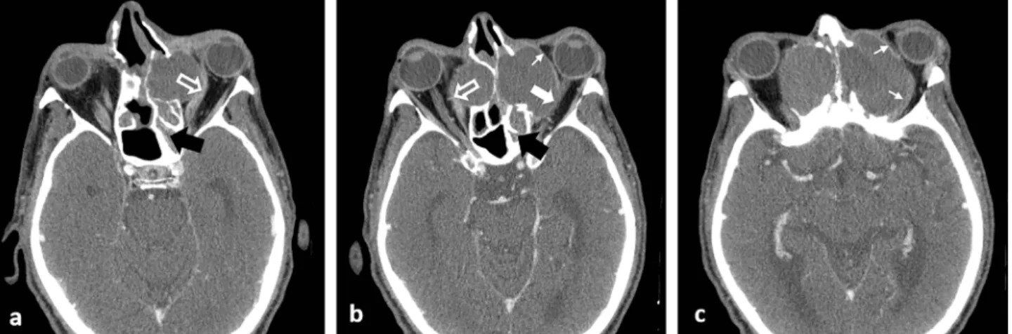
Fig. 3. a, b. Axial CT images of the frontal sinuses and c. sagittal CT reconstruction of the right frontal sinus showing thinning and erosion of the posterior wall of the right frontal sinus and the thin enhancing periosteum (thick arrows in a-c), separating the muco-coele from the anterior cranial fossa. Areas of thickening and erosion of the posterior wall of the left frontal sinus are also noted (thin Fig. 1. Axial CT images (a-c) of the sinuses and the orbit showing expansile, soft tissue lesions, with rim enhancement (thin white arrows in b, c), into the ethmoid sinuses (black arrows in a, b) and the orbit bilaterally. The mass compresses the left optic nerve (thick white arrow in b) and the medial rectus muscles (open white arrows in a, b) and causes outward displacement of the globe.
Fig. 2. Coronal CT reconstructions of the sinuses and the orbit showing an expansile, homogeneous mass with rim enhancement (thin arrow in a), filling the frontal sinuses and eroding the roof and the superomedial wall of the orbit. The mass compresses the
left optic nerve (thick white arrows in b, c), the superior rectus (open black arrowhead in c), medial rectus (open white arrows in b, c) and the superior oblique (black arrowhead in c) muscles and causes downward and outward displacement of the globe.
nus neoplasms, inverted papilloma, Rathke’s cleft cyst, dermoid cyst, hypophyseal adenoma, cranio-pharyngioma, optic glioma, schwannoma, chondro-ma-chondromyxoma and lymphoma [1, 2, 9].
Surgical excision is the treatment of choice. In the past, mucocoeles were removed with external
approach-es or an osteoplastic flap, however recently, most pa -tients are treated with endoscopic techniques, allowing lower morbidity and better cosmetic result [1, 3, 6]. The treatment is based on marsupialisation and enlarging the normal drainage pathways. Detection of recurrence requires a long follow-up with CT or MRI [1, 3].
thin enhancing periosteum was the only remaining structure to separate the mucocoele from the intrac-ranial cavity (Fig. 3). The typical imaging features established the diagnosis of an immense mucocoele.
The mucocoele was evacuated by functional endo-scopic sinus surgery under systemic antibiotic proph-ylaxis. The post-operative ophthalmologic examina-tion revealed progression of the visual acuity to 5/10 in the left eye and stability in the right eye.
R
Conflict of interest
The authors declared no conflicts of interest.
1. Lee TJ, Li SP, Fu CH, et al. Extensive paranasal sinus mucoceles: a 15-year review of 82 cases. Am J Otolar-yngol 2009; 30: 234-238.
2. De Carvalho BV, De Campos Carvalho LI, De Brito Corrêa J, et al. Typical and atypical presentations of paranasal sinus mucocele at computed tomography.
Radiol Bras 2013; 46 (6): 372-375.
3. Capra GG, Carbone PN, Mullin DP. Paranasal sinus mucocele. Head Neck Pathol 2012; 6: 369-372.
4. Peral-Cagigal B, Barrientos-Lezcano J, Floriano-Blan-co R, et al. Frontal sinus muFloriano-Blan-cocele with intracranial and intraorbital extension. Med Oral Pathol Oral Cir Bu-cal 2006; 11: E527-E530.
5. Kim YS, Kim K, Lee JG, et al. Paranasal sinus mucoce-les with ophthalmologic manifestations: a 17-year re-view of 96 cases. Am J Rhinol Allergy 2011; 25: 272-275.
6. Devars du Mayne M, Moya-Plana A, Malinvaud D, et
al. Sinus mucocele: Natural history and long-term re-currence rate. Eur Ann Otorhinolaryngol Head Neck Dis
2012; 129(3): 125-130.
7. Yue CP, Mann KS, Chan FL. Optic canal syndrome due to posterior ethmoid sinus mucocele. J Neurosurg
1986; 65: 871-873.
8. Mizushima Y, Mumo T, Yasui T, et al. Paranasal sinus mucocele with visual disturbances whose causative
legion was hardly identified on computed tomog -raphy imaging: A case report. Acta Oto-Laryngologica Case Reports 2019; 4(1): 10-12.
9. Perugini S, Pasquinin U, Menichelli F, et al. Mucoce-les in the paranasal sinuses involving the orbit: CT signs in 43 Cases. Neuroradiology 1982; 23: 133-139.
10. Van Tassel P, Lee YY, Jing BS, et al. Mucoceles of the paranasal sinuses: MR imaging with CT correlation.
AJNRAm J Neuroradiol 1989; 10: 607-612.