Temporal and Demographic Trends in Glomerular
Disease Epidemiology in the Southeastern
United States, 1986–2015
Michelle M. O’Shaughnessy,*†‡Susan L. Hogan,†Caroline J. Poulton,†Ronald J. Falk,*†Harsharan K. Singh,*† Volker Nickeleit,*†and J. Charles Jennette*†
Abstract
Background and objectivesLarge-scale, contemporary studies exploring glomerular disease epidemiology in the United States are lacking. We aimed to determine 30-year temporal and demographic trends in renal biopsy glomerular disease diagnosis frequencies in the southeastern United States.
Design, setting, participants, & measurementsIn this cross-sectional, observational study, we identified all patients with a native kidney biopsy specimen showing one of 18 widely recognized glomerular disease diagnoses referred to the University of North Carolina Chapel Hill Division of Nephropathology between 1986 and 2015. Biopsy era (1986–1995, 1996–2005, and 2006–2015) and demographics (age, sex, and race) were our primary and secondary predictors, respectively, and the relative frequency of each glomerular disease diagnosis was our primary outcome.
ResultsAmong 21,374 patients (mean age =48.3618.3 years old; 50.8% men; 56.8% white; 38.3% black; 2.8% Latino; 1.4% Asian; 0.8% other), the frequency of diabetic glomerulosclerosis in renal biopsy specimens increased dramatically over the three decades (5.5%, 11.4%, and 19.1% of diagnoses, respectively;Pfor trend,0.001). The frequency of FSGS initially increased but then declined (22.6%, 27.2%, and 24.7%, respectively;Pfor trend =0.64). The frequencies of other common glomerular disease subtypes remained stable (IgA nephropathy and ANCA/ pauci-immune GN) or declined (minimal change disease, membranous nephropathy, membranoproliferative GN, and lupus nephritis). These temporal trends were largely preserved within all demographic subgroups, although cross-sectional frequency distributions differed according to age, sex, and race.
ConclusionsWe identified significant changes in relative renal biopsy frequencies of many glomerular disease subtypes over three decades. Temporal trends were consistently observed within all major demographic groups, although relative predominance of individual glomerular disease subtypes differed according to patient age, sex, and race. We propose that exploration of behavioral and environmental exposures that likely underlie these findings should be the focus of future hypothesis-driven research.
Clin J Am Soc Nephrol12: 614–623, 2017. doi: https://doi.org/10.2215/CJN.10871016
Introduction
Epidemiologic studies reveal important insights into factors associated with glomerular disease develop-ment or progression and inform predictions of the relative likelihood of individual glomerular disease diagnoses for a given patient. The disproportionally high risks for FSGS in blacks and IgA nephropathy in Asians encouraged the discovery of racially deter-mined genetic risk variants (1,2). Associations between population sanitation standards or socioeconomic sta-tus and risks for certain GN subtypes suggest an etio-logic role for environmental and lifestyle factors in disease pathogenesis (3,4). Thus, identifying temporal changes in glomerular disease epidemiology within a geographic region might reliably inform future hypothesis-driven studies and public health interventions.
Within the United States, prior studies exploring glomerular disease epidemiology identified a marked
increase in the frequency of FSGS at the end of the 20th century (5–9). Whether this trend continued into the 21st century has not been established, although a small study (n=204) from Chicago suggested that the frequency of FSGS (2000–2011) might now be lower than that of membranous nephropathy among blacks (10). Temporal trends are less consistent across studies for other glomerular disease subtypes, ex-plained by differences in population demographics (e.g., white [9], military [11], or urban-dwelling pa-tients [7]) or clinical inclusion criteria (e.g., nephrotic syndrome [7,8] versus any glomerular disease [9,12]). Frequencies of especially rare glomerular diseases are seldom reported.
The Division of Nephropathology at the University of North Carolina (UNC) at Chapel Hill has provided a nephropathology service to the UNC and academic and community practices throughout the southeastern
*Division of Nephropathology, Department of Pathology and Laboratory Medicine,
and†Division of
Nephrology, Department of Medicine and the Kidney Center, University of North Carolina School of Medicine, Chapel Hill, North Carolina;
and‡Division of
Nephrology, Stanford University School of Medicine, Palo Alto, California
Correspondence: Dr. Michelle M. O’Shaughnessy, Stanford University Division of Nephrology, 777 Welch Road Suite DE, Palo Alto, CA 94304. Email: moshaugh@ stanford.edu
United States since the 1970s. By examining native kidney biopsy cases from patients submitted between 1986 and 2015, we aimed to describe temporal trends in glomerular disease frequencies over three decades and explore the influence of demographic factors on glomerular disease frequency distributions.
Materials and Methods
Patient Population
All native kidney biopsy specimens referred to the Division of Nephropathology at the UNC (1986–2015) with one of 18 widely recognized diagnostic categories of glomerular disease were considered for study inclusion. The referral population was derived predominantly from residents of North Carolina and its neighboring states, in-cluding Virginia, West Virginia, Tennessee, South Caro-lina, and Georgia. If a patient had multiple biopsies with a glomerular disease diagnosis, only the first was retained for this study. If more than one glomerular dis-ease diagnosis was made from a single biopsy specimen, that which seemed to be the major cause for the renal dysfunction prompting the biopsy was chosen as the study diagnosis (i.e., if the primary diagnosis was a glomerular disease, then we retained this diagnosis; otherwise, we searched for the presence of a glomerular disease as a sec-ondary diagnosis, such that a single predominant glomer-ular disease was elucidated for each patient).
Data Source
All renal biopsy specimens were processed by standard light, immunofluorescence, and electron microscopy pro-cedures. Diagnoses were those made by experienced nephropathologists involved in the clinical care of patients. For analysis, all Columbia variants of FSGS (13), all Ehren-reich and Churg stages of membranous nephropathy (14), and all International Society of Nephrology/Renal Pathol-ogy Society classes of lupus nephritis (15) were grouped into respective FSGS, membranous nephropathy, and lu-pus nephritis categories. Dense deposit disease (membra-noproliferative GN [MPGN] type 2) was analyzed separately from other forms of MPGN. Immune complex MPGN and C3 GN with an MPGN pattern of injury (in-cluding so-called types 1 and 3 MPGN) were included in the MPGN category, because the distinction between these two disease entities was only recently recognized (16). The uncommon C1q and IgM mesangial nephropathies were subsumed in minimal change disease and FSGS categories on the basis of the light microscopic pattern of injury. A diagnosis of pauci-immune necrotizing and crescentic GN was on the basis of pathologic phenotype without requir-ing serologic ANCA positivity. Antiglomerular basement membrane GN that was also ANCA positive was included in the antiglomerular basement membrane GN category.
Demographic data were abstracted from biopsy referral forms completed by referring nephrologists or available medical records.
Exposures, Outcomes, and Covariates
Temporal era was our primary exposure, categorized in to three consecutive 10-year time intervals (1986–1995, 1996–2005, and 2006–2015) for tabular presentation and
data analysis and six consecutive 5-year time intervals (1986–1990, 1991–1995, 1996–2000, 2001–2005, 2006–2010, and 2011–2015) for plotting. Glomerular disease subtype frequency was our primary outcome. As secondary out-comes, we also examined temporal trends in glomerular disease frequencies within demographic subgroups as well as by typical mode of clinical presentation for a given sub-type (nephrotic versus nephritic syndrome [17]). To ex-plore combined influences of age, sex, and race, we evaluated glomerular disease frequency distributions across age categories stratified by patient sex and race.
Statistical Analyses
Categorical variables were expressed as frequencies (percentages) and compared using chi-squared or Fisher exact testing as appropriate. When analyzing differences across study eras,Pfor trend values are reported. Contin-uous variables were expressed as means (SDs) or medians (interquartile ranges) and compared using ANOVA or Kruskall–Wallis testing as appropriate. Statistical analyses were performed using SAS Enterprise Guide, version 6.1 (SAS, Cary, NC). A two-sided Pvalue of,0.05 was con-sidered statistically significant when analyzing demo-graphic data, and a Bonferroni correction for multiple comparisons (0.05/18=0.0027) was applied when analyz-ing trends across 18 glomerular disease subtypes.
Institutional review board (IRB) approval for the study was obtained from the UNC Biomedical IRB (Study 97–0523).
Results
Patient Population
In total, 38,472 kidney biopsies (33,391 native and 5081 transplant) were evaluated between 1986 and 2015. Biopsy frequencies increased annually from 390 (1986) to 1923 (2015). Of these, 22,516 native kidney specimens had one of the 18 study diagnoses. From these, 1142 repeat biopsies in 1016 patients were excluded, leaving a final study population of 21,374 patients with one of the 18 glomerular disease subtypes of interest diagnosed on an initial native kidney biopsy.
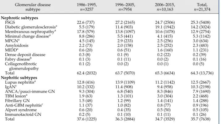
Temporal Trends in Glomerular Disease Frequency Renal biopsy frequencies by study era and glomerular disease subtype are provided in Figure 1 and Table 2. In the earliest era (1986–1995), FSGS predominated (22.6% of
studied patients) followed by membranous nephropathy (17.8%). In the middle (1996–2005) and later (2006–2015) eras, FSGS still predominated (27.2% and 24.7%, respec-tively) but was followed by lupus nephritis (13.9%) and Table 1. Temporal trends in patient demographics among the study cohort of patients with specified glomerular disease diagnoses
Demographic Characteristics
1986–1995, n=3257
1996–2005, n=7954
2006–2015, n=10,163
Total,
n=21,374 PValue
Age, mean (SD) 45.1 (19.1) 46.9 (18.2) 50.4 (17.9) 48.3 (18.3) ,0.001
Age category, yr, % (n) ,0.001
0–17 8.3 (265) 5.3 (417) 3.3 (334) 4.8 (1016)
18–39 32.0 (1020) 30.6 (2415) 24.8 (2518) 28.0 (5953)
40–64 40.2 (1282) 44.5 (3515) 47.0 (4772) 45.1 (9569)
.64 19.6 (624) 19.6 (1547) 24.9 (2525) 22.1 (4696)
Missing age, % 2.0 0.8 0.1 0.7
Men, % (n) 51.9 (1689) 50.1 (3949) 51.1 (5185) 50.8 (10,823) 0.17
Missing sex, % 0.1 0.8 0.1 0.3
Race, % (n) ,0.001
White 64.1 (1610) 56.6 (3782) 54.7 (4714) 56.8 (10,106)
Black 34.5 (867) 39.9 (2662) 38.2 (3292) 38.3 (6821)
Latino 0.6 (14) 1.8 (123) 4.2 (360) 2.8 (497)
Asian 0.0 (1) 1.0 (69) 2.0 (170) 1.4 (240)
Other 0.8 (19) 0.6 (42) 0.9 (81) 0.8 (142)
Missing race % 22.9 16.0 15.2 16.7
Percentages (except missing percentages) represent column percentages among patients with complete data.
membranous nephropathy (13.8%) in the middle era and diabetic glomerulosclerosis (19.1%) in the most recent era. Significant temporal changes in glomerular disease sub-type relative frequencies were observed (Figure 1, Table 2). Most notable was a steady increase in the frequency of diabetic glomerulosclerosis over the three study decades (5.5%, 11.4%, and 19.1% of diagnoses, respectively;Pfor trend,0.001). The frequency of FSGS increased initially, but then, it plateaued and ultimately declined (22.6%, 27.2%, and 24.7%, respectively; P=0.64). The frequencies of membranous nephropathy (17.8%, 13.8%, and 10.6%, respectively; P,0.001) and minimal change disease (8.8%, 5.5%, and 4.1%, respectively; P,0.001) declined substantially over the study interval, whereas those of lu-pus nephritis (12.8%, 13.9%, and 11.2%, respectively; P,0.001) and MPGN types 1 or 3 (4.5%, 2.9%, and 2.5%, respectively; P,0.001) declined more modestly. The fre-quencies of IgA nephropathy (10.2%, 11.4%, and 9.4%; P=0.004) and ANCA/pauci-immune GN (9.3%, 6.8%, and 8.3%;P=0.14) remained stable. As a sensitivity analy-sis, we re-examined the frequencies of remaining subtypes after excluding diabetic glomerulosclerosis to ensure that large shifts in the frequency of this diagnosis did not un-duly influence frequency distributions among the remain-ing 17 subtypes. Findremain-ings were not materially different (Figure 1B).
Among more rare subtypes (biopsy frequency ,3%), some significant temporal trends were also observed (Ta-ble 2). Significant increases were observed in thin base-ment membrane lesion and monoclonal Ig deposition disease frequencies.
Temporal Trends in Glomerular Disease Frequencies by Age, Sex, and Race
Temporal trends in men or women mirrored those in the overall cohort (Supplemental Figure 1). However, differ-ences between sexes were observed at a cross-sectional level. For example, lupus nephritis was more frequent in women than men (20.5% versus 4.7%, respectively), whereas the opposite was true for IgA nephropathy (7.7% versus 12.8%, respectively). Sex distributions by glo-merular disease subtype are shown in Supplemental Fig-ure 2 and Table 3.
For black or white patients, temporal trends were also similar to those observed in the full cohort (Supplemental Figure 1). Comparing races cross-sectionally, however, lu-pus nephritis (20.9% versus 6.3%, respectively) and FSGS (33.6% versus 20.4%, respectively) were more frequent and IgAN (2.3% versus 14.9%, respectively) and ANCA/pauci-immune GN (3.3% versus 11.4%, respectively) less fre-quent among blacks. Racial distributions, by subtype, are presented in Supplemental Figure 2 and Table 3.
Table 2. Temporal trends in the renal biopsy frequencies of glomerular disease subtypes among the study cohort of patients with specified glomerular disease diagnoses
Glomerular disease subtype
1986–1995, n=3257
1996–2005, n=7954
2006–2015, n=10,163
Total, n=21,374
Nephrotic subtypes
FSGS 22.6 (737) 27.2 (2165) 24.7 (2506) 25.3 (5408)
Diabetic glomerulosclerosisa 5.5 (179) 11.4 (903) 19.1 (1942) 14.2 (3024)
Membranous nephropathya 17.8 (579) 13.8 (1097) 10.6 (1078) 12.9 (2754)
Minimal change diseasea 8.8 (286) 5.5 (441) 4.1 (415) 5.3 (1142)
MPGNa 4.5 (145) 2.9 (233) 2.5 (256) 3.0 (634)
Amyloidosis 2.2 (73) 2.0 (158) 2.5 (252) 2.3 (483)
MIDDa 0.6 (20) 0.6 (51) 1.6 (160) 1.1 (231)
Dense deposit disease 0.3 (8) 0.1 (9) 0.2 (22) 0.2 (39)
Fabry diseaseb 0.1 (3) 0.1 (11) 0.0 (2) 0.1 (16)
Collagenofibrotic glomerulopathy
0.1 (2) 0.0 (2) 0.0 (1) 0.0 (5)
Total 62.4 (2032) 63.7 (5070) 65.3 (6634) 64.3 (13,736)
Nephritic subtypes
Lupus nephritisa 12.8 (416) 13.9 (1109) 11.2 (1142) 12.5 (2667)
IgANc 10.2 (332) 11.4 (908) 9.4 (958) 10.3 (2198)
ANCA/pauci-immune GN 9.3 (304) 6.8 (540) 8.3 (846) 7.9 (1690)
TBM lesiona 1.9 (63) 1.3 (101) 3.0 (304) 2.2 (468)
Fibrillary GN 1.5 (48) 1.2 (99) 1.4 (141) 1.4 (288)
Anti-GBM nephritisc 1.1 (37) 1.0 (82) 0.8 (77) 0.9 (196)
Alport syndrome 0.6 (20) 0.4 (35) 0.5 (50) 0.5 (105)
Immunotactoid GN 0.2 (5) 0.1 (10) 0.1 (11) 0.1 (26)
Total 37.6 (1225) 36.3 (2884) 34.7 (3529) 35.7 (7638)
All values represent column percentages (n). MPGN, membranoproliferative GN (nondense deposit disease); MIDD, monoclonal
immune deposition disease; IgAN, IgA nephropathy; TBM, thin basement membrane; GBM, glomerular basement membrane.
aChi-squared test for trend,P,0.003.
bFisher exact test,P,0.05 butP.0.003.
Temporal trends by age category (children, young adults, middle-aged adults, and older adults) are also presented in Supplemental Figure 1. Again, no marked deviations from overall trends were observed, with the exception of a low frequency of diabetic glomerulosclero-sis in children. However, cross-sectional differences were again apparent; lupus nephritis was almost as common as FSGS in young adults (25.7% versus 27.3%, respectively) but rare in older adults (2.2%), whereas ANCA/pauci-im-mune GN was especially frequent in older adults (17.9%) but rare in children (3.9%) and young adults (2.7%). Av-erage ages by glomerular disease subtype are shown in Table 3.
Differences in Glomerular Disease Frequencies by Age Category, Sex, and Race
To further explore the variations in disease frequencies observed between age groups, we examined changes in glomerular disease frequencies across the age spectrum (Figures 2 and 3). Very young (0–9 years old) patients were most likely to have minimal change disease, very old (.79 years old) patients were most likely to have ANCA/pauci-immune GN, and younger or middle-aged adults were most likely to have FSGS. Stratifying by sex and race re-vealed additional insights; for example, IgAN was most frequent in young adult white men, whereas lupus nephri-tis peaked in young adult black women.
When glomerular disease subtype frequencies were evaluated as a proportion of all glomerular disease diag-noses in a given age group, the predominance of certain subtypes within particular age groups became even more apparent (Figure 4). Among patients typically presenting with nephrotic syndrome, the likelihood of a renal biopsy diagnosis of minimal change disease declined precipi-tously after early childhood, whereas diabetic
glomerulosclerosis was less likely to be diagnosed in older patients, and FSGS was frequent throughout. Among pa-tients typically presenting with nephritic features, the likelihood of IgA nephropathy declined with advancing age, lupus nephritis peaked in young adulthood, and ANCA/pauci-immune GN was strikingly common in older adults.
Discussion
In this study of 21,374 patients with a biopsy-confirmed glomerular disease diagnosis residing predominantly in the southeastern United States, we identified significant temporal shifts in the epidemiology of many glomerular disease subtypes over the past three decades (1986–2105). Most striking was a marked increase in renal biopsy fre-quency of diabetic glomerulosclerosis from 5.5% of pa-tients in the earliest decade to 19.1% of papa-tients most recently. This finding was consistently observed within all studied age, sex, and racial groups, with the exception of children. Contemporaneously, we observed an initial increase in frequency of FSGS at the end of the 20th cen-tury, as previously reported in other United States cohorts (5–9), followed by a plateau and decline in its frequency more recently, afinding not previously described. At the same time, we observed significant declines in relative fre-quencies of some other glomerular disease subtypes (e.g., membranous nephropathy, minimal change disease, mem-branoproliferative GN [excluding dense deposit disease], and lupus nephritis) along with stable frequencies of oth-ers (e.g., IgA nephropathy and ANCA/pauci-immune GN). These temporal trends were not explained by large shifts in the demographic composition of our study pop-ulation and were consistently observed in most age, sex, and racial subgroups.
Interpretation of our study is complicated by the steadily increasing biopsy referral rate to the UNC over the study interval. Although we report trends in relative disease frequencies (i.e., as a proportion of all biopsies with a glo-merular disease diagnosis), we note that, in some cases, a decline in relative disease frequency was accompanied by an increase in absolute disease frequency. The underlying reasons for the rising background biopsy rate may include (1) a declining threshold to biopsy patients; (2) an increase in the referral population size, mirroring that occurring in North Carolina (19,20); (3) increasing numbers of nephrol-ogists referring biopsies to the UNC; or (4) true increases in disease incidence. This latter possibility is supported by the fact that diseases with a stronger environmental/life-style component to their pathogenesis (e.g., FSGS and di-abetic glomerulosclerosis) underwent more marked increases in disease frequency than those with a more clearly established genetic or autoimmune pathogenesis (e.g., ANCA/pauci-immune GN or IgA nephropathy). It is notable that, for some glomerular disease subtypes (e.g., membranous nephropathy or minimal change disease), the absolute numbers of patients declined in thefinal decade, supporting a true decline in disease incidence. Conversely, the rising frequency of diabetic glomerulosclerosis mark-edly exceeded the increasing background biopsy referral rate, supporting a true increase in the incidence of this
biopsy diagnosis and echoing findings from the general population that the incidence of diabetes mellitus has in-creased almost twofold between 1995 and 2010 (21).
In an effort to disentangle the influence of shifts in population demographics from that of changes in envi-ronmental, lifestyle, or practice pattern factors, we evalu-ated for temporal changes in the sex, age, or racial composition of our study cohort in addition to examining temporal trends in glomerular disease frequencies within several demographic subgroups. In summary, we did not identify convincing evidence that demographic shifts in our study population were responsible for the changes in glomerular disease frequencies that we observed and suggest that changes in lifestyle/environmental factors or clinical practice are more likely to underlie ourfindings. Compared with prior studies exploring glomerular disease epidemiology in the United States, our study has some notable differences. First, white and black races were both well represented in our cohort, differing from studies that focused almost exclusively on white patients (9). Sec-ond, we included all age groups, unlike studies focusing only on adults (12,22). Third, we examined systemic cau-ses of glomerular disease (e.g., lupus nephritis, ANCA/ pauci-immune GN, and diabetic glomerulosclerosis) in ad-dition to so-called primary glomerular diseases to capture a wide spectrum of patients undergoing kidney
Male Female
White Black
biopsy for evaluation of suspected glomerular disease. This differs from prior studies limited to patients with ne-phrotic syndrome (7,8), proteinuria in excess of 2 g/24 h (5), or a more restricted set of primary glomerular disease diagnoses (6,8,22,23). Both of the prior studies that included a wide spectrum of primary and secondary merular diseases (9,12) excluded patients with diabetic glo-merulosclerosis, prompting our decision to analyze our data with and without diabetic glomerulosclerosis to facilitate comparisons with these studies (our conclusions were unal-tered). Fourth, our study is the largest and most contempo-rary to date; with the exception of two much smaller studies published earlier this year (12,22), recent reports of United States glomerular disease epidemiology are lacking.
Despite these differences, many of ourfindings support those previously reported. The marked increase in FSGS frequency previously identified at the end of the 20th century (5–9), particularly among blacks, was again seen in our patient cohort. However, our study is the first to show a plateau and subsequent decline in FSGS frequency more recently; unlike a smaller study (n=204) that reported a predominance of membranous nephropathy over FSGS among all racial groups in a contemporary (2001–2011) patient cohort (10), FSGS remained the most frequent glomerular disease subtype in all racial groups in our study. Whether the recent decline in FSGS frequency represents a reduction in patients with primary/idiopathic cases, patients with secondary (e.g., obesity-related) cases,
or the likelihood to biopsy a patient with proteinuria could not be discerned from our data.
The rapid and steady increase in the frequency of diabetic glomerulosclerosis that we observed has not, to our knowledge, previously been reported. We propose that this finding largely reflects a true increase in disease incidence, mirroring the increase in diabetes mellitus in-cidence observed in the United States over the same time interval (24). Additionally, an increasing tendency to bi-opsy older patients or search for nondiabetic glomerular diseases among patients with diabetes (25,26) might un-derlie thesefindings.
Thus, we consider declines in the relative frequencies of these diagnoses to be most likely due to true declines in disease incidence. Descriptions of temporal trends in several more rare glomerular disease subtypes are also novel to our study. In addition to reporting temporal trends, we also confirm some previously described demographic predispositions to glomerular disease development (e.g., a higher risk for IgA nephropathy in younger white or Asian patients [23] and a higher risk for lupus nephritis in younger black patients or women [27]) in a large and contemporary United States cohort. We also comprehensively analyzed glomerular dis-ease frequencies according to several combinations of de-mographic factors and typical modes of glomerular disease presentation (nephrotic versus nephritic), which may serve as useful resources to clinicians evaluating pa-tients in the clinic or researchers aiming to target high-risk patient groups for inclusion in future translational re-search studies or interventional trials.
Our study has several limitations. We had too few Latino and Asian patients to enable analysis of temporal trends or demographic associations in these racial/ethnic groups. We could not precisely determine our referral population size and thus, report relative (not absolute) incidences. We estimate that the Division of Nephropathology at the UNC biopsies derive from a population of approximately 10 million people, although accurate estimates of population sizes over time could not be determined.
To conclude, we identified in a large, contemporary United States population that the frequencies of many glomerular disease subtypes have shifted considerably over the past three decades. We provide evidence that changes in population demographics (age, sex, or race) contributed minimally to thesefindings and instead, pro-pose that environmental and lifestyle changes most likely underlie them. Of particular concern was the dramatic increase in the frequency of diabetic glomerulosclerosis that we observed given the adverse outcomes (28) and in-creased health care costs (29) associated with this diabetic complication. On a more encouraging note, the relative (and in some cases, absolute) frequencies of some other glomerular disease subtypes declined. We propose that further exploration of the underlying reasons for these ep-idemiologic shifts is warranted and that these efforts might reveal novel insights into disease pathogenesis or therapeutic opportunities.
Acknowledgments
The study was coordinated by the Glomerular Disease Collabo-rative Network, which has been codirected by R.J.F. and J.C.J. since 1985, comprises several hundred nephrologists primarily in the southeastern United States, and facilitates clinicopathologic re-search focused on glomerular diseases. We thank these nephrolo-gists and their patients for providing the renal biopsy specimens and patient data that are in this report.
M.M.O was supported by a fellowship award from Mallinckrodt Pharmaceuticals for the duration of this study.
Mallinckrodt Pharmaceuticals had no role in study design; col-lection, analysis, or interpretation of data; writing of the report; or the decision to submit the report for publication.
Disclosures None.
References
1. Genovese G, Friedman DJ, Ross MD, Lecordier L, Uzureau P, Freedman BI, Bowden DW, Langefeld CD, Oleksyk TK, Uscinski Knob AL, Bernhardy AJ, Hicks PJ, Nelson GW, Vanhollebeke B, Winkler CA, Kopp JB, Pays E, Pollak MR: Association of trypa-nolytic ApoL1 variants with kidney disease in African Americans.
Science329: 841–845, 2010
2. Gharavi AG, Kiryluk K, Choi M, Li Y, Hou P, Xie J, Sanna-Cherchi S, Men CJ, Julian BA, Wyatt RJ, Novak J, He JC, Wang H, Lv J, Zhu L, Wang W, Wang Z, Yasuno K, Gunel M, Mane S, Umlauf S, Tikhonova I, Beerman I, Savoldi S, Magistroni R, Ghiggeri GM, Bodria M, Lugani F, Ravani P, Ponticelli C, Allegri L, Boscutti G, Frasca G, Amore A, Peruzzi L, Coppo R, Izzi C, Viola BF, Prati E, Salvadori M, Mignani R, Gesualdo L, Bertinetto F, Mesiano P, Amoroso A, Scolari F, Chen N, Zhang H, Lifton RP: Genome-wide association study identifies susceptibility loci for IgA
ne-phropathy.Nat Genet43: 321–327, 2011
3. Hurtado A, Johnson RJ: Hygiene hypothesis and prevalence of
glomerulonephritis.Kidney Int Suppl68: S62–S67, 2005
4. McQuarrie EP, Mackinnon B, McNeice V, Fox JG, Geddes CC: The incidence of biopsy-proven IgA nephropathy is associated
with multiple socioeconomic deprivation.Kidney Int85: 198–
203, 2014
5. Braden GL, Mulhern JG, O’Shea MH, Nash SV, Ucci AA Jr., Germain MJ: Changing incidence of glomerular diseases in
adults.Am J Kidney Dis35: 878–883, 2000
6. Dragovic D, Rosenstock JL, Wahl SJ, Panagopoulos G, DeVita MV, Michelis MF: Increasing incidence of focal segmental glo-merulosclerosis and an examination of demographic patterns.
Clin Nephrol63: 1–7, 2005
7. Haas M, Meehan SM, Karrison TG, Spargo BH: Changing etiol-ogies of unexplained adult nephrotic syndrome: A comparison of
renal biopsy findings from 1976-1979 and 1995-1997.Am J
Kidney Dis30: 621–631, 1997
8. Korbet SM, Genchi RM, Borok RZ, Schwartz MM: The racial
prevalence of glomerular lesions in nephrotic adults.Am J Kidney
Dis27: 647–651, 1996
9. Swaminathan S, Leung N, Lager DJ, Melton LJ 3rd, Bergstralh EJ, Rohlinger A, Fervenza FC: Changing incidence of glomerular disease in Olmsted County, Minnesota: A 30-year renal biopsy
study.Clin J Am Soc Nephrol1: 483–487, 2006
10. Kraus MA, Punj S, Cimbaluk D, Hart PD: Resurgence of mem-branous nephropathy in African Americans in inner city Chicago.
Clin Kidney J6: 373–378, 2013
11. Pontier PJ, Patel TG: Racial differences in the prevalence and
presentation of glomerular disease in adults.Clin Nephrol42:
79–84, 1994
12. Murugapandian S, Mansour I, Hudeeb M, Hamed K, Hammode E, Bijin B, Daheshpour S, Thajudeen B, Kadambi P: Epidemiology of glomerular disease in southern arizona: Review of 10-year
renal biopsy data.Medicine (Baltimore)95: e3633, 2016
13. D’Agati VD, Fogo AB, Bruijn JA, Jennette JC: Pathologic classifi-cation of focal segmental glomerulosclerosis: A working
pro-posal.Am J Kidney Dis43: 368–382, 2004
14. Ehrenreich TC, Churg J: Pathology of membranous nephropathy.
Pathol Annu3: 145–186, 1968
15. Weening JJ, D’Agati VD, Schwartz MM, Seshan SV, Alpers CE, Appel GB, Balow JE, Bruijn JA, Cook T, Ferrario F, Fogo AB, Ginzler EM, Hebert L, Hill G, Hill P, Jennette JC, Kong NC, Lesavre P, Lockshin M, Looi LM, Makino H, Moura LA, Nagata M; International Society of Nephrology Working Group on the Classification of Lupus Nephritis; Renal Pathology Society Working Group on the Classification of Lupus Nephritis: The classification of glomerulonephritis in systemic lupus
eryth-ematosus revisited.Kidney Int65: 521–530, 2004
16. Servais A, Fre´meaux-Bacchi V, Lequintrec M, Salomon R, Blouin J, Knebelmann B, Gru¨nfeld JP, Lesavre P, Noe¨l LH, Fakhouri F: Primary glomerulonephritis with isolated C3 de-posits: A new entity which shares common genetic risk factors
with haemolytic uraemic syndrome.J Med Genet44: 193–199,
2007
17. Jennette J, Falk R: Glomerular clinicopathologic syndromes. In:
National Kidney Foundation’s Primer on Kidney Disease, edited
18. US Census Bureau: Census Interactive Population Search, 2010. Available at: http://www.census.gov/2010census/popmap/ipmtext. php?fl=37. Accessed January 10, 2017
19. US Census Bureau: Population Change and Distribution, 1990– 2000. Available at: http://www.census.gov/prod/2001pubs/ c2kbr01-2.pdf. Accessed January 10, 2017
20. US Census Bureau: Population Distribution and Change, 2000– 2010. Available at: http://www.census.gov/prod/cen2010/briefs/ c2010br-01.pdf. Accessed January 10, 2017
21. Centers for Disease Control and Prevention (CDC): Increasing prevalence of diagnosed diabetes–United States and Puerto Rico,
1995-2010.MMWR Morb Mortal Wkly Rep61: 918–921, 2012
22. Sim JJ, Batech M, Hever A, Harrison TN, Avelar T, Kanter MH, Jacobsen SJ: Distribution of biopsy-proven presumed primary glo-merulonephropathies in 2000-2011 among a racially and
ethni-cally diverse US population.Am J Kidney Dis68: 533–544, 2016
23. Nair R, Walker PD: Is IgA nephropathy the commonest primary
glomerulopathy among young adults in the USA?Kidney Int69:
1455–1458, 2006
24. Menke A, Casagrande S, Geiss L, Cowie CC: Prevalence of and trends in diabetes among adults in the united states, 1988-2012.
JAMA314: 1021–1029, 2015
25. Pham TT, Sim JJ, Kujubu DA, Liu IL, Kumar VA: Prevalence of
nondiabetic renal disease in diabetic patients.Am J Nephrol27:
322–328, 2007
26. Yaqub S, Kashif W, Hussain SA: Non-diabetic renal disease in
patients with type-2 diabetes mellitus.Saudi J Kidney Dis Transpl
23: 1000–1007, 2012
27. Feldman CH, Hiraki LT, Liu J, Fischer MA, Solomon DH, Alarco´n GS, Winkelmayer WC, Costenbader KH: Epidemiology and so-ciodemographics of systemic lupus erythematosus and lupus nephritis among US adults with Medicaid coverage, 2000-2004.
Arthritis Rheum65: 753–763, 2013
28. Tancredi M, Rosengren A, Svensson AM, Kosiborod M, Pivodic A, Gudbjo¨rnsdottir S, Wedel H, Clements M, Dahlqvist S, Lind M:
Excess mortality among persons with type 2 diabetes.N Engl J
Med373: 1720–1732, 2015
29. Slabaugh SL, Curtis BH, Clore G, Fu H, Schuster DP: Factors associated with increased healthcare costs in Medicare Advantage patients with type 2 diabetes enrolled in a large
representative health insurance plan in the US.J Med Econ18:
106–112, 2015
Received:October 17, 2016Accepted:December 21, 2016
Published online ahead of print. Publication date available at www. cjasn.org.
See related editorial,“Temporal Trends in the Epidemiology of
Biopsy-Proven Glomerular Diseases: An Alarming Increase in
Diabetic Glomerulosclerosis,”on pages 556–558.