MBoC
|
ARTICLE
Polo kinase Cdc5 associates with centromeres
to facilitate the removal of centromeric cohesin
during mitosis
ABSTRACT Sister chromatid cohesion is essential for tension-sensing mechanisms that mon-itor bipolar attachment of replicated chromatids in metaphase. Cohesion is mediated by the association of cohesins along the length of sister chromatid arms. In contrast, centromeric cohesin generates intrastrand cohesion and sister centromeres, while highly cohesin en-riched, are separated by >800 nm at metaphase in yeast. Removal of cohesin is necessary for sister chromatid separation during anaphase, and this is regulated by evolutionarily con-served polo-like kinase (Cdc5 in yeast, Plk1 in humans). Here we address how high levels of cohesins at centromeric chromatin are removed. Cdc5 associates with centromeric chromatin and cohesin-associated regions. Maximum enrichment of Cdc5 in centromeric chromatin occurs during the metaphase-to-anaphase transition and coincides with the removal of chro-mosome-associated cohesin. Cdc5 interacts with cohesin in vivo, and cohesin is required for association of Cdc5 at centromeric chromatin. Cohesin removal from centromeric chromatin requires Cdc5 but removal at distal chromosomal arm sites does not. Our results define a novel role for Cdc5 in regulating removal of centromeric cohesins and faithful chromosome segregation.
INTRODUCTION
Accurate chromosome segregation is important for the mainte-nance of genome stability during growth and proliferation of
organ-isms (Lengauer et al., 1998; Maddox et al., 2012). Errors in chromo-some segregation result in aneuploidy, which has been linked with several human diseases, such as developmental disorders and many cancers (Santaguida and Amon, 2015; Singh and Gerton, 2015). The kinetochore, composed of centromeric DNA (CEN) and associated proteins, is an essential component of the chromosomal segrega-tion machinery (Burrack and Berman, 2012; Choy et al., 2012; Maddox et al., 2012). The assembly of kinetochore proteins at bud-ding yeast CEN (core centromere composed of ∼125 base pairs of defined DNA sequences) results in a highly ordered, unique, and topologically distinct chromatin structure in the chromosomes (Clarke and Carbon, 1980; Bloom and Carbon, 1982; Bloom et al., 1984; Saunders et al., 1990), the structural integrity of which is re-quired for accurate chromosome segregation (Newlon, 1988; Verdaasdonk and Bloom, 2011; Haase et al., 2013; Mishra et al., 2013; Mishra et al., 2015). Budding yeast centromeric chromatin
Monitoring Editor Orna Cohen-Fix
National Institutes of Health
Received: Jan 4, 2016 Revised: Apr 27, 2016 Accepted: May 19, 2016
This article was published online ahead of print in MBoC in Press (http://www .molbiolcell.org/cgi/doi/10.1091/mbc.E16-01-0004) on May 25, 2016. †These authors contributed equally to this work.
‡Present address: Department of Medical Genetics, Faculty of Medicine, Turgut Ozal University, 06010 Ankara, Turkey.
*Address correspondence to: Munira A. Basrai ([email protected]).
© 2016 Mishra et al. This article is distributed by The American Society for Cell Biology under license from the author(s). Two months after publication it is avail-able to the public under an Attribution–Noncommercial–Share Alike 3.0 Unported Creative Commons License (http://creativecommons.org/licenses/by-nc-sa/3.0). “ASCB®,” “The American Society for Cell Biology®,” and “Molecular Biology of the Cell®” are registered trademarks of The American Society for Cell Biology. Abbreviations used: CAR, cohesin-associated region; CEN, centromere; ChIP, chromatin immunoprecipitation; C-loop, centromere loop; GFP, green cent protein; qPCR, quantitative PCR; RC, reporter chromosome; RFP, red fluores-cent protein.
Prashant K. Mishraa, Sultan Ciftci-Yilmaza,†,‡, David Reynoldsb,†, Wei-Chun Aua, Lars Boeckmanna,
Lauren E. Dittmana, Ziad Jowhara, Tejaswini Pachpora, Elaine Yehc, Richard E. Bakerd,
M. Andrew Hoytb, Damien D’Amourse, Kerry Bloomc, and Munira A. Basraia,*
aGenetics Branch, National Cancer Institute, National Institutes of Health, Bethesda, MD 20892; bDepartment of
Biology, Johns Hopkins University, Baltimore, MD 21218; cDepartment of Biology, University of North Carolina, Chapel
Hill, NC 27599; dDepartment of Microbiology and Physiological Systems, University of Massachusetts Medical School,
Worcester, MA 01655; eInstitute for Research in Immunology and Cancer and Département de Pathologie et Biologie
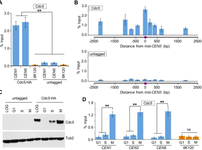
2287 cycle arrest was confirmed by fluorescence-activated cell sorting (FACS) assay (Supplemental Figure S1). Western blot analysis re-vealed a cell cycle–dependent expression pattern of Cdc5, with maximum protein levels observed in M-phase cells, only low levels detected in S-phase cells, and no detectable expression in G1 cells (Figure 1C), as previously observed (Hardy and Pautz, 1996; Charles et al., 1998). The CEN enrichment of Cdc5 was significantly higher in mitotic (M) cells (Figure 1D). No significant enrichment of Cdc5 at CEN was detected in G1- and S-phase cells; the ChIP signals in G1- and S-phase cells were largely similar to those detected at a nega-tive control region, 6K120 (Figure 1D). Control experiments per-formed with an untagged wild-type strain showed no significant enrichment of Cdc5 at either CEN or negative control region 6K120 (Supplemental Figure S2).
Next we examined the CEN association of Cdc5 during cell cycle progression using wild-type cells arrested in G1 phase by α-factor treatment and at various times after release from G1 arrest (Figure 2A). On the basis of cell and nuclear morphology, we categorized cells as G1, S, metaphase (ME), anaphase (A), and telophase (T) fol-lowing procedures described previously (Calvert and Lannigan, 2010). In agreement with previous results (Figure 1B), Western blot analysis revealed cell cycle–regulated expression of Cdc5, with increased levels observed in metaphase and anaphase cells (Figure 2B). ChIP results showed that CEN association of Cdc5 is low in cells soon after release from α-factor arrest (40 min), increasing as cells enter mitosis (60 min), maximum in anaphase (80 min), and decreasing as cells exit mitosis (100–120 min; Figure 2, A and C). We did not detect Cdc5 enrichment at a negative control region, 6K120 (Figure 2C bottom). On the basis of these results, we conclude that CEN association of Cdc5 is cell cycle dependent, with maximum enrichment in mitosis (metaphase and anaphase).
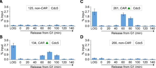
Because a previous study showed enrichment of Cdc5 at CARs (Rossio et al., 2010), we investigated whether Cdc5 localization at CARs is cell cycle dependent. We examined three CARs on chromo-some III (Figure 3A, green triangles), and two non-CARs (Figure 3A, black vertical lines) previously reported to either associate or fail to associate with cohesin components, respectively (Eckert et al., 2007; Ng et al., 2009). The CARs are 114 (CEN3), 134 (peri-CEN, 20 kb from CEN3), and 261 (chromosome arm, 147 kb from CEN3), and non-CARs are 125 (peri-CEN, 11 kb from CEN3) and 266 (chromo-some arm, 152 kb from CEN3; Figure 3A). Experiments performed with an untagged wild-type strain showed no significant enrichment of Cdc5 at any of the regions tested (Figure 3B). In logarithmically growing cells, Cdc5 associates specifically with CARs (Figure 3B). Cdc5 enrichment at CARs was significantly higher in mitotic cells (M) than in S-phase cells and was barely detectable in G1 (Figure 3C). The increased levels of Cdc5 at CARs in mitosis were verified by as-saying cells synchronized in G1 and released into pheromone-free medium. Consistent with the increased enrichment of Cdc5 at peri-CEN and arm CARs (134 and 261, respectively) in nocodazole-treated cells (Figure 3C), we observed higher levels of Cdc5 at these regions in cells undergoing mitosis (60–100 min postrelease; Figure 4). Taken together, these results show that Cdc5 enrichment at CEN and CARs is cell cycle dependent, with maximum enrichment during mitosis.
Induced expression of Cdc5 in G1 does not result in its recruitment to centromeric chromatin and CARs
The mitotic enrichment of Cdc5 at CEN and CAR sites correlates with the protein expression levels of Cdc5, and no significant enrich-ment of Cdc5 was observed at these sites in G1, when protein expression of Cdc5 was not detected (Figure 1, C and D). Therefore (CEN and pericentromeric DNA that extends ∼30–50 kb around the
CEN) exhibits a cruciform structure referred to as centromere loop (C-loop), which is analogous to that proposed in the looping model for vertebrate centromeres (Yeh et al., 2008; Verdaasdonk et al., 2012). The intramolecular linkages in the C-loop and the intermo-lecular linkages along the length of the chromosomes promote the biorientation of kinetochores and sister chromatids toward opposite spindle pole bodies for faithful chromosome segregation.
Cohesion along the length of the chromosomes is facilitated by chromosomal association of cohesins (Hornig and Uhlmann, 2004; Gerton, 2007; Brooker and Berkowitz, 2014). The cohesin complex is composed of four subunits: Scc1/Mcd1, Scc3, Smc1, and Smc3 (Hartman et al., 2000; Laloraya et al., 2000; Nasmyth, 2002; Glynn et al., 2004; Weber et al., 2004; Gerton, 2005; Bose and Gerton, 2010; Rossio et al., 2010; Marston, 2014; Roccuzzo et al., 2015). An enrichment of cohesins is seen at cohesin-associated regions (CARs) and at centromeric chromatin (Blat and Kleckner, 1999; Megee et al., 1999; Tanaka et al., 1999). Removal of cohesins from chromo-somes facilitates separation of sister chromatids at anaphase and cell cycle progression (Hartman et al., 2000; Marston, 2014; Guacci et al., 2015). Evolutionarily conserved Cdc5 has been shown to reg-ulate several aspects of mitosis (Zitouni et al., 2014; Archambault et al., 2015), including sister chromatid separation, by phosphorylat-ing Scc1/Mcd1 (Alexandru et al., 2001) to promote its proteolytic cleavage by separase (Uhlmann et al., 2000). Mutation of phosphor-ylatable serines to alanines in Scc1/Mcd1 or depletion of Cdc5 re-sults in anaphase delay (Alexandru et al., 2001; Hornig and Uhl-mann, 2004). It has been proposed that preferential phosphorylation of chromatin-bound Scc1/Mcd1 by Cdc5 accelerates its cleavage compared with soluble cohesin (Hornig and Uhlmann, 2004); however, not all Scc1/Mcd1 is cleaved by separase in anaphase (Uhlmann et al., 1999).
Here we report that Cdc5 associates with centromeric chromatin and CARs in a cell cycle–dependent manner. Enrichment of Cdc5 in mitosis overlaps with the timing of removal of cohesin from chromo-somes. Cohesin proteins interact with Cdc5 in vivo and are required for the enrichment of Cdc5 at centromeric chromatin during mitosis. We demonstrate the persistence of centromeric cohesin in ana-phase cells of a cdc5 mutant. Our results show that Cdc5 associates with centromeric chromatin to regulate the removal of cohesin from this region and promote faithful chromosome segregation.
RESULTS
Cdc5 associates with centromeric chromatin in a cell cycle–dependent manner
To determine whether Cdc5 associates with centromeric chromatin, we performed chromatin immunoprecipitation (ChIP) experiments using a wild-type strain carrying hemagglutinin (HA)-tagged Cdc5 expressed from its own promoter. ChIP–quantitative PCR (qPCR) showed that Cdc5 associates with core CEN (CEN1 and CEN5) in logarithmically growing cells (Figure 1A). No significant enrichment of Cdc5 was observed at the negative control region 6K120 (Rossio et al., 2010) or in an untagged wild-type strain (Figure 1A). We next examined Cdc5 enrichment in chromatin regions flanking CEN. ChIP-qPCR showed that Cdc5 is strongly enriched at the central core region of CEN3 and flanking chromatin (Figure 1B). No signifi-cant enrichment was detected in an untagged wild-type strain used as a control (Figure 1, A and B).
Cohesin proteins interact with Cdc5 and regulate the mitotic enrichment of Cdc5 at centromeric chromatin and CARs
The association of Cdc5 at CEN and CARs (Figures 1–4) coincides with the high levels of cohesins shown to associate at these re-gions (Megee et al., 1999; Laloraya et al., 2000; Glynn et al., 2004; Weber et al., 2004; Eckert et al., 2007); hence we asked whether cohesin interacts with Cdc5 in vivo and investigated its role in Cdc5 centromeric association. In budding yeast, the cohe-sin complex is composed of four subunits: Scc1/Mcd1, Scc3, Smc1, and Smc3 (Nasmyth, 2002). We constructed strains ex-pressing Cdc5 and TAP-tagged Mcd1 or Cdc5 and green fluores-cent protein (GFP)–tagged Smc3 from their endogenous promot-ers. Immunoprecipitation experiments showed in vivo interaction of Cdc5 with Mcd1 and Smc3 (Figure 6). No interaction was ob-served in control experiments using a strain with untagged Mcd1 or Smc3 (Figure 6).
we sought to determine whether recruitment of Cdc5 at CEN and CAR sites is regulated by the cell cycle or is an outcome of its cell cycle–dependent expression pattern. We constructed a strain ex-pressing HA-tagged Cdc5 from a galactose-inducible promoter (GAL1-3HA-CDC5) and performed ChIP experiments using cells growing logarithmically and cells arrested in G1 with α-factor treat-ment. The galactose-induced expression of Cdc5 in logarithmically growing and G1 cells was determined by Western blotting (Figure 5A). FACS analysis confirmed the synchronization of cells in G1 (Figure 5B). ChIP-qPCR revealed significant enrichment of Cdc5 at CEN3 and CARs (134, 261) in logarithmically growing cells, but no significant enrichment of Cdc5 was detected at these regions in G1 cells (Figure 5C). The ChIP signals in G1 cells were largely similar to those detected at the negative control region 6K120 or at CEN3, CARs, and 6K120 region in an untagged wild-type strain (Figure 5C). On the basis of these results, we conclude that the enrichment of Cdc5 at CEN and CARs is cell cycle regulated.
2289 phase transition (Figures 2 and 4), we posited that Cdc5 might be required for the removal of chromatin-bound cohesin. In wild-type cells, cohesin is lost from both centromeric chromatin and chromo-somal arms at anaphase onset (Marston, 2014). To characterize the Cdc5 dependence of cohesin loss, we monitored the localization of Smc3-GFP in anaphase cells of wild-type and cdc5-99 strains grown at permissive temperature (25°C) and after a 2.5-h shift to the non-permissive temperature of 37°C. The well-characterized tempera-ture-sensitive cdc5-99 mutant exhibits a defect in mitotic exit at the nonpermissive temperature of 37°C (St-Pierre et al., 2009). Spindle pole body (SPB) component Spc29–red fluorescent protein (RFP) was used as a marker for centromeric chromatin due to the close physical proximity of the SPB to the kinetochores in budding yeast. Using the distance between SPBs as a measure for spindle length, we quantitated Smc3-GFP foci in anaphase cells with a spindle length of 3–8 μm. In addition to the diffuse Smc3-GFP fluorescence observed over the separated chromatin masses in both wild-type and cdc5-99 cells, distinct Smc3-GFP foci were observed in the cdc5-99 mutant (Figure 8A). No detectable Smc3-GFP foci were ever observed in wild-type cells in anaphase. Quantification of the cdc5-99 cells with Smc3-GFP foci at one or both of the spindle poles was significantly higher (32 vs. 13%, p < 0.05) at 37°C (n= 144) than at 25°C (n = 125; Figure 8B). We next examined the localization Next we examined whether Mcd1 is required for enrichment of
Cdc5 at CEN and CARs. Because cohesin subunits are essential for cell viability, we used mcd1-1, a well-characterized temperature-sen-sitive mutant that arrests in M phase at the nonpermissive tempera-ture (37°C) and exhibits defects in establishment and maintenance of cohesion (Guacci et al., 1997; Noble et al., 2006; Skibbens et al., 2010; Eng et al., 2014, 2015). The expression of Cdc5 was similar in wild-type and mcd1-1 strains grown at 25 and 37°C, as measured by Western blot analysis (Figure 7A). The enrichment of Cdc5 at CEN and CARs in nocodazole-treated (M-phase arrest) wild-type and mcd1-1 cells grown at 25°C and after shift to 37°C was assayed by ChIP. The cell cycle arrest was verified by FACS assay (Figure 7B). ChIP-qPCR showed that Cdc5 levels at CEN3, as well as CARs lo-cated at peri-CEN (134) and the chromosomal arm (261), are re-duced threefold to fourfold specifically in the mcd1-1 strain at 37°C (Figure 7C). There was no significant enrichment of Cdc5 at negative control region 6K120 under any condition (Figure 7C). We conclude that cohesin is required for enrichment of Cdc5 at CEN and CARs.
cdc5-99 strains exhibit persistence of cohesin at CEN
and peri-CEN regions in anaphase
Given our findings that association of Cdc5 at centromeric chromatin and CARs is maximal in cells undergoing the
metaphase-to-ana-FIGURE 2: CEN association of Cdc5 is cell cycle regulated, with maximum enrichment in cells undergoing mitosis. A wild-type strain containing Cdc5-HA (YMB9264) was grown at 30°C, arrested in G1 with α-factor, washed, and released into pheromone-free medium. α-Factor was added again 80 min after release to block the cells at the subsequent G1. Samples were taken at various time points (minutes) after G1 release. (A) FACS analysis revealed cell cycle progression. Cell cycle stages were determined based on cell morphology and nuclear position by microscopic examination of 100 cells for each time point. Different stages of the cell cycle: G1, S phase (S), metaphase (ME), anaphase (A), and telophase (T). (B) Expression of Cdc5 is cell cycle regulated. Western blot analysis was carried out on whole-cell protein extracts prepared from samples taken at various times after release from G1 arrest as in A. Blots were probed with
α-HA (Cdc5) and α-Tub2 (loading control) antibodies. (C) Centromeric levels of Cdc5 are highest in mitotic cells. ChIP for Cdc5 was carried out using α-HA antibodies. Enrichment of Cdc5 at CEN3 (top) and a negative control region
number of cdc5-99 cells with Mcd1-GFP foci at one or both of the spindle poles was sig-nificantly higher (24 vs. 3%, p < 0.05) at 37°C (n= 197) than at 25°C (n= 94; Figure 8D).
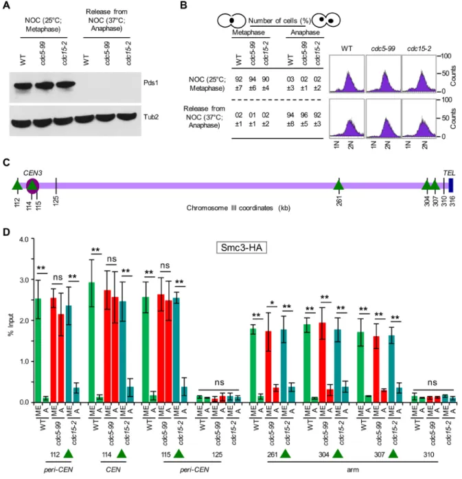
The proximity of Smc3-GFP and Mcd1-GFP foci to Spc29-RFP in cdc5-99 strains suggested the persistence of cohesin in centromeric chromatin; hence we used ChIP to assay the association of Smc3-HA at CARs close to (CEN) or distant from the cen-tromere (arm), using either metaphase or anaphase cells from wild-type, cdc5-99, and cdc15-2 strains. Metaphase cells were ob-tained by growing strains at 25°C in no-codazole, and anaphase cells were obtained after release from nocodazole at the non-permissive temperature of 37°C (Figure 9). We used cdc15-2 as a control because this strain arrests at a stage similar to cdc5 mu-tants during mitosis (Surana et al., 1993; St-Pierre et al., 2009). The cell cycle stage was determined by monitoring Pds1 levels (Cohen-Fix et al., 1996) by Western blotting, DNA content by FACS, and nuclear mor-phology by microscopy (Calvert and Lannigan, 2010). High levels of Pds1 are present in metaphase; however, levels of Pds1 are undetectable in anaphase, as deg-radation of Pds1 is required for anaphase onset (Cohen-Fix et al., 1996; Cohen-Fix and Koshland, 1999). Consistent with this, high levels of Pds1 were observed in meta-phase but not in anameta-phase cells of wild-type, cdc5-99, and cdc15-2 strains (Figure 9, A and B). Large-budded cells with the nucleus at the neck or separated nuclei indicate metaphase and anaphase cells, respectively (Figure 9B). ChIP-qPCR showed enrichment of Smc3-HA at CEN (114), CARs flanking the centromere (112, 115), and the arms (261, 304, 307) in metaphase cells of wild-type, cdc5-99, and cdc15-2 strains (Figure 9, C and D). Smc3-HA levels were significantly reduced at both the centromeric chromatin (CEN [114], peri-CEN CARs [112, 115]) and the chromosomal arms (261, 304, 307) in anaphase cells of wild-type and cdc15-2 strains, and these levels were largely similar to those at non-CARs (125, 310; Figure 9D). In contrast, in anaphase cells of the cdc5-99 strain, Smc3-HA levels were significantly re-duced only at CARs at chromosomal arms (261, 304, 307) and remained high at cen-tromeric chromatin (CEN [114] and peri-CEN CARs [112, 115]). The levels of Smc3-HA at CEN and peri-CEN CARs in anaphase cells of cdc5-99 strains were not statistically different from those observed at these chromosomal regions in metaphase cells (Figure 9D). No significant enrichment of Scm3-HA was detected at non-CARs (125, 310) at either metaphase or ana-phase (Figure 9D). These results suggest that Cdc5 is required for the removal of cohesin from centromeric chromatin during mitosis. pattern of another cohesin component, Mcd1-GFP, in anaphase cells
of wild-type and cdc5-99 strains using growth conditions and proce-dure as described earlier. The persistence of Mcd1-GFP foci was ob-served in anaphase cells of cdc5-99 strain, whereas no detectable Mcd1-GFP foci were observed in wild-type cells (Figure 8C). The
FIGURE 3: Cdc5 associates with CARs in a cell cycle–dependent manner, with maximum enrichment in mitosis. (A) Schematic showing CARs and non-CARs on chromosome III. Cohesins have been shown to either associate (CARs) or fail to associate (non-CARs) at these regions in previous studies (Eckert et al., 2007; Ng et al., 2009). Centromere (CEN3, violet circle), CAR (green triangle), and non-CAR (black vertical line) sites. CEN is located at 114 kb; peri-CEN CAR 134 (20 kb from CEN), chromosome arm CAR 261 (147 kb from CEN), peri-CEN non-CAR 125 (11 kb from CEN), and chromosome arm non-CAR 266 (152 kb from CEN) were examined. (B) Cdc5 associates with CARs. ChIP was performed for Cdc5 in a wild-type strain tagged with Cdc5-HA (YMB9264) and an untagged control strain (YMB9263). Cells were grown to
2291
FIGURE 5: Induced expression of Cdc5 in G1 does not result in its recruitment to CEN and CARs. A wild-type strain containing GAL1-3HA-CDC5 (YMB9441) and untagged control (JG595) were grown at 30°C in yeast extract/peptone with 2% galactose and raffinose and arrested in G1 with α-factor for 2 h. Samples were collected for protein extraction, DNA content, and ChIP analyses. (A) Induced expression of Cdc5 (GAL1-3HA-CDC5) in G1. Western blot analysis was carried out on whole-cell protein extracts prepared from cultures growing logarithmically or arrested in G1 with α-factor treatment. Blots were probed with α-HA (Cdc5) and α-Tub2 (loading control) antibodies. (B) FACS analysis revealed synchronization of cells in G1. (C) Cdc5 does not associate with CEN and CARs in G1. ChIP for Cdc5 (GAL1-3HA-CDC5) was carried out using α-HA antibodies. Enrichment of Cdc5 at CEN3, CARs (134, 261), and a negative control region
6K120 was determined by qPCR and is shown as percentage input. Average from three biological replicates ± SE.
**p < 0.01; ns, not statistically significant, Student’s t test.
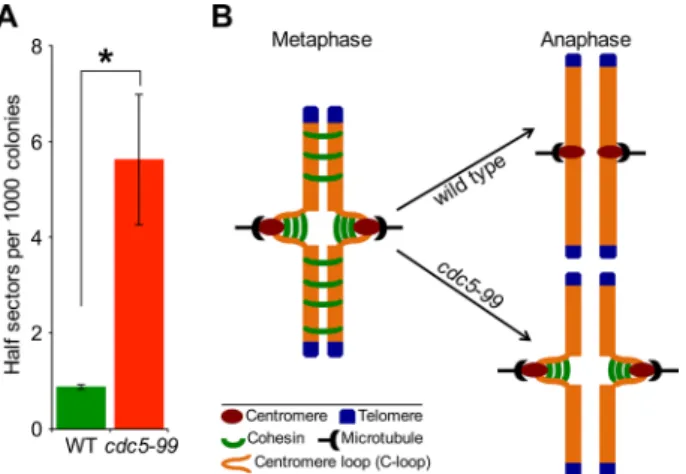
mined the frequency of loss of the RC using a colony color assay as described previously (Spencer et al., 1990). Loss of the RC results in formation of red sectors in an otherwise white colony. Colonies that are at least half red indicate loss of the RC in the first cell division. The frequency of RC loss in cdc5-99 strains was about sixfold higher than in the wild-type strain (Figure 11A), similar to that reported for kinetochore mutants (Kastenmayer et al., 2005; Ma et al., 2012).
DISCUSSION
Evolutionarily conserved polo-like kinase Cdc5 and its homologues play a range of crucial and conserved roles in regulation of cell cycle events (Alexandru et al., 2001; Lee et al., 2005; St-Pierre et al., 2009; Rossio et al., 2010; Ratsima et al., 2011; Botchkarev et al., 2014; Walters et al., 2014; Zitouni et al., 2014; Archambault et al., 2015). Among its many roles, Cdc5 is required for the phosphorylation and subsequent cleav-age of Scc1/Mcd1 in order to promote sister chromatid separation (Alexandru et al., 2001; Hornig and Uhlmann, 2004). Our results pro-vide mechanistic insight into the role of Cdc5 in the removal of cen-tromeric cohesin during mitosis. We show that Cdc5 associates with centromeric chromatin and is required for the efficient removal of cohesin at core CEN and peri-CEN regions during mitosis. These conclusions are based on results showing that Cdc5: 1) associates with CEN and CARs in a cell cycle–dependent manner, with maxi-mum enrichment in mitosis; 2) interacts with cohesins in vivo; 3) re-quires cohesin for its mitotic enrichment at CEN and CARs; 4) is re-quired for the removal of cohesin from centromeric chromatin in anaphase; and 5) is required for faithful chromosome segregation.
Enrichment of Cdc5 at centromeric chromatin was highest in mi-totic cells undergoing the transition from metaphase to anaphase. Even though Cdc5 is expressed in S-phase and anaphase cells, we observed significantly lower levels of chromatin-associated Cdc5 during the S phase of the cell cycle. Moreover, ectopic expression of Cdc5 in G1 does not result in its recruitment to CEN and CARs, sug-gesting that the binding of Cdc5 to these chromosomal regions is regulated by the cell cycle. Association of Cdc5 flanking CEN6 and CARs was reported in a ChIP-on-chip study evaluating the role of Rsc2 in mitotic exit (Rossio et al., 2010). Our comprehensive ChIP analysis shows that the cell cycle–regulated centromeric enrichment of Cdc5 coincides with its role in removal of cohesin from this region. We discovered that cohesin components Scc1/Mcd1 interact with Cdc5 in vivo, and the interaction is required for enrichment of Cdc5 at CEN and CARs. It is of note that high-throughput proteomic stud-ies also identified cohesin components when Cdc5 was used as bait in affinity-capture mass spectroscopy experiments (Ho et al., 2002; Snead et al., 2007; Breitkreutz et al., 2010). The cohesin-dependent enrichment of Cdc5 at CEN and CARs reflects a dynamic enzyme– substrate relationship between Cdc5 and cohesin components, re-sulting in preferential removal of chromatin-bound cohesin. Of most importance, our results suggest a role for Cdc5 in removal of centro-meric cohesins, most likely from the intramolecular linkages in the C-loop (Yeh et al., 2008; Hu et al., 2011), but not cohesins in the arms of chromosomes (Figure 11B). The C-loop model resolved a paradox in which sister kinetochores can separate up to 800 nm in A previous study showed that SMC subunits of cohesin complex
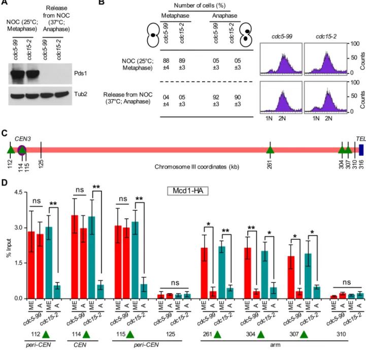
(e.g., Smc1) remain associated with chromosomes for ∼10 min after the degradation of Scc1/Mcd1 in anaphase (Tanaka et al., 1999). Hence we investigated whether Cdc5 is also required for removal of Mcd1 from centromeric chromatin. We examined the association of Mcd1-HA at CEN and CARs in cdc5-99 and cdc15-2 grown at 25°C in nocodazole (metaphase) and after release from nocodazole at 37°C (anaphase). We detected high levels of Pds1 in metaphase but not in anaphase cells of cdc5-99, and cdc15-2 strains (Figure 10, A and B). The levels of Pds1 were slightly lower in cdc15-2 than in the cdc5-99 strain (Figure 10A). ChIP-qPCR showed enrichment of Mcd1-HA at CEN (114), CARs flanking the centromere (112, 115), and the arms (261, 304, 307) in metaphase cells of cdc5-99 and cdc15-2 strains (Figure 10, C and D). Mcd1-HA levels were signifi-cantly reduced at CEN (114), peri-CEN CARs (112, 115), and chro-mosomal arms CARs (261, 304, 307) in anaphase cells of cdc15-2, and these levels were largely similar to those at non-CARs (125, 31; Figure 10D). Consistent with the results for Smc3-HA, significantly reduced levels of Mcd1-HA were observed at CARs in chromosomal arms (261, 304, 307), whereas Mcd1-HA levels remained high at centromeric chromatin (CEN [114] and peri-CEN CARs [112, 115]) in anaphase cells of cdc5-99 strains. The levels of Mcd1-HA at CEN and peri-CEN CARs in anaphase cells of cdc5-99 strains were statis-tically not different from those observed at these chromosomal re-gions in metaphase cells (Figure 10D). No significant enrichment of Mcd1-HA was detected at non-CARs (125, 310) at either metaphase or anaphase (Figure 10D). The persistence of both Smc3 and Mcd1 in CEN and peri-CEN chromatin in anaphase cells of the cdc5-99 strain supports a role for Cdc5 in the removal of cohesin from cen-tromeric chromatin.
Cdc5 is required for faithful chromosome segregation The cell cycle–regulated centromeric association of Cdc5 and its role in removal of centromeric cohesin led us to examine whether defects in Cdc5 would result in increased chromosome loss. We constructed a cdc5-99 strain carrying a reporter chromosome (RC) and
deter-FIGURE 6: Cdc5 interacts with cohesin proteins Mcd1 and Smc3 in vivo. (A) Cdc5 interacts in vivo with Mcd1. Wild-type strain (MAY8878) expressing TAP-tagged Mcd1 (CDC5 MCD1-TAP) and a control strain (BY4741) expressing untagged Mcd1 (CDC5 MCD1) from their native promoters were grown at 30°C. Cell extracts were prepared and used in immunoprecipitation experiments using α-Cdc5 monoclonal and α-TAP antibodies. Eluted proteins were analyzed by Western blotting with α-Cdc5, α-TAP (Mcd1), and α-Tub2 (loading control) antibodies. IN, input; IP, immunoprecipitated samples. (B) Cdc5 interacts in vivo with Smc3. Wild-type strain
2293 et al., 2009; Botchkarev et al., 2014; Walters et al., 2014; Zitouni et al., 2014; Roccuzzo et al., 2015), chromosome segregation errors observed in cdc5-99 strain may not be due to solely defects in co-hesin removal. Previous studies showed that Cdc5 interacts with and phosphorylates kinetochore proteins in vivo (e.g., Slk19) to promote proper microtubule attachment and spindle function (Stegmeier et al., 2002; Snead et al., 2007; Park et al., 2008; Liang et al., 2009; Richmond et al., 2013). Plk1, the human homologue of Cdc5, also localizes to the CEN (Arnaud et al., 1998; Kishi et al., 2009), where it likely phosphorylates Rad21 (the human Scc1 homologue; Waizenegger et al., 2000). In addition, Plk1 phosphorylates kineto-chore protein Mis18BP1, which in turn licenses the recruitment of new CENP-A to kinetochores in the G1 phase of the cell cycle (McKinley and Cheeseman, 2014). Of note, Cse4 (the budding yeast CENP-A homologue) was identified as an in vivo interacting partner of Cdc5 in a genome-wide proteomic study (Snead et al., 2007). Future studies should help us to address whether Cdc5-mediated phosphorylation of kinetochore proteins affects the removal of cen-tromeric cohesins.
In summary, our studies provide novel insights into the role of Cdc5 in removal of centromeric cohesin but not cohesin from vivo despite high levels of cohesin at centromeres. The persistence
of Smc3 and Mcd1 in anaphase of cdc5-99 cells at the nonpermis-sive temperature may reflect the presence of intramolecularly linked cohesin in the C-loop, whereas the absence of cohesin in chromo-some arms shows that Cdc5 is dispensable for removal of intermo-lecularly linked cohesins. Cdc5 has been shown to regulate removal of cohesin from chromosome arms for segregation of homologous chromosomes in meiosis I but to be dispensable for meiosis II (Brar et al., 2006; Katis et al., 2010; Attner et al., 2013). Attner et al. (2013) concluded that additional kinases or substrates and/or the differen-tial nature of centromeric versus chromosome arm cohesin regulate the removal of centromeric cohesin in meiosis II. On the basis of our results, we conclude that Cdc5 represents the first example of dif-ferential regulation of centromeric versus arm cohesin during mitosis in budding yeast.
The mitotic enrichment of Cdc5 at CEN suggests a role for Cdc5 in kinetochore function because of major changes in spatial geom-etry and spindle tension that occur at the kinetochore during mito-sis. Consistent with this hypothesis, we observed errors in chromo-some segregation in the cdc5-99 strain. Because Cdc5 is involved in many biological processes (Archambault and Glover, 2009; St-Pierre
sin antagonist WAPL, whereas removal of centromeric cohesin at the metaphase-to-anaphase transition by Plk1 is achieved by a separase-dependent mechanism (Waizeneg-ger et al., 2000; Hauf et al., 2001; Gandhi et al., 2006; Kueng et al., 2006; Haarhuis et al., 2013). Mutations and overexpression of Plk1 are observed in many cancers (Davies et al., 2005; Stephens et al., 2005; Kan et al., 2010), displaying chromosomal instability and aneuploidy (Degenhardt and Lampkin, 2010). Hence elucidation of molecular mechanisms by which polo kinases regulate cohesin removal and faithful chromosome segregation might help us identify and de-velop novel therapeutic targets for cancer therapy.
MATERIALS AND METHODS
Strains, plasmids, and culture conditions
Saccharomyces cerevisiae strains and plas-mids used in this study are listed in Table 1. Strains were grown in 1% yeast extract/2% Bacto-peptone/2% glucose (YPD) or in yeast synthetic medium containing 2% glucose and supplements to allow for the selection of plasmids used.
Assay for the loss of reporter chromosome
Loss of a nonessential RC was measured using a colony color assay in which RC loss results in red sectors in an otherwise white colony (Spencer et al., 1990). Wild-type and cdc5-99 strains were grown to loga-rithmic phase in selective medium to main-tain the RC. Cultures were diluted and plated on complete synthetic medium with limiting adenine at 30ºC. The frequency of chromosome loss was measured by count-ing the colonies that were at least half red, indicating loss of the RC at the first cell division. At least 3000 colonies of three in-dividual transformants were examined for each strain.
ChIP and qPCR experiments
ChIP assays were performed with three bio-logical replicates as described previously (Mishra et al., 2007, 2011). Antibodies used to capture protein–DNA complexes were α -HA (A2095; Sigma-Aldrich, St. Louis, MO) and α-Myc (A7470; Sigma-Aldrich). ChIP-qPCR was carried out using SYBR Green Master Mix in a 7500 Fast Real Time PCR System (Applied Biosystems, Foster City, CA) following conditions used previously (Mishra et al., 2011). The enrichment values were determined as percentage input using the ΔΔCT method (Livak and Schmittgen, 2001). Primer sequences used in this study are pre-sented in Table 2.
chromosomal arms. Differential regulation of cohesin removal has also been reported in mammalian cells, where removal of cohesin from chromosomal arms in prometaphase is regulated by the
2295
FIGURE 9: Cdc5 is required for the removal of Smc3 from centromeric chromatin. WT (YMB9693), cdc5-99 (YMB9709), and cdc15-2 (YMB9703) expressing 6HA and Pds1-18Myc from their endogenous promoters were used. Smc3-6HA was an internally tagged version of Smc3 (six copies of HA inserted at amino acid residue 607) using plasmid pVG466 (a gift from Vincent Guacci). Strains were grown in YPD at permissive temperature (25°C) with nocodazole (NOC, 20 μg/ml) for 2 h to synchronize cells in metaphase. Cells were then shifted to nonpermissive temperature (37°C) for 1 h, followed by release from NOC into YPD at 37°C for 45 min to enrich cells in anaphase. Samples were collected for Western blot analysis for Pds1, nuclear morphology, DNA content, and ChIP analyses. Three biological replicates were performed for each strain. (A) Western blot analysis showing high levels of Pds1 only in metaphase cells of WT,
cdc5-99, and cdc15-2 strains. Antibodies used were α-Myc (Pds1) and α-Tub2 (loading control). (B) Cell cycle stages
FIGURE 10: Cdc5 is required for the removal of Mcd1 from centromeric chromatin. cdc5-99 (YMB9711), and cdc15-2 (YMB9712) strains expressing Mcd1-3HA, and Pds1-18Myc from their endogenous promoters were used. Strains KT046 (Tong and Skibbens, 2014) and D1469 carrying MCD1-3HA were used to construct strains YMB9711 and YMB9712, respectively. Strains were grown in YPD at permissive temperature (25°C) with nocodazole (NOC; 20 μg/ml) for 2 h to synchronize cells in metaphase. Cells were then released into YPD at nonpermissive temperature (37°C) for 2.5 h to enrich cells in anaphase. Samples were collected for Western blot analysis, nuclear morphology, DNA content, and ChIP analyses. Three biological replicates were performed for each strain. (A) Western blotting showing high levels of Pds1 only in metaphase cells of cdc5-99 and cdc15-2 strains. Antibodies used were α-Myc (Pds1) and α-Tub2 (loading control). (B) Cell cycle stages (metaphase and anaphase) were determined based on nuclear position and cell morphology by microscopic examination of 100 cells for each strain per replicate. DNA content was determined by FACS. (C) Schematic representation of CARs and non-CARs on chromosome III as detailed in Figure 9C. (D) Mcd1 persists at CEN and peri-CEN chromatin in cdc5-99 strains. ChIP experiments were performed using chromatin prepared from metaphase (ME) or anaphase (A) cells from strains as described using α-HA antibodies. Enrichment of Mcd1 at CEN3 (114), peri-CEN CARs (112, 115), and chromosomal arm CARs (261, 304, 307) were determined by qPCR and is shown as percentage input. The non-CAR peri-CEN loci 125 and 310 in chromosome arms were used as negative controls. Average from three biological replicates ± SE. **p <0.01; *p < 0.05; ns, not significant, Student’s t test.
Immunoprecipitation and Western blotting
Immunoprecipitation assays were performed as described previ-ously (Mishra et al., 2011). Protein extracts were prepared
2297 SDS–polyacrylamide gels and transferred to nitrocellulose membrane. Primary antibodies used were α-HA (clone 12CA5; Roche Molecular Systems, Pleasanton, CA), α-GFP (A11122; Life Technologies, Carlsbad, CA), α-TAP (CAB1001; Thermo Scientific, Waltham, MA), α-Cdc5 (11H12 and 4F10; Medimabs, Montreal, Canada), α-Myc (Z-5, sc-789; Santa Cruz Bio-technology, Dallas, TX), and α-Tub2 (Mishra et al., 2011). Secondary antibodies were horseradish peroxidase (HRP)– conjugated donkey α-rabbit immunoglobulin G (IgG; NA934V, Amersham Biosciences, Amersham, United Kingdom), and HRP-conjugated sheep α-mouse IgG (NA931V; Amersham Biosciences).
Microscopy
Cultures grown to logarithmic phase at permissive temperature (25°C) and after a shift to nonpermissive temperature (37°C) were used for all imaging experiments. Anaphase cells that ex-hibited a spindle length of 3–8 μm were selected based on the position of spindle pole bodies (Spc29-RFP; Chen et al., 2000; Maddox et al., 2000). Cells were imaged for Smc3-GFP, Mcd1-GFP, and Spc29-RFP signals at room temperature on a Nikon TE-2000E inverted microscope equipped with a 1.4 numerical aperture/100× Plan-Apo objective (Nikon Instruments, New York, NY) as described previously (Haase et al., 2013; Salmon et al., 2013).
FIGURE 11: Cdc5 is required for faithful chromosome segregation. (A) Cdc5 is required for faithful chromosome segregation. Frequency of RC loss in wild-type (WT; YPH363) and cdc5-99 (YMB9438) strains was determined as described in Materials and Methods. At least 3000 colonies from three independent transformants were counted. Values are mean ± SE. *p < 0.05, Student’s t test. (B) Schematic model for the role of Cdc5 in cohesin removal from centromeric chromatin during mitosis. In WT cells, cohesin is removed from chromosomal arms and centromeric chromatin during metaphase-to-anaphase transition, whereas in cdc5-99 strains, cohesin is removed from chromosomal arms but persists at centromeric chromatin.
Strain name Genotype Reference
YMB9431 MATa ura3-1 leu2-3112 his3-11,15 trp1-1 ade2-1 can1-100 SMC3-GFP::URA3 This study
YMB9263 MATa ura3-52 lys2-801 ade2-101 trp1Δ63 his3Δ200 leu2Δ1 CSE4-13MYC::LEU2 CEN-TRP1 This study YMB9264 MATa ura3-52 lys2-801 ade2-101 trp1Δ63 his3Δ200 leu2Δ1 CSE4-13MYC::LEU2
CEN-CDC5-3HA::TRP1 (p344)
This study
JG595 MATa ura3-1 leu2,3-112 his3-1 trp1-1 ade2-1 can1-100 Δbar1 CSE4-12MYC::URA3 SCM3-3FLAG::kanMX
Camahort et al. (2007)
YMB9441 MATa ura3-1 leu2,3-112 his3-1 trp1-1 ade2-1 can1-100 Δbar1 CSE4-12Myc::URA3 SCM3-3FLAG::kanMX GAL1-3HA-CDC5::TRP1
This study
YMB9693 MATa ura3-1 leu2-3112 his3-11,15 trp1-1 ade2-1 can1-100 SMC3-N607-6HA::URA3 PDS1-18MYC::LEU2 This study
YMB9695 MATa MCD1-GFP leu2-3112 ura3-52 his3-11,15 bar1 GAL+ SPC29-RFP::Hyg This study
YMB9696 MATa MCD1-GFP leu2-3112 ura3-52 his3-11,15 bar1 GAL+ SPC29-RFP::Hyg cdc5-99::HIS3MX6 This study YMB9703 MATa ura3-1 leu2-3112 his3-11,15 trp1-1 ade2-1 can1-100 SMC3-N607-6HA::URA3
PDS1-18MYC::LEU2 cdc15-2
This study
YMB9709 MATa ura3-1 leu2-3112 his3-11,15 trp1-1 ade2-1 can1-100 SMC3-N607-6HA::URA3 PDS1-18MYC::LEU2 cdc5-99::HIS3MX6
This study
YMB9711 MATa ade2-1 his3-11,15 leu2-3112 trp1-1 ura3-1 can1-100 MCD1-3HA::TRP1 PDS1-18MYC::LEU2 cdc5-99::HIS3MX6
This study
YMB9712 MATα leu2-3112 trp1-1can1-100 ura3-1 ade2-1 his3-11,15 [phi+] rad5-535 MCD1-3HA::HPHMX6 cdc15-2::TRP1 PDS1-18MYC::LEU2
This study
YMB8702 MATa ura3-1 leu2-3112 his3-11,15 trp1-1 ade2-1 can1-100 This study
KBY6097-1 MATα ura3-1 leu2-3112 his3-11,15 trp1-1 ade2-1 can1-100 SMC3-GFP::URA3 cdc5-99::HIS3MX6 SPC29-RFP::Hyg
This study
KBY6098-1 MATa ura3-1 leu2-3112 his3-11,15 trp1-1 ade2-1 can1-100 SMC3-GFP::URA3 SPC29-RFP::Hyg This study YMB9438 MATα ura3-52 lys2-801 leu2-Δ1 ade2-101 his3-Δ200 [CFIII (CEN3.L) CFVII (RAD2.d) URA3 SUP11]
cdc5-99::HIS3
This study
YPH363 MATα ura3-52 lys2-801 leu2-Δ1 ade2-101 his3-Δ200 [CFIII (CEN3.L) CFVII (RAD2.d) URA3 SUP11] Spencer et al. (1990)
TABLE 1: Strains and plasmids used in this study.
Strain name Genotype Reference
BY4741 MATa ura3Δ0 leu2Δ0 his3Δ1 met15Δ0 Open Biosystems,
Lafayette, CO
MAY8878 MATa his3Δ1 leu2Δ0 met15Δ0 ura3Δ0 MCD1-TAP::HIS3 This study
YPH499 MATa ura3-52 lys2-801 ade2-101 trp1Δ63 his3Δ200 leu2Δ1 Sikorski and
Hieter (1989)
MAY8775 MATa ura3-52 ade2-101 his3-11,15 leu2-3112 CDC5-13MYC This study
MAY8851 MATa ura3-52 ade2-101 his3-11,15 leu2-3112 CDC5-13MYC mcd1-1 This study
VG3507-2A MATa MCD1-GFP leu2-3112 ura3-52 his3-11,15 bar1 GAL+ V. Guacci,
University of California, Berkeley, CA
KT046 MATa ade2-1 his3-11,15 leu2-3112 trp1-1 ura3-1 can1-100 MCD1-3HA::TRP1 Tong and
Skibbens (2014) D1469 MATα leu2-3112 trp1-1can1-100 ura3-1 ade2-1 his3-11,15 [phi+] rad5-535 MCD1-3HA::HPHMX6
cdc15-2::TRP1
D. D’Amours, Université de Montréal, Montréal, QC
Plasmids Description Reference
p344 CEN-CDC5-3HA::TRP1 D. D’Amours
pVG466 SMC3-N607-6HA::URA3 V. Guacci
pSB205 PDS1-18MYC::LEU2 S. Biggins,
Fred Hutchinson Cancer Research Center, Seattle, WA
TABLE 1: Strains and plasmids used in this study. Continued
Locus Forward (5′–3′) Reverse (5′–3′) Reference
CEN1 CTCGATTTGCATAAGTGTGCC GTGCTTAAGAGTTCTGTACCAC Choy et al. (2011)
CEN3 GATCAGCGCCAAACAATATGG AACTTCCACCAGTAAACGTTTC Choy et al. (2011)
CEN5 AAGAACTATGAATCTGTAAATGACTGATTCAAT CTTGCACTAAACAAGACTTTATACTACGTTTAG Choy et al. (2011)
6K120 AACGTCACTTTTTTTCCAGGG GCAAAGCTAGCTAACGAACAA This study CEN3-L1 ATATTGTTTGGCGCTGATCGCC TTGATGAACTTTTCAAAGATGAC Choy et al. (2011) CEN3-L2 TCATCTTTGAAAAGTTCATCAAGG GATAACAAAGCATGGTATGGCG Choy et al. (2011) CEN3-L3 GCCATACCATGCTTTGTTATCGTC TATTATGCTCCCCTGGATTTTATGCG Choy et al. (2011) CEN3-L4 AGCATTAGAGCCACTGTCATTTC ATAATTAAGATACGAATGTGTTCGTTG Mishra et al. (2013) CEN3-R1 TTTACTGGTGGAAGTTTTGCTCA GTCAACGAGTCCTCTCTGGCTA Choy et al. (2011) CEN3-R2 GAGAGGACTCGTTGACGTAGAA GAATATGATAATGGTTACACCAGTAGG Choy et al. (2011) CEN3-R3 TGTAACCATTATCATATTCATGAC GATTTAATGCACGTTATGTTTCG Choy et al. (2011) CEN3-R4 ACTGACAGCACCATTAATCAATCA TATCCTCAGTAGAGGGCAAAGTT Mishra et al. (2013)
125 TGCCAAGTTGTGCTTTTTAGTTGAG TTGATGTGTTGATTGTTCGGTCAC Eckert et al. (2007),
Ng et al. (2009)
134 CCGATGGTTAGGATTTCCAACG GGTTTTCAGAACAGAATGGGGC Eckert et al. (2007),
Ng et al. (2009)
261 TTGCCACAGCCACAGATATAACTG GATGGACAAAGCGTTGTATCCG Eckert et al. (2007),
Ng et al. (2009)
266 CAGACTTCCTCCAAGCAACAGG TCCTCCTTAGCCTTTTCTTCAGC Eckert et al. (2007),
Ng et al. (2009)
304 CCCCTTCCAAAGACCTGACA CCAGCCGTCGATCCTAAAGA Laloraya et al. (2000)
307 GTAGCGCTCTCAACTACCCT TCGTATACTGTTAGGGTCTGCA Laloraya et al. (2000)
310 TCTCGGAATTTATCATGACCCAT AAACCCTGCACACATTTCGT Laloraya et al. (2000)
2299 Cohen-Fix O, Koshland D (1999). Pds1p of budding yeast has dual roles:
inhibition of anaphase initiation and regulation of mitotic exit. Genes Dev 13, 1950–1959.
Cohen-Fix O, Peters JM, Kirschner MW, Koshland D (1996). Anaphase initia-tion in Saccharomyces cerevisiae is controlled by the APC-dependent degradation of the anaphase inhibitor Pds1p. Genes Dev 10, 3081–3093. Davies H, Hunter C, Smith R, Stephens P, Greenman C, Bignell G, Teague
J, Butler A, Edkins S, Stevens C, et al. (2005). Somatic mutations of the protein kinase gene family in human lung cancer. Cancer Res 65, 7591–7595.
Degenhardt Y, Lampkin T (2010). Targeting Polo-like kinase in cancer therapy. Clin Cancer Res 16, 384–389.
Eckert CA, Gravdahl DJ, Megee PC (2007). The enhancement of pericen-tromeric cohesin association by conserved kinetochore components promotes high-fidelity chromosome segregation and is sensitive to microtubule-based tension. Genes Dev 21, 278–291.
Eng T, Guacci V, Koshland D (2014). ROCC, a conserved region in cohesin’s Mcd1 subunit, is essential for the proper regulation of the maintenance of cohesion and establishment of condensation. Mol Biol Cell 25, 2351–2364.
Eng T, Guacci V, Koshland D (2015). Interallelic complementation provides functional evidence for cohesin-cohesin interactions on DNA. Mol Biol Cell 26, 4224–4235.
Gandhi R, Gillespie PJ, Hirano T (2006). Human Wapl is a cohesin-binding protein that promotes sister-chromatid resolution in mitotic prophase. Curr Biol 16, 2406–2417.
Gerton J (2005). Chromosome cohesion: a cycle of holding together and falling apart. PLoS Biol 3, e94.
Gerton JL (2007). Enhancing togetherness: kinetochores and cohesion. Genes Dev 21, 238–241.
Glynn EF, Megee PC, Yu HG, Mistrot C, Unal E, Koshland DE, DeRisi JL, Gerton JL (2004). Genome-wide mapping of the cohesin complex in the yeast Saccharomyces cerevisiae. PLoS Biol 2, E259.
Guacci V, Koshland D, Strunnikov A (1997). A direct link between sister chromatid cohesion and chromosome condensation revealed through the analysis of MCD1 in S. cerevisiae. Cell 91, 47–57.
Guacci V, Stricklin J, Bloom MS, Guo X, Bhatter M, Koshland D (2015). A novel mechanism for the establishment of sister chromatid cohesion by the ECO1 acetyltransferase. Mol Biol Cell 26, 117–133.
Haarhuis JH, Elbatsh AM, van den Broek B, Camps D, Erkan H, Jalink K, Medema RH, Rowland BD (2013). WAPL-mediated removal of cohesin protects against segregation errors and aneuploidy. Curr Biol 23, 2071–2077.
Haase J, Mishra PK, Stephens A, Haggerty R, Quammen C, Taylor RM 2nd, Yeh E, Basrai MA, Bloom K (2013). A 3D map of the yeast kinetochore reveals the presence of core and accessory centromere-specific histone. Curr Biol 23, 1939–1944.
Hardy CF, Pautz A (1996). A novel role for Cdc5p in DNA replication. Mol Cell Biol 16, 6775–6782.
Hartman T, Stead K, Koshland D, Guacci V (2000). Pds5p is an essential chromosomal protein required for both sister chromatid cohesion and condensation in Saccharomyces cerevisiae. J Cell Biol 151, 613–626. Hauf S, Waizenegger IC, Peters JM (2001). Cohesin cleavage by separase
required for anaphase and cytokinesis in human cells. Science 293, 1320–1323.
Ho Y, Gruhler A, Heilbut A, Bader GD, Moore L, Adams SL, Millar A, Taylor P, Bennett K, Boutilier K, et al. (2002). Systematic identification of protein complexes in Saccharomyces cerevisiae by mass spectrometry. Nature 415, 180–183.
Hornig NC, Uhlmann F (2004). Preferential cleavage of chromatin-bound cohesin after targeted phosphorylation by Polo-like kinase. EMBO J 23, 3144–3153.
Hu B, Itoh T, Mishra A, Katoh Y, Chan KL, Upcher W, Godlee C, Roig MB, Shirahige K, Nasmyth K (2011). ATP hydrolysis is required for relocating cohesin from sites occupied by its Scc2/4 loading complex. Curr Biol 21, 12–24.
Kan Z, Jaiswal BS, Stinson J, Janakiraman V, Bhatt D, Stern HM, Yue P, Haverty PM, Bourgon R, Zheng J, et al. (2010). Diverse somatic muta-tion patterns and pathway alteramuta-tions in human cancers. Nature 466, 869–873.
Kastenmayer JP, Lee MS, Hong AL, Spencer FA, Basrai MA (2005). The C-terminal half of Saccharomyces cerevisiae Mad1p mediates spindle checkpoint function, chromosome transmission fidelity and CEN as-sociation. Genetics 170, 509–517.
Katis VL, Lipp JJ, Imre R, Bogdanova A, Okaz E, Habermann B, Mechtler K, Nasmyth K, Zachariae W (2010). Rec8 phosphorylation by casein kinase
ACKNOWLEDGMENTS
We are highly thankful to Vincent Guacci and Robert Skibbens for strains, plasmids, and helpful suggestions. We thank Sue Biggins for plasmids and Jennifer Gerton for strains, Kathy McKinnon of the National Cancer Institute Vaccine Branch FACS Core for assistance with FACS and the Basrai laboratory for discussions. P.K.M., S.C.Y., W.C.A., L.B., L.E.D., Z.J., T.P., and M.A.B. were supported by the Intramural Research Program of the National Cancer Institute, Na-tional Institutes of Health; M.A.H. and D.R. by the NaNa-tional Science Foundation; E.Y. and K.B. by the National Institutes of Health (R37 GM32238); and D.D. by the Cancer Research Society and the Cana-dian Institutes of Health Research (MOP 82912).
REFERENCES
Alexandru G, Uhlmann F, Mechtler K, Poupart MA, Nasmyth K (2001). Phos-phorylation of the cohesin subunit Scc1 by Polo/Cdc5 kinase regulates sister chromatid separation in yeast. Cell 105, 459–472.
Archambault V, Glover DM (2009). Polo-like kinases: conservation and divergence in their functions and regulation. Nat Rev Mol Cell Biol 10, 265–275.
Archambault V, Lepine G, Kachaner D (2015). Understanding the Polo kinase machine. Oncogene 34, 4799–4807.
Arnaud L, Pines J, Nigg EA (1998). GFP tagging reveals human Polo-like kinase 1 at the kinetochore/centromere region of mitotic chromosomes. Chromosoma 107, 424–429.
Attner MA, Miller MP, Ee LS, Elkin SK, Amon A (2013). Polo kinase Cdc5 is a central regulator of meiosis I. Proc Natl Acad Sci USA 110, 14278–14283. Blat Y, Kleckner N (1999). Cohesins bind to preferential sites along yeast
chromosome III, with differential regulation along arms versus the cen-tric region. Cell 98, 249–259.
Bloom KS, Amaya E, Carbon J, Clarke L, Hill A, Yeh E (1984). Chromatin conformation of yeast centromeres. J Cell Biol 99, 1559–1568.
Bloom KS, Carbon J (1982). Yeast centromere DNA is in a unique and highly ordered structure in chromosomes and small circular minichromosomes. Cell 29, 305–317.
Bose T, Gerton JL (2010). Cohesinopathies, gene expression, and chromatin organization. J Cell Biol 189, 201–210.
Botchkarev VV Jr, Rossio V, Yoshida S (2014). The budding yeast Polo-like kinase Cdc5 is released from the nucleus during anaphase for timely mitotic exit. Cell Cycle 13, 3260–3270.
Brar GA, Kiburz BM, Zhang Y, Kim JE, White F, Amon A (2006). Rec8 phos-phorylation and recombination promote the step-wise loss of cohesins in meiosis. Nature 441, 532–536.
Breitkreutz A, Choi H, Sharom JR, Boucher L, Neduva V, Larsen B, Lin ZY, Breitkreutz BJ, Stark C, Liu G, et al. (2010). A global protein kinase and phosphatase interaction network in yeast. Science 328, 1043–1046. Brooker AS, Berkowitz KM (2014). The roles of cohesins in mitosis, meiosis,
and human health and disease. Methods Mol Biol 1170, 229–266. Burrack LS, Berman J (2012). Flexibility of centromere and kinetochore
structures. Trends Genet 28, 204–212.
Calvert ME, Lannigan J (2010). Yeast cell cycle analysis: combining DNA staining with cell and nuclear morphology. Curr Protoc Cytom Chapter 9, Unit 9.32.1–16.
Camahort R, Li B, Florens L, Swanson SK, Washburn MP, Gerton JL (2007). Scm3 is essential to recruit the histone h3 variant cse4 to centromeres and to maintain a functional kinetochore. Mol Cell 26, 853–865. Charles JF, Jaspersen SL, Tinker-Kulberg RL, Hwang L, Szidon A, Morgan
DO (1998). The Polo-related kinase Cdc5 activates and is destroyed by the mitotic cyclin destruction machinery in S. cerevisiae. Curr Biol 8, 497–507.
Chen Y, Baker RE, Keith KC, Harris K, Stoler S, Fitzgerald-Hayes M (2000). The N terminus of the centromere H3-like protein Cse4p performs an essential function distinct from that of the histone fold domain. Mol Cell Biol 20, 7037–7048.
Choy JS, Acuna R, Au WC, Basrai MA (2011). A role for histone H4K16 hypoacetylation in Saccharomyces cerevisiae kinetochore function. Genetics 189, 11–21.
Choy JS, Mishra PK, Au WC, Basrai MA (2012). Insights into assembly and regulation of centromeric chromatin in Saccharomyces cerevisiae. Bio-chim Biophys Acta 1819, 776–783.
Rossio V, Galati E, Ferrari M, Pellicioli A, Sutani T, Shirahige K, Lucchini G, Piatti S (2010). The RSC chromatin-remodeling complex influences mitotic exit and adaptation to the spindle assembly checkpoint by con-trolling the Cdc14 phosphatase. J Cell Biol 191, 981–997.
Salmon ED, Shaw SL, Waters JC, Waterman-Storer CM, Maddox PS, Yeh E, Bloom K (2013). A high-resolution multimode digital microscope system. Methods Cell Biol 114, 179–210.
Santaguida S, Amon A (2015). Short- and long-term effects of chromosome mis-segregation and aneuploidy. Nat Rev Mol Cell Biol 16, 473–485. Saunders MJ, Yeh E, Grunstein M, Bloom K (1990). Nucleosome depletion
alters the chromatin structure of Saccharomyces cerevisiae centromeres. Mol Cell Biol 10, 5721–5727.
Sikorski RS, Hieter P (1989). A system of shuttle vectors and yeast host strains designed for efficient manipulation of DNA in Saccharomyces cerevisiae. Genetics 122, 19–27.
Singh VP, Gerton JL (2015). Cohesin and human disease: lessons from mouse models. Curr Opin Cell Biol 37, 9–17.
Skibbens RV, Marzillier J, Eastman L (2010). Cohesins coordinate gene transcriptions of related function within Saccharomyces cerevisiae. Cell Cycle 9, 1601–1606.
Snead JL, Sullivan M, Lowery DM, Cohen MS, Zhang C, Randle DH, Taunton J, Yaffe MB, Morgan DO, Shokat KM (2007). A coupled chemical-genetic and bioinformatic approach to Polo-like kinase pathway explora-tion. Chem Biol 14, 1261–1272.
Spencer F, Gerring SL, Connelly C, Hieter P (1990). Mitotic chromosome transmission fidelity mutants in Saccharomyces cerevisiae. Genetics 124, 237–249.
Stegmeier F, Visintin R, Amon A (2002). Separase, polo kinase, the kineto-chore protein Slk19, and Spo12 function in a network that controls Cdc14 localization during early anaphase. Cell 108, 207–220. Stephens P, Edkins S, Davies H, Greenman C, Cox C, Hunter C, Bignell G,
Teague J, Smith R, Stevens C, et al. (2005). A screen of the complete protein kinase gene family identifies diverse patterns of somatic muta-tions in human breast cancer. Nat Genet 37, 590–592.
St-Pierre J, Douziech M, Bazile F, Pascariu M, Bonneil E, Sauve V, Ratsima H, D’Amours D (2009). Polo kinase regulates mitotic chromosome conden-sation by hyperactivation of condensin DNA supercoiling activity. Mol Cell 34, 416–426.
Surana U, Amon A, Dowzer C, McGrew J, Byers B, Nasmyth K (1993). De-struction of the CDC28/CLB mitotic kinase is not required for the meta-phase to anameta-phase transition in budding yeast. EMBO J 12, 1969–1978. Tanaka T, Cosma MP, Wirth K, Nasmyth K (1999). Identification of cohesin
association sites at centromeres and along chromosome arms. Cell 98, 847–858.
Tong K, Skibbens RV (2014). Cohesin without cohesion: a novel role for Pds5 in Saccharomyces cerevisiae. PLoS One 9, e100470.
Uhlmann F, Lottspeich F, Nasmyth K (1999). Sister-chromatid separation at anaphase onset is promoted by cleavage of the cohesin subunit Scc1. Nature 400, 37–42.
Uhlmann F, Wernic D, Poupart MA, Koonin EV, Nasmyth K (2000). Cleavage of cohesin by the CD clan protease separin triggers anaphase in yeast. Cell 103, 375–386.
Verdaasdonk JS, Bloom K (2011). Centromeres: unique chromatin structures that drive chromosome segregation. Nat Rev Mol Cell Biol 12, 320–332. Verdaasdonk JS, Gardner R, Stephens AD, Yeh E, Bloom K (2012).
Tension-dependent nucleosome remodeling at the pericentromere in yeast. Mol Biol Cell 23, 2560–2570.
Waizenegger IC, Hauf S, Meinke A, Peters JM (2000). Two distinct pathways remove mammalian cohesin from chromosome arms in prophase and from centromeres in anaphase. Cell 103, 399–410.
Walters AD, May CK, Dauster ES, Cinquin BP, Smith EA, Robellet X, D’Amours D, Larabell CA, Cohen-Fix O (2014). The yeast polo kinase Cdc5 regulates the shape of the mitotic nucleus. Curr Biol 24, 2861–2867.
Weber SA, Gerton JL, Polancic JE, DeRisi JL, Koshland D, Megee PC (2004). The kinetochore is an enhancer of pericentric cohesin binding. PLoS Biol 2, E260.
Yeh E, Haase J, Paliulis LV, Joglekar A, Bond L, Bouck D, Salmon ED, Bloom KS (2008). Pericentric chromatin is organized into an intramolecular loop in mitosis. Curr Biol 18, 81–90.
Zitouni S, Nabais C, Jana SC, Guerrero A, Bettencourt-Dias M (2014). Polo-like kinases: structural variations lead to multiple functions. Nat Rev Mol Cell Biol 15, 433–452.
1 and Cdc7-Dbf4 kinase regulates cohesin cleavage by separase during meiosis. Dev Cell 18, 397–409.
Kishi K, van Vugt MA, Okamoto K, Hayashi Y, Yaffe MB (2009). Functional dynamics of Polo-like kinase 1 at the centrosome. Mol Cell Biol 29, 3134–3150.
Kueng S, Hegemann B, Peters BH, Lipp JJ, Schleiffer A, Mechtler K, Peters JM (2006). Wapl controls the dynamic association of cohesin with chro-matin. Cell 127, 955–967.
Laloraya S, Guacci V, Koshland D (2000). Chromosomal addresses of the cohesin component Mcd1p. J Cell Biol 151, 1047–1056.
Lee KS, Park JE, Asano S, Park CJ (2005). Yeast polo-like kinases: function-ally conserved multitask mitotic regulators. Oncogene 24, 217–229. Lengauer C, Kinzler KW, Vogelstein B (1998). Genetic instabilities in human
cancers. Nature 396, 643–649.
Liang F, Jin F, Liu H, Wang Y (2009). The molecular function of the yeast polo-like kinase Cdc5 in Cdc14 release during early anaphase. Mol Biol Cell 20, 3671–3679.
Livak KJ, Schmittgen TD (2001). Analysis of relative gene expression data using real-time quantitative PCR and the 2(-Delta Delta C(T)) method. Methods 25, 402–408.
Ma L, Ho K, Piggott N, Luo Z, Measday V (2012). Interactions between the kinetochore complex and the protein kinase A pathway in Saccharomy-ces cerevisiae. G3 (Bethesda) 2, 831–841.
Maddox PS, Bloom KS, Salmon ED (2000). The polarity and dynamics of microtubule assembly in the budding yeast Saccharomyces cerevisiae. Nat Cell Biol 2, 36–41.
Maddox PS, Corbett KD, Desai A (2012). Structure, assembly and reading of centromeric chromatin. Curr Opin Genet Dev 22, 139–147.
Marston AL (2014). Chromosome segregation in budding yeast: sister chro-matid cohesion and related mechanisms. Genetics 196, 31–63. McKinley KL, Cheeseman IM (2014). Polo-like kinase 1 licenses CENP-A
deposition at centromeres. Cell 158, 397–411.
Megee PC, Mistrot C, Guacci V, Koshland D (1999). The centromeric sister chromatid cohesion site directs Mcd1p binding to adjacent sequences. Mol Cell 4, 445–450.
Mishra PK, Au WC, Choy JS, Kuich PH, Baker RE, Foltz DR, Basrai MA (2011). Misregulation of Scm3p/HJURP causes chromosome instability in Saccharomyces cerevisiae and human cells. PLoS Genet 7, e1002303. Mishra PK, Baum M, Carbon J (2007). Centromere size and position in
Candida albicans are evolutionarily conserved independent of DNA sequence heterogeneity. Mol Genet Genomics 278, 455–465. Mishra PK, Guo J, Dittman LE, Haase J, Yeh E, Bloom K, Basrai MA (2015).
Pat1 protects centromere-specific histone H3 variant Cse4 from Psh1-mediated ubiquitination. Mol Biol Cell 26, 2067–2079.
Mishra PK, Ottmann AR, Basrai MA (2013). Structural integrity of centro-meric chromatin and faithful chromosome segregation requires Pat1. Genetics 195, 369–379.
Nasmyth K (2002). Segregating sister genomes: the molecular biology of chromosome separation. Science 297, 559–565.
Newlon CS (1988). Yeast chromosome replication and segregation. Micro-biol Rev 52, 568–601.
Ng TM, Waples WG, Lavoie BD, Biggins S (2009). Pericentromeric sister chromatid cohesion promotes kinetochore biorientation. Mol Biol Cell 20, 3818–3827.
Noble D, Kenna MA, Dix M, Skibbens RV, Unal E, Guacci V (2006). Intersec-tion between the regulators of sister chromatid cohesion establishment and maintenance in budding yeast indicates a multi-step mechanism. Cell Cycle 5, 2528–2536.
Park CJ, Park JE, Karpova TS, Soung NK, Yu LR, Song S, Lee KH, Xia X, Kang E, Dabanoglu I, et al. (2008). Requirement for the budding yeast polo kinase Cdc5 in proper microtubule growth and dynamics. Eukary-otic Cell 7, 444–453.
Ratsima H, Ladouceur AM, Pascariu M, Sauve V, Salloum Z, Maddox PS, D’Amours D (2011). Independent modulation of the kinase and polo-box activities of Cdc5 protein unravels unique roles in the maintenance of genome stability. Proc Natl Acad Sci USA 108, E914–E923. Richmond D, Rizkallah R, Liang F, Hurt MM, Wang Y (2013). Slk19 clusters
kinetochores and facilitates chromosome bipolar attachment. Mol Biol Cell 24, 566–577.


![Figure 10D). Consistent with the results for Smc3-HA, significantly reduced levels of Mcd1-HA were observed at CARs in chromosomal arms (261, 304, 307), whereas Mcd1-HA levels remained high at centromeric chromatin (CEN [114] and peri-CEN CARs [112, 115](https://thumb-us.123doks.com/thumbv2/123dok_us/8315836.2203206/7.877.56.565.72.282/figure-consistent-significantly-observed-chromosomal-remained-centromeric-chromatin.webp)