EVOLUTION AND PREVENTION OF ANTIBIOTIC RESISTANCE: SMALL MOLECULE INHIBITORS OF BACTERIAL RECOMBINATION ENZYMES
Eliza J.R. Peterson
A dissertation submitted to the faculty of the University of North Carolina at Chapel Hill in partial fulfillment of the requirements for the degree of Doctor of Philosophy in the
Department of Biochemistry and Biophysics
Chapel Hill 2013
Approved by:
Scott F. Singleton, Ph.D.
Ann H. Erickson, Ph.D.
Miriam Braunstein, Ph.D.
Charles W. Carter Jr, Ph.D
Abstract
ELIZA J.R. PETERSON: Evolution and Prevention of Antibiotic Resistance: Small Molecule Inhibitors of Bacterial Recombination Enzymes
(Under the direction of Scott F. Singleton)
Acknowledgements
I am grateful for the opportunity I have had to pursue my doctorate degree at the University of North Carolina and want to thank those who been instrumental in my time here. First, I thank my advisor, Dr. Scott Singleton, for his support and guidance throughout my graduate career. Scott has taught me more than I realize and in particular how to be a careful and critical scientist. His combination of intellect, professionalism, and kindness is incredible. I especially appreciate his willingness to let me explore areas of interest to me. I cannot remember a time he said “no” to any of my ideas, including attending a conference in Brazil!
I extend thanks to the members of my thesis committee, Dr. Ann Erickson, Dr. Miriam Braunstein, Dr. Charlie Carter and Dr. Ed Collins. Their thoughtful suggestions and wise advice were instrumental to my progress in graduate school. More thanks go out to important collaborators and scientific mentors at the Center for Integrative Chemical Biology and Drug Discovery, Dr. Dmitri Kireev and Bill Janzen. Their knowledge and expertise were an amazing resource during my training. I have enjoyed the company of past and present members of the Singleton research group and several deserve special mention for their help and friendship: Dr. Anna Gromova, Dr. Justin Richards, Dr. John Bauman, Dr. Mike Jones, Dr. Lisa Heimbach, Morgan Chapman and Demet Guntas.
them every success and happiness in the future. To Blaire Steinwand, thank you for sharing this journey with me. It is difficult to imagine graduate school without the laughs, tears, and bicycle rides we shared together.
Preface
Parts of this work were done in collaboration with other talented scientists.
Chapter 2 represents work done in collaboration with the Center for Integrative Chemical
Biology and Drug Discovery at the University of North Carolina. I am the lead author on
the paper and with the help of Dr. Scott Singleton, I designed and performed all
experiments and contributed to a majority of the writing. William Janzen provided
valuable guidance during RecA Transcreener assay optimization and Dr. Dmitri Kireev
assisted with analysis and clustering of active compounds. The paper was published
previous to writing this dissertation with the following citation:
Peterson EJ, Janzen WP, Kireev D, Singleton SF. High-throughput screening for RecA inhibitors using a transcreener adenosine 5'-O-diphosphate assay. Assay Drug Dev Technol. 2012. Jun;10(3):260-8).
Permission to include the article in its entirety in a PhD dissertation was retained from
Mary Ann Liebert, Inc (publisher of Assay Drug Dev Technol).
Chapter 3 represents an unpublished analysis of RecA HTS data performed
primarily by myself. Other members of the Singleton lab contributed to producing the
data used in the analysis. Morgan Chapman performed most of the SOS induction
experiments and Demet Guntas produced the majority of antibiotic potentiation data. Dr.
John Bauman, a former member of the lab, also assisted in producing some data as well.
Chapter 4 also presents unpublished data performed primarily by myself. Some
Clemson University. I received guidance from Dr. Nichola Garbett and Prof. Jonathan
Chaires at University of Louisville in developing the competition dialysis assays. I also
received valuable assistance from Victoria Madden, a research analyst at UNC’s
Microscopy Services Laboratory, with imaging using Transmission Electron Microscopy
Finally, Chapter 5 represents a publication for which I was sole first author. We
again collaborated with CICBDD and authors WPJ and DK provided the same assistance
as previous (Chapter 2). Dr. Marika Midon and Dr. Alfred Pingoud provided guidance
with the EndA plasmid conversion assay and characterizing the EndA variants. Andrea
Moon and Dr. Lars Pedersen purified the EndA (H160G) protein that was used in all assays for EndA activity. With Dr. Singleton’s help, I designed the experiments,
executed them, and then wrote the paper. The paper was published previous to writing
this thesis with the following citation:
Peterson EJ, Kireev D, Moon AF, Midon M, Janzen WP, Pingoud A, Pedersen LC, Singleton SF. Inhibitors of Streptococcus pneumoniae surface endonuclease EndA discovered by high-throughput screening using a PicoGreen fluorescence assay. J Biomol Screen. 2013 Mar;18(3):247-57.
Permission to include the article in its entirety in a PhD dissertation was retained from
Sage publications (publisher of JBS). Additional EndA screening of 960 compounds
from an EndA-focused set assembled by virtual screening methods was also performed
but not included in Chapter 5. These results are in preparation for submission to the
Journal of Medicinal Chemistry. Dr. Dmitri Kireev designed and performed all the virtual screening approaches. My contributions to the work included all design and
execution of EndA activity assays. Dr. Dmitri Kireev, co-first author on the paper, has
Table of Contents
List of Figures ... xi
List of Tables ... xiv
List of Abbreviation ... xv
Chapter 1 ... 1
1.1. Challenges of antibacterial discovery ... 1
1.2. A revised model for accelerated evolution of antibiotic resistance... 3
1.3. The role of recombination enzymes in accelerated evolution of antibiotic resistance ... 5
1.3.1. Induction of the SOS response and competence for transformation by antibiotics ... 6
1.3.2. RecA and the SOS response ... 9
1.3.3. EndA and competence for transformation ... 10
1.4. Other roles of RecA and EndA in bacterial pathogenicity ... 12
1.4.1. RecA ... 12
1.4.2. EndA ... 12
1.5. Intervening in accelerated evolution by small molecule inhibition of recombination enzymes... 14
1.6. Our strategy to develop small molecule inhibitors of recombination enzymes ... 16
Chapter 2 ... 20
2.1. Introduction ... 20
2.2. Assay development and optimization ... 23
2.3. Screen design and pilot study of LOPAC library ... 26
2.4. Screening 113,477 compounds for RecA inhibition ... 29
2.6. Conclusions ... 34
2.7. Materials and Methods ... 36
Chapter 3 ... 43
3.1. Introduction ... 43
3.2. Evaluation of compound attrition in hit generation stage ... 44
3.3. Relationship between enzyme inhibition and in vitro bacterial activity ... 48
3.3.1 Antibiotic potentiation ... 48
3.3.2 Inhibition of SOS activation ... 52
3.4. Identification of prospective compounds for follow-up as “hits” for RecA inhibition ... 56
3.5. Conclusion ... 58
3.6. Materials and Methods ... 59
Chapter 4 ... 62
4.1. Introduction ... 62
4.2. Structural modifications aimed at selective inhibition of RecA ... 63
4.3. Investigating DNA binding by styrylquinazolines and progenitor compounds using thermal denaturation ... 68
4.4. Evaluation of direct ligand DNA interactions using competition dialysis ... 72
4.5. Evaluation of small molecule colloidal aggregate formation ... 76
4.6. Conclusions ... 81
4.7. Materials and methods ... 82
Chapter 5 ... 86
5.1. Introduction ... 86
5.2. Assay development and validation... 89
5.3. Pilot screen of LOPAC library ... 97
5.6. Conclusions ... 108
5.7. Materials and methods ... 110
Chapter 6 ... 116
Appendix A ... 120
A.1. Introduction ... 120
A.2. Multiple virtual screening approaches ... 122
A.3. Ad hoc substructure and pharmacophore search ... 124
A.4. Similarity search and Naïve Bayes learning ... 126
A.5. Structure-base screening ... 129
A.6. Hit analysis ... 130
A.7. Conclusions ... 134
A.8. Materials and methods ... 134
List of Figures
Figure 1.1. Dangerous antibiotic-related trends ... 3
Figure 1.2. The conventional and revised pathways leading to resistant bacteria ... 5
Figure 1.3. The molecular models for antibiotic accelerated evolution of resistance ... 6
Figure 1.4. The transport of DNA during competence for transformation ... 12
Figure 1.5. The structure of NETs and the pathogenic role of EndA in destroying NETs ... 13
Figure 2.1. Cartoons depicting the inactive RecA monomers and active RecA-DNA filament (RDF) ... 23
Figure 2.2. Assay optimization with respect to (A) ATP/ADP signal, (B) time and enzyme dependence, and (C) DMSO tolerance determined in 384-well format ... 26
Figure 2.3. Assay validation over multiple plates and multiple days ... 26
Figure 2.4. Results from the LOPAC pilot screen ... 28
Figure 2.5. RecA HTS results ... 31
Figure 2.6. Reproducible concentration–response measurements for validated inhibitor UNC10036220 from the 100k Diversity Collection screen ... 33
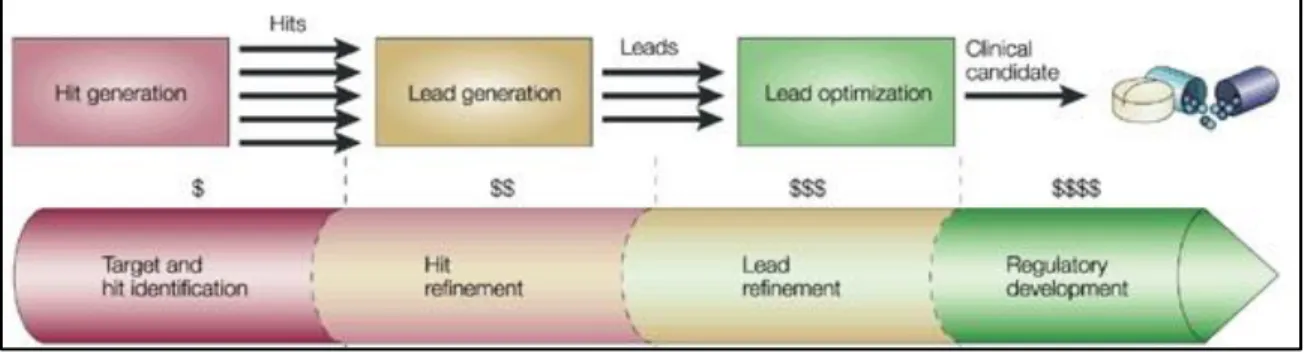
Figure 3.1. Stages in the drug discovery process ... 45
Figure 3.2. Compound attrition during hit generation stage of RecA inhibitor discovery program ... 46
Figure 3.3. Compounds potentiate ciprofloxacin bacterial killing ... 52
Figure 3.4. -galactosidase strains used in SOS activation assay ... 54
Figure 3.5. Compounds inhibitory activity in E. coli SOS gene reporter assay ... 55
Figure 3.6. Dose-dependent inhibition of SOS activation by RecA inhibitior UN10001584 ... 56
Figure 3.7. 5-Alkylidene-4thiazolidinone series as prospective compounds for follow-up ... 57
Figure 4.2. Pharmacologically active aminoquinolines ... 67 Figure 4.3. Ligands assayed for thermal denaturation shift ... 70
Figure 4.4. UV melting profiles of poly(dT)poly(dA) in the absence of ligand (A) and in the presence of BRITE-345133 at base pair to drug ratio (rbd) of 2.5 (B) at a
rate of 1 °C/min ... 71 Figure 4.5. UV melting temperatures of dsDNA in the presence of ligands ... 71 Figure 4.6. Schematic diagram of the competition dialysis experiment ... 73
Figure 4.7 Results obtained by the competition dialysis method for
styrylquinazoline and progenitor compounds ... 76 Figure 4.8. Proposed mechanism of nonspecific inhibition by compound
aggregates ... 79 Figure 4.9. Results obtained by static light scattering (SLS) for styrylquinazolines
and progenitor compounds ... 80 Figure 4.10. Transmission electron micrographs of styrylquinazolines ... 81
Figure 5.1. Cartoon of PicoGreen screening assay for discovery of EndA
inhibitors ... 94
Figure 5.2. Assay optimization with respect to (A) DNA substrate in the presence
of PicoGreen and (B) imidazole ... 94 Figure 5.3. Titration of EndA(H160G) in optimized assay buffer ... 95 Figure 5.4. Assay optimization with respect to time and enzyme dependence ... 95
Figure 5.5. Bar graph depicting initial velocities of EndA reaction in the presence
of various amounts of DMSO ... 96
Figure 5.6. Optimization of TritonX-100 and BSA in the PicoGreen nuclease
assay ... 96
Figure 5.7. EndA PicoGreen assay validation over multiple plates and multiple
Figure 5.10. Kinase Focus Set HTS results ... 103 Figure 5.11. Reproducible concentration-response measurements of a validated
compound from the Kinase Focus Set screen ... 104
Figure 5.12. Evaluation of reordered Kinase Focus Set hits by DNA binding
counter-screen assay ... 107 Figure 5.13. Biochemical characterization of a promising EndA inhibitor ... 108 Figure A.1. Ad hoc and substructure methods of virtual screening for EndA
List of Tables
Table 1.1. ... 8
Table 2.1. RecA HTS Assay Protocol ... 28
Table 2.2. Summary of Successful HTS for RecA Inhibitors ... 32
Table 4.1. Nucleic acid samples used in competition dialysis experiments ... 75
Table 4.2. Absorption maxima and extinction coefficients of styrylquinazoline and progenitor compounds ... 75
Table 4.3. CAC by static light scattering for styrylquinazolines and progenitor compounds ... 80
List of Abbreviation ADP Adenosine 5-diphosphate
ATP Adenosine 5-triphosphate
CAC Critical aggregation concentration
CICBDD Center for integrated chemical biology and drug discovery D buffer Detection buffer
DMSO Dimethyl sulfoxide DNA Deoxyribonucleic acid DSB Double-strand DNA break dsDNA Double-stranded DNA DLS Dynamic light scattering EDTA Ethylenediaminetetraacetic acid FP Fluorescence polarization
FQRP Fluoroquinolone-resistant Pseudomonas aeruginosa GSK GlaxoSmithKline
HAI Hospital acquired infections HBA H-bond acceptor
HBD H-bond donor
HEPES 4-(2-hydroxyethyl) piperazine-1-ethanesulfonic acid HTS High throughput screening
IV Intravenous
NET Neutrophil Extracellular Trap NPF Nucleoprotein filament
OH Hydroxy radical
PAINS Pan assay interference compounds PMBN Polymixin B nonapeptide
PSA Polar surface area R buffer Reaction buffer RDF RecA-DNA filament
SAR Structure-activity relationship SD Standard deviation
SDS Sodium dodecyl sulfate SLS Static light scattering
SOS programmed response in most bacteria to cope with the effects of DNA damage
ssDNA Single-stranded DNA
TEM Transmission electron microscopy
UV Ultraviolet
VRE Vancomycin-resistant Enterococci VS Virtual screening
Chapter 1 Introduction 1.1. Challenges of antibacterial discovery
The introduction of penicillin in the 1930s opened up the era of antibiotics1and is heralded as one of the greatest discoveries in the history of medicine. Without question, since its introduction in the early twentieth century, antibacterial chemotherapy has substantially reduced the burden of disease caused by bacterial infections. 2 However, the widespread emergence of antibiotic resistance in bacteria has occurred as a consequential response and severely compromises the therapeutic efficacy of the antibiotics once seen as huge successes. Today, antibiotic resistance is recognized as a major challenge for the health of the human population.3
evident that antibiotic resistance in pathogenic bacteria has enormous human and
economic consequences.
Infections with resistant bacteria were first reported over 60 years ago, 8 and since
the 1980s, the incidence of resistant bacteria has increased dramatically (Figure 1.1). As resistance increases, it limits the treatment options available to healthcare providers. This
requires the development of new antibacterial agents, but the discovery of such is not proving to be an easy task. The “golden era” when novel secondary metabolites were
easily identified is over and modern approaches to develop antibiotic drugs have had only
limited success. 9 Only 13 new oral or intravenous antibiotics have been approved since
1998 (Figure 1.1). 7 This low output of novel drugs has resulted in the downsizing or ending of many industrial research efforts. Taken together, the alarming rise in antibiotic
resistant bacterial strains and the diminishing supply of new antibiotics are disturbing
trends that are intersecting with dangerous consequences (Figure 1.1). The goal behind the work undertaken in this dissertation aimed to increase our knowledge of bacterial
recombination enzymes in the development of antibiotic-resistant bacterial infectious
diseases and potentially present RecA and EndA as novel therapeutic targets for
Figure 1.1. Dangerous antibiotic-related trends. Left panel: The % incidence increase in nosocomial infections involving methicillin-resistant Staphylococcus aureus (MRSA), vancomycin-resistant Enterococci (VRE) and fluoroquinolone-resistant Pseudomonas aeruginosa
(FQRP) in the past 30 years. Right Panel: The decreasing number of approved antibiotics in a similar time span.
1.2. A revised model for accelerated evolution of antibiotic resistance
Antibiotic resistant strains of bacteria emerge when genetic changes allow
individual bacteria to escape antibiotic killing, thereby surviving and propagating their
resistance to subsequent generations. Bacteria become resistant to antibiotics by
acquiring one of several common molecular strategies: (1) drug inactivation, (2)
modification of the drug target, and (3) decreasing the amount of drug reaching the target
(usually by modifying influx/efflux pump expression). 10 In all of these cases, a heritable change in the bacteria’s DNA is required. Such alterations to the bacterial genome occur
through local changes in the DNA sequence (de novo mutation), intrachromosomal
shuffling of DNA sequences (recombination), or the acquisition of exogenous DNA from
plasmids, transposons or pathogenicity islands or through natural transformation
mediated via competence).
Regardless of the mechanism, genetic change has conventionally been viewed as
a purely passive process (e.g., mutations occur randomly, Figure 1.2, top panel). However, recent studies demonstrate that bacteria actively promote their own genetic
diversification and, importantly, the rate of genetic change increases dramatically when
bacteria are exposed to antibiotics (Figure 1.2, bottom panel). 11-14 These accelerated genetic changes can lead to the evolution11 and spread15 of antibiotic resistance as well as
the production and dissemination of virulence factors. 16-19 In this way, antibiotics not
only select for resistant strains of bacteria by killing their sensitive neighbors, but also
accelerate the emergence of such resistant bacteria in the first place. Interestingly, a report nearly 50 years ago observed that antibiotics “catalyze” bacteria to take on “the
resistant character.” 20
There was little follow-up at the time, but today it has been clearly
shown that antibiotic-induced stress increases the frequency and efficacy of evolutionary
activities11,14,21 and that key bacterial recombination enzymes are important for the
Figure 1.2. The conventional and revised pathways leading to resistant bacteria. Top panel: the conventional model depicts antibiotics selecting for preexisting resistant variants (red cells). Bottom panel: in contrast, the revised model shows antibiotics accelerating the presence of resistant variants by inducing accelerated mutagenesis (resistant blue cells) and genetransfer (green cells that acquired green resistance genes).
1.3. The role of recombination enzymes in accelerated evolution of antibiotic resistance
As introduced above, bacterial genetic diversification is accelerated in the
presence of antibiotics. Changes in the bacterial genome occur through induced mutation,
intrachromosomal recombination, and the acquisition of foreign DNA. Moreover, these
genetic changes originate from bacterial processes that couple antibiotic stress with
accelerated bacterial evolution. These processes are programmed stress responses that allow bacteria to cope with the effects of DNA damage caused by antibacterial agents,
known as the SOS response and competence for transformation (a.k.a., SOS and
competence). While SOS and competence differ by bacterial species and other details,
they both are induced by antibiotics and lead to activities that promote genetic diversity.
26
In addition, both pathways are tightly regulated by the bacteria and depend on the
function of select recombination enzymes, such as RecA and EndA. The following three
distinct activities of (2) RecA and (3) EndA promote genetic diversity and resistance
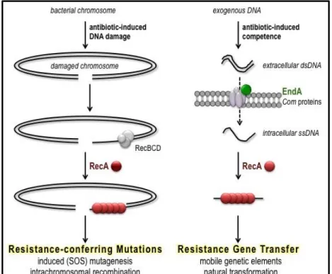
development in response to antibiotic-induced stress (Figure 1.3).
Figure 1.3. The molecular models for antibiotic accelerated evolution of resistance. Left panel: antibiotic-induced DNA damage activates RecA leading to induced (SOS) mutagenesis and increased intrachromosomal recombination. Right panel: antibiotic induction of competence for transformation via EndA-RecA leads to horizontal gene transfer.
1.3.1. Induction of the SOS response and competence for transformation by antibiotics
While many naturally occurring phenomena can cause bacterial stress and initiate the
SOS response and competence for transformation, it has been shown that antibiotic treatment
serves as a powerful instigator of SOS and competence (Table 1.1). 11,14,15,21,22,25,27-33 For example, ciprofloxacin is a fluoroquinolone-based inhibitor of bacterial type II DNA
topoisomerases, including gyrase and topoisomerase IV, which targets a protein-DNA
The protein complex, RecBCD processes DSB to ssDNA and stalled replication forks
inherently expose ssDNA. This ssDNA substrate, resulting from exposure to ciprofloxacin,
activates RecA, which in turn induces the SOS response. 27,35,36 Similarly, fluoroquinolone
antibiotics were also found to induce competence for transformation in Streptococcus pneumoniae and ciprofloxacin was reported to increase transformation frequency by threefold to fourfold in the human pathogen.14
The ability of antibiotic chemotherapy to stimulate SOS and competence extends
beyond ciprofloxacin and fluoroquinolones to other classes of antibiotics (Table 1.1).
Among these, β-lactams have been shown to activate SOS expression through a
two-component signaling pathway, 30 and aminoglycosides cause an accumulation of
misfolded proteins that induces competence in S. pneumoniae. 37 From this, it is apparent that antibiotic-induced stress leads to SOS and competence induction, but that the
mechanism(s) and signal(s) may differ by antibiotic class and by bacterial species. In 2007, a unified mechanism of antibiotic killing was proposed, suggesting that all bactericidal antibiotics, regardless of their primary target, cause metabolic stress that leads to the production of reactive oxygen species (ROS). 21,38 In turn, the highly reactive radicals oxidate biomolecules and lead to general cell death. Subsequent studies reported that antibiotic-dependent ROS production oxidizes the guanine nucleotide pool, leading to double-strand DNA breaks and death. 39 This common mechanism of antibiotic killing via ROS production became widely accepted, but was recently refuted by several groups.
40-42 The contradicting studies found that antibiotic treatment did not accelerate the
evidence that ROS do not play a role in killing bacteria by antibiotics and that it seems unlikely that all bactericidal antibiotics kill by the same mechanism. 40 Of course, this does not preclude that antibiotics trigger bacterial stress responses, but describes that activation of SOS and competence by antibiotics does not involve ROS and that the mechanism(s) involved remain elusive.
1.3.2. RecA and the SOS response
The bacterial SOS response is a graded response to DNA damage that initially
up-regulates DNA-excision repair and recombination repair processes, then eventually
promotes cell-cycle arrest and global mutagenesis if the DNA damage persists. The
LexA repressor is a dimeric protein that binds in the promoter region of SOS genes and
prevents access of RNA polymerase. 43 The active RecA nucleoprotein filament (NPF)
causes LexA to undergo autoproteolytic cleavage, a signaling-like function of RecA that
is dependent on the formation of an ATP·RecA·ssDNA nucleoprotein filament (NPF).
44,45
The RecA NPF is composed of hundreds to thousands of RecA monomers assembled on ssDNA. 46 The RecA NPF governs the SOS response, as its activation and ability to
interact with LexA requires RecA recognition and binding to ssDNA, a rather rare
substrate that only occurs when DNA damage has occurred or when DNA synthesis is
interrupted and stalled replication forks are observed.47
The cleavage of LexA sequentially de-represses up to 40 genes involved in DNA repair and mutagenic translesion DNA synthesis. 48-51 In the early and middle stages of the SOS response the bacterium overexpresses proteins, including RecA, involved in DNA repair and recombination. If this attempt at DNA repair is not successful, the late stage of the SOS response expresses low-fidelity polymerases that introduce mutations. 43 The mutagenic action of these polymerases creates resistance-conferring mutations.
11,13,52-54 As such, the RecA-mediated SOS response to repair DNA damage, arising in
the course of antibiotic exposure, facilitates the de novo development and transmission of antibiotic resistance genes.
was first identified as “Recombinase A”, the most widely studied and biologically
important recombinase enzyme in bacteria. 55 The activated RecA NPF facilitates
recombinational repair of damaged DNA. 56 In addition, RecA-mediated recombination
can incorporate foreign genes from exogenous homologous DNA. 22 This homologous
genetic recombination is a frequent source of nucleotide changes that enable bacterial
evolution including the acquisition of resistance.54,57-59
In summary, through undetermined mechanism(s), antibiotics induce bacterial
stress and damage DNA in ways that produce ssDNS and activate RecA. Once active,
RecA-mediated processes serve to repair DNA damage while also promoting genomic
diversification. Therefore, RecA facilitates accelerated bacterial evolution that occurs in
response to antibiotic treatment, including the evolution and transmission of antibiotic
resistance.
1.3.3. EndA and competence for transformation
The ability to respond to and repair DNA damage is an essential process required by all forms of cellular life. However, some important pathogens lack the classical SOS response as defined by increased mutation after DNA damage and regulation by RecA-LexA (activator-repressor). In place of an SOS response, bacteria such as Neisseria gonorrhoeae and Streptococcus pneumonia have evolved to be naturally competent for DNA transformation. During transformation, bacteria take up DNA from the environment and incorporate it into is genome.60 This process is up-regulated when the bacteria are under antibiotic-induced stress, and may be exploited for acquisition of resistance. 26
environmental stressors, including the presence of antibiotics. 14,61,62 Like SOS, the induction of competence results in the transcription of 105-124 genes that function as the regulatory and structural elements associated with transformation. 63 Intriguingly, in S. pneumoniae, RecA has been shown to be instrumental in activating competence for transformation when the bacteria are under antibiotic-induced stress. 14 Once competence is induced, gene products from the com regulon associate to form a multiprotein complex that carries out DNA uptake and transport. 64 Within this complex, the surface endonuclease EndA degrades the non-transforming strand of DNA (Figure 1.5). 65 The nuclease activity of EndA is necessary for DNA transport and thus required for efficient bacterial transformation in pneumococcus. It has been demonstrated that S. pneumoniae strains with deactivating endA mutations exhibit transformation efficiencies that are less than 1% of those of wild-type (wt) strains. 64,66-68 Through the activities of EndA, bacteria take-up exogenous DNA and eventually integrate it into the chromosome by RecA-mediated recombination. 14
Figure 1.4. The transport of DNA during competence for transformation. Along with other Com proteins, EndA is part of the uptake apparatus necessary for DNA transport. EndA is responsible for degrading the non-transforming strand of DNA.
1.4. Other roles of RecA and EndA in bacterial pathogenicity 1.4.1. RecA
In addition to their roles in accelerated bacterial evolution, RecA and EndA also play important roles in establishing infection and in virulence. As established above, the activities of RecA facilitate SOS mutagenesis and genetic recombination, but also are reportedly important for disease development following infections by various pathogens. E. coli, 69 Salmonella enterica, 70 Vibrio cholera, 71 Helicobacter pylori, 72 and S. aureus73 strains that are recA-null have demonstrated significantly attenuated virulence (102-106 fold). RecA-mediated recombination has also been shown to play a role in the induction of toxin biosynthesis, 74 virulence factor production73 and the genetic diversification of biofilms. 75 Therefore, in many diverse bacterial species the activity of RecA plays an important role in bacterial pathogenicity.
1.4.2. EndA
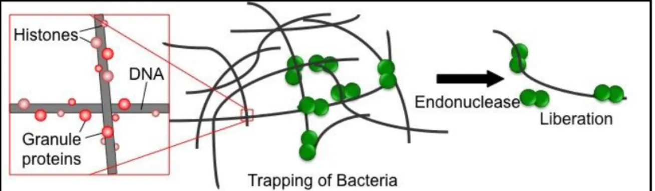
host continuously deploys an array of innate and acquired immune defenses to prevent pneumococci from traversing epithelial barriers. 76 Recently, neutrophils have been shown to produce neutrophil extracellular traps (NETs) that entrap and kill pathogens in the extracellular environment. 77 NETs consist of neutrophil DNA as a backbone with embedded histones and granule proteins. In 2006, Beiter and colleagues78 showed that EndA can degrade the DNA scaffolding of NETs, thereby facilitating pneumococcal escape (Figure 1.6). In mouse models of infection, endA-expressing S. pneumoniae strains were able to destroy NETs and disseminate through the mouse, while mutant strains deficient in endA remained in the upper respiratory. 78 These observations identify EndA as a virulence factor that counteracts host-mediated trapping by NETs and increases S. pneumoniae pathogenicity.
1.5. Intervening in accelerated evolution by small molecule inhibition of recombination enzymes
The information presented above establishes that genetic diversification in bacteria is accelerated in the presence of antibiotics. Bacterial genetic diversity originates from the SOS response and transformation via competence, processes that are regulated by the bacterium and depend on the function of RecA and EndA. With the revelation that accelerated bacterial evolution requires the activity of RecA and EndA, there is opportunity to intervene with properly designed inhibitors. The ability to selectively control the activities of RecA and EndA would permit the dissection of the stress response pathways (i.e., SOS and competence for transformation), and their contributions to accelerated bacterial evolution. Moreover, small molecule inhibitors of RecA and/or EndA would enable a greater understanding of the role of these enzymes in bacterial pathogenicity and the development and transmission of antibiotic resistance. Finally, by demonstrating that accelerated evolution in bacteria can be attenuated by inhibition of RecA and EndA, there is potential that these enzymes will emerge as intriguing targets for the discovery of novel drugs to combat antibiotic resistance.
(1) An inhibitor of RecA may serve as an adjuvant to traditional antibiotic chemotherapy by reducing the rate at which resistance genes emerge or are transferred. Bacteria deficient in recA have been shown to have a reduced or nonexistent capacity for developing drug resistance. RecA stimulates the induction of the SOS response,
promoting de novo mutagenesis and the creation of resistance. RecA also participates in
horizontal gene transfer, a process by which resistant genes are shared among bacteria of
the same and different species. Inhibition of one or both of these activities will delay or
completely prevent antibiotic resistance evolution and transmission.
(2) An inhibitor of RecA may serve as an adjuvant to bactericidal antibiotic chemotherapy by potentiating the toxicity of the primary antibiotic. Bactericidal antibiotic exposure induces the recA-controlled SOS response, thereby up-regulating DNA repair processes. It has been shown that RecA-null bacteria are susceptible to
much lower doses of a wide variety of antibiotics, compared to their wt counterparts21.
Thus, it seems reasonable that a small molecule inhibitor of RecA would synergistically
enhance the killing effect of currently therapeutically useful antibiotics. The use of lower
effective doses of antibiotics may extend the effective life of an antibacterial compound
or the rescue of shelved compounds.
flexibility and the easy capacity to acquire resistance, while the second activity affords the pneumococcus the ability to establish infection. Therefore, we propose that an inhibitor of EndA activities may lead to one or more of the following outcomes:
(1) An inhibitor of EndA may serve as an adjuvant to traditional antibiotic chemotherapy by reducing the rate at which resistance genes emerge or are transferred. Deletion of endA diminishes the efficiency of transformation in S. pneumoniae. Through its role in DNA uptake during competence for transformation, EndA contributes to accelerated genetic changes that can result in the transfer of antibiotic resistance genes. Inhibition of EndA will decrease or prevent antibiotic resistance development and spread.
(2) An inhibitor of EndA may serve as an adjuvant to antipneumococcal chemotherapy by attenuating infection. EndA facilitates pneumococcal escape of NETs by degrading the DNA scaffold. Mutant S. pneumoniae strains lacking EndA activity do not destroy NETs and show decreased virulence in mouse models of infection. Thus, it seems reasonable that a small molecule inhibitor of EndA would synergistically attenuate infection along with a current therapeutic. A combination strategy with an EndA inhibitor has the potential to extend the efficacy of approved drugs and provide better treatment against S. pneumoniae.
1.6. Our strategy to develop small molecule inhibitors of recombination enzymes Upon antibiotic exposure, genes conferring drug resistance are created by
increased genetic mutation and/or increased uptake and incorporation of resistance genes
into the genome. These processes effectively accelerate the rate at which bacterial
context, the bacterial RecA and EndA proteins have emerged as intriguing targets for the
small molecule suppression of antibiotic resistance. Moreover, inhibiting RecA and
EndA has the potential to potentiate current antibiotics and attenuate pneumococcal
infection, respectively. To identify inhibitors of RecA and EndA, we developed
high-throughput screening (HTS) programs that resulted in the discovery of several classes of
molecules that exert an inhibitory effect on these enzymes.
The search for inhibitors of any enzyme begins with assays that are capable of detecting the influence of a prospective small-molecule modulator on the activity of that enzyme. For our HTS programs, we developed novel biochemical assays capable of detecting the inhibition of either RecA or EndA. The activities of RecA require the formation of the RecA NPF that hydrolyzes ATP. Thus, monitoring ATPase activity can be used as a diagnostic for small molecule inhibition of RecA by in vitro screening. We wanted to improved upon previously reported RecA ATPase assays that had sensitivity limitations and required high concentrations of enzyme and ATP substrate. 80-83 We accomplished this by adapting the Transcreener ADP2 fluorescence polarization (FP) assay from BellBrook Laboratories to detect RecA ATP hydrolysis. The development and optimization of the RecA Transcreener assay for HTS along with initial screening results are described in Chapter 1. The work presented in this chapter has been peer-reviewed and published in ASSAY and Drug Development Technologies (2012 Jun;10(3):260-8).
HTS program. This stage produces large amounts of data and a thorough analysis of this data is discussed in Chapter 3.
The hit-finding process identified eight series of compounds with potential for
further development as RecA inhibitors. Chapter 4 describes the structural progression of
one of these series, the styrylquinazolines, and our work towards defining the mode of
action of styrylquinazolines and select progenitors. This chapter examines mechanistic
and physiochemical properties that are important to the identification of RecA inhibitors
with desired biological activity.
Finally, we developed a distinct but similar HTS program for the identification of EndA inhibitors. EndA-mediated digestion of double stranded DNA (dsDNA) was assessed in biochemical screening by fluorescence changes in the DNA dye ligand, PicoGreen. The novel EndA PicoGreen assay was employed to screen thousands of small molecules from both random and directed libraries and measure their ability to inhibit EndA nuclease activity. The development of the EndA PicoGreen assay, preliminary screening results and evaluation of active compounds are detailed in Chapter 5. We have published the EndA PicoGreen HTS results in Journal of Biomolecular Screening 2013 Mar;18(3):247-57 and the virtual screening of EndA, included in Appendix I, will be submitted for publication to the Journal of Medicinal Chemistry.
The long-range goal of this project is to discover novel small molecules that will
prevent the de novo development and transmission of antibiotic resistance genes. The
projects outlined in this dissertation provide methods for detecting the inhibition of RecA
and EndA and have resulted in the discovery of several classes of molecules that exert an
accelerated evolution that leads to antibiotic resistance is an intriguing and fresh approach
to antibacterial discovery. The development of RecA and EndA inhibitors to combat
resistant bacterial infections complements traditional antibiotic discovery programs and has
Chapter 2
High-Throughput Screening for RecA Inhibitors Using a Transcreener Adenosine 5'-O-Diphosphate Assay1
2.1. Introduction
New antibacterial strategies will be required to overcome the looming public
health threat posed bythe combination of an increasing prevalence of antibiotic-resistant
bacterial pathogens with a dwindling pipeline of new antibiotics. 7,84 Significant scientific
and environmental challenges remain in the discovery and development of novel
mechanism antibiotics. 85 One alternative to conventional antibiotic discovery would be
the development of adjuvants to enhance the outcomes of antibacterial therapy. Recent
studies demonstrate that bacterial strains with inactive RecA enzyme are more
susceptible than wild-type strains to killing by antibacterial agents. 21,30,54,86 Moreover,
loss of RecA function also attenuates the rates of induced mutagenesis and
intrachromosomal recombination upon antibiotic exposure, thereby slowing the
development of antibiotic resistance. 54,86,87 RecA inactivation also diminishes the
efficiency of horizontal gene transfer, hindering the acquisition and dissemination of
antibiotic resistance genes. 15,88,89 Given this evidence, we hypothesized that
small-molecule RecA inhibitors could sensitize bacteria to conventional antibiotics and
attenuate the frequency with which resistance genes develop and are transmitted. 23 The
1 The text of this chapter is taken from a published manuscript that was coauthored with
discovery of potent and selective RecA inhibitors that modulate the target in living
bacteria would be an important step in establishing RecA as a druggable target in the
management of bacterial infectious diseases.
RecA’s importance in the bacterial survival of and response to antibacterial
exposure arise from its cardinal roles in mediating the SOS response and facilitating
DNA strand exchange. All RecA activities require the formation of a helical
homopolymeric filament comprising multiple ATP-bound RecA monomers coating
single-stranded DNA (ssDNA). 46 Once formed, this RecA-DNA filament (RDF)
hydrolyzes ATP. Thus, monitoring ATPase activity can be used as a diagnostic for small
molecule inhibition of RecA by in vitro screening. 80-83 These previously reported assay
technologies had sensitivity limitations, requiring high concentrations of enzyme ([RecA]
≥ 0.5 M) and substrate ([ATP] ≥ 0.75 mM). Under such “forcing” conditions, RecA
exists almost exclusively in an active, DNA-bound conformational state and inhibitors
selective for this conformation (Figure 2.1A) were primarily identified.
The development of a more sensitive screening assay with the ability to detect
low ATP turnover under reduced RecA and ATP concentrations was desired. Under such “non-forcing” conditions, RecA could sample both its inactive and active conformational
states, facilitating the identification of additional small molecule inhibitors without bias
with respect to RecA conformational preference (Figure 2.1B). Moreover, a reduced enzyme concentration would lower the stoichiometric limit on measured IC50 values and
reduced ATP levels would allow the identification of ATP competitive inhibitors.
Assay optimization under these desired conditions represented a challenge
activation is modest and its turnover number for ATP hydrolysis is low (kcat 0.5 s-1).
The robust assessment of RecA’s ATPase activity under such conditions required a
sensitive detection methodology.
The Transcreener ADP2 FP assay from BellBrook Laboratories (Madison, WI)
is a screening assay kit that allows ADP detection using an ADP-antibody/tracer system
with fluorescence polarization (FP) readout. Briefly, the Transcreener ADP2 FP assay
utilizes an antibody that selectively binds ADP to quantify the ADP produced from an
ATPase reaction. ADP conjugated to AlexaFluor-633 dye is used as a tracer and the
amount of tracer that is displaced from the antibody is proportional to the amount of ADP
generated in the reaction. This highly sensitive, homogeneous assay technology has been
used to study protein kinases,90lipid kinases,91and heat shock proteins, 92 Moreover, the
assays have proven to be suitable for high-throughput screening (HTS). 93 In the present
study, we report the adaptation of the Transcreener ADP2 FP assay to detect the ATP
hydrolysis activity of purified E. coli RecA protein. We demonstrate the ability of the assay technology to be optimized for lower enzyme and ATP concentrations and its use
in HTS of a diverse collection of drug-like small molecules, leading to the identification
Figure 2.1. Cartoons depicting the inactive RecA monomers and active RecA-DNA filament (RDF).(A) Assay conditions, including high concentrations of RecA, DNA, and ATP, shift the filament assembly and activation equilibrium such that RecA exists almost exclusively in an active, DNAbound conformational state. Inhibitors identified under such forcing conditions will be strongly biased toward binders of this conformational state. (B) The desired assay conditions include 10-fold lower concentrations of RecA and ATP, and RecA samples both its inactive and active conformational states during the assay. Such nonforcing conditions would facilitate the identification of additional inhibitors without bias with respect to RecA conformational preference as well as ATP competitive inhibitors.
2.2. Assay development and optimization
Previous reaction conditions used for measuring RecA’s ATPase activity required
relatively high concentrations of RecA and ATP. To lower enzyme and substrate
concentrations, we optimized the conditions of the Transcreener ADP2 FP assay to our
desired levels of enzyme and ATP. We also assessed the robustness, reproducibility, and
HTS compatibility of the Transcreener assay to ensure its success in screening collections
An ADP/ATP standard curve was produced (Figure 2.2A), as described in the Transcreener ADP2 FP assay technical manual. 94 The experiment simulates the
production of ADP during the course of a RecA-catalyzed ATP hydrolysis reaction and
the expected decrease in FP signal (mP) was observed. The ATP concentration required
for half-maximal steady-state velocity, or S0.5 for cooperative enzymes such as RecA, 95
depends on the reaction conditions and published values range from 2.5 to 200 M. 23,96
In the presence of poly(dT) as the activating DNA, RecA’s ATPase activity was
measured with initial ATP concentrations from 25 to 100 M. It was found that 75 M
was the lowest concentration that resulted in linear (steady-state) kinetics for nearly 40
min with an acceptable signal-to-background ratio. Subsequently, under the optimized
ATPase screening conditions described below, the apparent S0.5 was measured to be 88
μM (data not shown). Because a substrate concentration equal to or slightly less than the
KM (S0.5) value in HTS campaigns allows the identification of all types of inhibitors,
including those that are competitive with substrate,97 we selected 75 M as the ATP
concentration and optimized the other assay parameters.
In the presence of 75 M ATP and a saturating concentration of poly(dT) (5 M-nts), RecA’s ATPase activity increased monotonically with initial RecA concentration
(data not shown). A RecA concentration of 30 nM was selected for further experiments
because this was the lowest RecA concentration to produce approximately linear enzyme
activity over a reasonable time range and sufficient conversion to achieve an adequate
signal window. Optimized conditions included 30 nM RecA, 75 M ATP, and 5 M-nts
poly(dT). Employing these conditions, the ADP concentration produced at 37 C was
Transcreener assay demonstrated linear ADP-production kinetics to 40 min and we
elected to use 30 min as the reaction time to remain comfortably within the assay’s linear
dynamic range.
It is common for small-molecule library samples to be dissolved in DMSO in
preparation for screening. Therefore, the effect of DMSO on RecA activity was
investigated at several different DMSO concentrations, up to 10% (vol/vol) (Figure 2.2C). There was no significant reduction in RecA ATPase activity by DMSO up to the highest concentration tested and the final DMSO concentration used for screening was
0.5-1% (vol/vol).
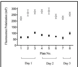
To assess the reproducibility of the Transcreener assay, a number of test 384-well
plates were evaluated for intercolumn, interplate, and interday variability. Over a span of
three days, eight replicate assay plates were run under the optimized conditions described
above and used to compile the mean control signals (Figure 2.3) for Z factor calculation98. The maximum activity controls (128 per plate, even columns of a 384-well
plate) consisted of wells containing RecA, DNA, ATP and 1% DMSO in buffer. The
background or inhibited controls (128 per plate, odd columns of a 384-well plate)
consisted of wells containing RecA, DNA, ATP and 50 mM EDTA in buffer. The FP
signal was read at 30 min and the overall quality of the Transcreener assay was assessed
Figure 2.2. Assay optimization with respect to (A) ATP/ADP signal, (B) time and enzyme dependence, and (C) DMSO tolerance determined in 384-well format. (A) ATP/ADP titration, with a constant adenosine concentration of 75 µM. (B) Reaction time course in the () presence or () absence of 30 nM RecA. (C) FP (mP) signal after 30-min RecA ATPase reaction in the presence of various amounts of DMSO. The data presented are mean standard deviation (SD) of triplicate wells (n = 3).
Figure 2.3. Assay validation over multiple plates and multiple days. Each data point represents the mean FP (mP) signal of 128 wells from 3 different experiments on 3 different days (2 or 3 plates per experiment); the error bars indicate the SD of each mean value. The () represent the mean values for the background or inhibited controls (50 mM EDTA final) and the () represent the mean values for the maximum activity controls (1% DMSO final).
2.3. Screen design and pilot study of LOPAC library
The LOPAC comprises 1,280 biologically active compounds and was assessed in
triplicate for RecA inhibition at a final compound concentration of 15 M. The LOPAC
pilot screen was characterized by an average Z factor of 0.76, and 19 compounds were identified with average relative inhibition activities 75% inhibition as an average of
three replicates (Figure 2.4A). This active threshold of 75% inhibition represented 6 standard deviations (SD) above the mean and provided a hit rate of 1.5%. As shown in
Figure 2.4B, the pilot screen also demonstrated strong correlation between replicates. Analysis of the three replicates revealed five compounds as false positives and an
estimated false positive rate of 0.4%.
The LOPAC was screened previously against RecA using a different ATPase
assay16 under different conditions and 9 of the 19 active compounds were identified as
RecA inhibitors by both assays. The high correspondence of the active compounds
confirmed the Transcreener assay as a reliable method for identifying RecA inhibitors
from large compound libraries. Importantly, however, the fact that previously
unidentified RecA inhibitors were identified using the Transcreener assay substantiated
our assay design and demonstrated the ability to explore new chemical space for RecA
Table 2.1. RecA HTS Assay Protocol
Step Parameter Value Description
1 Library compound 1 L 10x in R Buffer containing 10% (v/v) DMSO; 15 µM final
2 Controls 1 L 10x; 1% DMSO and 50 mM EDTA final 3 RecA 4.5 L 2x in R Buffer; 30 nM final
4 Pre-incubation 20 min 37 C
5 ATP & poly(dT) 4.5 L 2x in R buffer; 75 M ATP & 5 M poly(dT) final
6 Incubation 30 min 37 C
7 Antibody & tracer 10 L 2x in D Buffer; 37.5 g/mL Ab & 2 nM tracer final 8 Assay readout after 30 min EnVision Multilabel Reader using far red FP filter set Step Notes
1 Multimek transfer to 384-well plates (tip wash 5x with 20 L of dH20)
2 10% aq DMSO added to columns 1-2; 500 mM EDTA added to columns 23-24 3 8-tip Multidrop dispense reagent to all wells
4 plates uncovered in non-shaking incubator 5 8-tip Multidrop dispense reagent to all wells 6 plates uncovered in non-shaking incubator 7 8-tip Multidrop dispense reagent to all wells
8 plates kept uncovered at 25 C in dark for 30 min until read on multilabel reader using the PerkinElmer Cy5 FP dual emission label (620/40 nm excitation filter, 688/45 nm emission filter and D658/fp688 dual mirror)
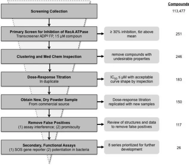
2.4. Screening 113,477 compounds for RecA inhibition
Although the pilot screen provided a number of novel inhibitors of RecA, our
main objective in screening the LOPAC was to validate the Transcreener assay as robust
and reproducible for the evaluation of additional libraries containing compounds with
greater potential for development as small-molecule therapeutics. Towards this end, we
screened 113,477 compounds from collections at The Center for Integrative Chemical
Biology and Drug Discovery (CICBDD) at the University of North Carolina at Chapel
Hill. Compounds from the 100k Diversity Collection and the Kinase Focus Set were
screened against RecA in singleton at 15 M from 1 mM DMSO stocks using the
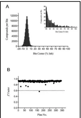
Transcreener assay. The Diversity Collection screening results demonstrated a normal
distribution centered on a mean of 0% inhibition with a SD of 5% (Figure 2.5A). Similarly, the Kinase Focus Set screen gave a mean of -0.9% and an SD of 7% (data not
shown). Overall, the average Z factor was 0.92 for the 359 analyzed plates (Figure 2.5B). A single plate fell below the 0.6 minimum Z factor accepted value and was repeated prior to inclusion in data analysis.
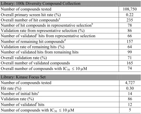
As summarized in Table 2.2, the 100k Diversity Collection primary HTS resulted in a total of 235 compounds with greater than 30% inhibition. The activity threshold was
set at 30% inhibition and defined as 6 SD above the mean. The 30% inhibition cut-off
was stringent by screening standards and yielded a sufficient number of hits for follow-up
(0.22% hit rate). From the 235 initial hit compounds, 78 compounds were selected based
on their structural representativity99 for IC50 determination as described in Materials and
Methods. The remaining 157 (of 235) hits were labeled as the “remaining hits” and given
We performed a concentration-response study of all the available HTS initial hits
using the Transcreener assay. From the representative selection, 77 compounds were
available in powder stock for evaluation. The concentration-response measurements
were performed in triplicate and 66 compounds were characterized by IC50 ≤ 35 M
(Table 2.2). The validation rate of the representative selection set was 86%, and nearly half of the inhibitors were characterized by IC50 ≤ 10 M.
Of the remaining hits, 154 compounds were obtained in powder form and were
similarly evaluated by IC50 measurement in triplicate. The concentration-response curves
allowed 84 compounds to be characterized by IC50 ≤ 35 M (Table 2.2). The validation
rate of the remaining hits (55%) was lower than that of the representative selection set,
but still confirmed over half as validated biochemical RecA inhibitors. The high
validation rate of the representative selection set was a result of our structural grouping
analysis of the HTS results to prioritize the more potent inhibitors.
The Kinase Focus Set was collected by the CICBDD and comprised 4,727
kinase-directed drug-like small molecules not found in the 100k Diversity Collection. Given
that both kinases and RecA are ATP-dependent enzymes, a compound library with small
molecules directed at kinases was screened with hopes it might present compounds active
against RecA ATPase activity. The screen yielded 14 compounds with 43% inhibition
(6 SD above the mean). The 0.3% hit rate was similar to the Diversity Collection
screen. The IC50 determination of the 14 initial hits was performed in duplicate from
powder stocks and validated 12 compounds as having IC50 ≤ 35 M (Table 2.2).
tested. A reproducible IC50 of 5 1 M confirms the robustness and reproducibility of
the assay.
Table 2.2. Summary of Successful HTS for RecA Inhibitors
aAll compounds with 30% inhibition (6xSD above the mean)
bCompounds with 30% inhibition and selected based on their scaffold group. See text for selection details.
cCompounds with IC
50 35 M with visual inspection of curve shape and max % inhibition 60%
dRemaining compounds with 30% inhibition
eCompounds with 43% inhibition (6xSD above the mean) fCompounds with IC
50 35 M
Library: 100k Diversity Compound Collection
Number of compounds tested 108,750
Overall primary screen hit rate (%) 0.22
Overall number of hit compoundsa 235
Number of hit compounds in representative selectionb 78 Validation rate from representative selection (%) 86 Number of validatedc hits from representative selection 66
Number of remaining hit compoundsd 157
Validation rate of remaining hits (%) 64
Number of validated hits from remaining hits 99
Overall validation rate (%) 71
Overall number of validated compounds 165
Overall number of compounds with IC50 10 M 74
Library: Kinase Focus Set
Number of compounds tested 4,727
Hit rate (%) 0.30
Number of initial hitse 14
Validation rate (%) 86
Number of validatedf hits 12
Figure 2.6. Reproducible concentration–response measurements for validated inhibitor UNC10036220 from the 100k Diversity Collection screen. The curve representing the mean IC50 value of 5 1 µM was consistent with data from three independent
runs (circle, square and triangle symbols, respectively).
2.5. Follow-up evaluation of select inhibitors
Further evaluation revealed that many of the inhibitors were active in biological
assays with live E. coli and were characterized by biochemical mechanisms of inhibition that differed from those of known RecA inhibitors. Of the 165 validated hits from the
100k Diversity Collection, 35 were active in one or more bacteriological assays for SOS
activation or fluoroquinolone antibacterial potentiation. We then selected 15 inhibitors
that were active in bioassays with live bacteria, were characterized by IC50 values < 10
M in the Transcreener assay, or both. For the 15 selected inhibitors, ATPase rates were
measured as a function of substrate ATP concentration at different inhibitor
concentrations. Analysis of resulting velocity-ATP concentration curves unexpectedly
revealed that none of the compounds exhibited competition with ATP; however, 13
compounds were noncompetitive inhibitors and 2 were mixed type inhibitors with > 1.
100
Similar characterization of RecA inhibitors identified by previous assays80-83 revealed
with restricted specificity for the active conformational state of the RecA filament.
Although we expected some of the tested compounds from the Transcreener assay to
competitively bind the ATP site, the discovery of inhibitors that target both free RecA
enzyme and the substrate-bound conformation of RecA confirmed the successful
identification of RecA inhibitors with varied specificity for RecA conformation. A full
accounting of these results will be reported elsewhere.
2.6. Conclusions
RecA facilitates important biological processes that allow bacteria to survive and
respond to antibacterial exposure. Because RecA is ubiquitous101 and highly
conserved102 among bacteria, but has only distant human homologs,46,103-107potent and
selective RecA inhibitors may serve as novel chemotherapeutic adjuvants to enhance
conventional antibiotics. Although we have previously developed biochemical assay
technologies that allowed target-based screening to identify RecA inhibitors, the prior
assay conditions restricted both the available conformational state of RecA and the
inhibitor mechanistic types that could be explored. A major conclusion of this work is
that the Transcreener ADP2 FP assay method could be adapted for a robust and
reproducible HTS (Z′ = 0.92) of 113,477 small molecules for inhibition of RecA ATPase activity. The sensitivity of the Transcreener ADP2 FP technology allowed the enzyme
and substrate concentrations to be reduced by more than an order of magnitude. From the
compounds identified as active in the primary HTS assay, 246 compounds were
evaluated in concentration-response format and 79 inhibitors characterized by IC50 ≤ 10
mechanistic types and selectivities for RecA conformational states were identified. These
novel RecA inhibitors represent a variety of chemotypes and 33 unique scaffold groups
(group size > 1) that will serve as synthetically tractable hit series for medicinal
chemistry efforts aimed at optimizing biochemical and bacteriological inhibition of RecA
activities.
In part, the continued search for new RecA inhibitors is motivated by the
increasingly urgent public health threat posed by antibiotic-resistant pathogens. It is now
clear that the use and misuse of antibiotics has played major roles in the selection and
spread of resistant pathogens. The exposure of bacteria to antibacterial compounds results
in the selection of resistant variants that ultimately dominate the population. Importantly,
however, recent studies suggest that bacteria are not merely passively subjected to natural
selection but can actively promote genetic diversification. The origins of genetic variation
include local changes in the DNA sequence (mutation), intrachromosomal shuffling of
DNA sequences (recombination), and the acquisition of DNA sequences from other
organisms (horizontal gene transfer). Induced mutagenesis in response to antibiotic
exposure dramatically accelerates bacterial mutation. 11,52-54 Likewise, recombination
plays a major role in bacterial evolution,12,54,58,59and may be a more frequent source of
nucleotide changes in E. coli than de novo mutation. 57 Finally, horizontal gene transfer is an important source of genetic diversity in bacteria.15,22,108,109
The genetic diversification processes described above are regulated by the
bacterium and depend on the function of select proteins and enzymes, including RecA.
Indeed, RecA has multiple functions that contribute to induced mutation, intragenomic
stimulating autocleavage of the LexA repressor, activation of error-prone DNA
polymerase V by stimulating autocleavage of UmuD, and facilitation of homologous
genetic recombination. The biochemical dependency of these phenomena on RecA
activities suggests that potent, selective RecA inhibitors could be developed to attenuate
the development, acquisition and dissemination of resistance, affording an opportunity to
inhibit evolutionary processes and enhance antibacterial chemotherapy. Current experiments are underway to evaluate the inhibitors of RecA’s DNA-dependent ATPase
activity in bacteriological assays.
2.7. Materials and Methods 2.7.1 Materials and equipments
The Transcreener ADP2 FP assay kit (cat. No. 3010-10K) was purchased from
BellBrook Labs. Poly(dT) single-stranded DNA was purchased from The Midland
Certified Reagent Company (Midland, TX). RecA was purified and stored as previously
described.46 Unless otherwise stated, all other reagents used for buffers and assays were
purchased from Fisher Scientific International (Ipswich, MA).
2.7.2 LOPAC compounds collection
The Library of Pharmacologically Active Compounds (LOPAC) was purchased from Sigma-Aldrich as 10-mM stocks in DMSO. The library was previously prepared as
1-L samples in 384-well V-bottom polypropylene microplates (Greiner, Monroe, NC),
sealed by a ALPS 3000 microplate heat sealer (Thermo Fisher Scientific, Hudson, NH)
Mg (OAc)2, 1 mM DTT, 5% glycerol, and 0.01% TritonX-100) over 2 steps using a
Thermo Scientific Multidrop Combi Reagent Dispenser (Waltham, MA) and Multimek NSX-1536 assay workstation system fitted with a 384-well head (Nanoscreen,
Charleston, SC). Finally, 1 L of this stock was spotted into the wells of a 384-well black PerkinElmer Proxiplate (Waltham, MA) for assay use, as described below.
2.7.3. 100k Diversity screening compound collection
The 100k Diversity Collection of screening compounds was developed by
structural chemists from St. Jude and the CICBDD. Compounds were selected based on
structural diversity at the Murcko scaffold level. 99 Essentially, a compound’s Murcko
scaffold includes contiguous ring systems plus chains that link two or more rings. For
Murcko scaffolds with more than 20 compounds, 20 compounds were randomly selected
for that scaffold in order to maximize the diversity of scaffolds in the Diversity
Collection. Compounds were also filtered to eliminate reactive function groups (REOS
score > -2)110and include compounds that obey the “rule of five” 111 with slight deviations
in order to permit slightly larger and more lipophilic compounds. Based on the above
selection process, a set of 100k compounds was chosen and purchased from Enamine Ltd
(Kiev, Ukraine) in powder stock. At random, around 5% of the collection was examined
by LC/MS for identity and purity confirmation. The compound collection plates were
prepared by resuspending the powder stock to 1 mM in DMSO in barcoded glass vials
with sonication using a Covaris S2 (Covaris, Woburn, MA). Compounds were plated at
1 mM in 100% DMSO in 384-well V-bottom polypropylene microplates using a Tecan
Genesis 200 (Münnedorf, Switzerland). A Multimek spotted 1 L of the 1 mM