DNA Product Formation in Female Sprague
−
Dawley Rats Following
Polyhalogenated Aromatic Hydrocarbon (PHAH) Exposure
Lina Gao,
†,¶Esra Mutlu,
§,¶Leonard B. Collins,
†Nigel J. Walker,
§Hadley J. Hartwell,
†James R. Olson,
⊥Wei Sun,
‡Avram Gold,
†Louise M. Ball,
†and James A. Swenberg
*
,††Department of Environmental Sciences and Engineering and ‡Department of Biostatistics, University of North Carolina at Chapel
Hill, Chapel Hill, North Carolina 27599, United States
§National Toxicology Program, National Institute of Environmental Health Sciences, NIH, RTP, Durham, North Carolina 27709,
United States
⊥Department of Pharmacology and Toxicology, State University of New York at Buffalo, Buffalo, New York 14214, United States
ABSTRACT: DNA oxidation damage has been regarded as one of the possible mechanisms for the hepatic carcinogenesis of dioxin-like compounds (DLCs). In this study, we evaluated the toxic equivalency factor (TEF) from the standpoint of induced DNA oxidation products and their relationship to toxicity and carcinogenicity. Nine DNA oxidation products were analyzed in
the liver of female Sprague−Dawley rats exposed to
2,3,7,8-tetrachlorodibenzo-pdioxin (TCDD) alone or the tertiary
mixture of TCDD, 3,3′,4,4′,5-pentachlorobiphenyl (PCB 126),
and 2,3,4,7,8-pentachlorodibenzofuran (PeCDF) by gavage for
14, 31, and 53 weeks (5 days/week) by LC−MS/MS:
8-oxo-7,8-dihydro-2′-deoxyguanosine (8-oxo-dGuo); 1,N6-etheno-2′-deoxyadenosine (1,N6-εdAdo);N2,3-ethenoguanine (N2,3-εG);
7-(2-oxoethly)guanine (7-OEG); 1,N2-etheno-2′-deoxyguanosine (1,N2-εdGuo); malondialdehyde (M
1dGuo); acrolein (AcrdGuo);
crotonaldehyde (CrdGuo); and 4-hydroxynonenal (HNEdGuo) derived 2′-deoxyguanosine adducts. Exposure to TCDD (100
ng/kg/day) significantly induced 1,N6-εdAdo at 31 and 53 weeks, while no increase of 8-oxo-dGuo was observed. Significant
increases were observed for 8-oxo-dGuo and 1,N6-εdAdo at all time points following exposure to the tertiary mixture (TEQ 100 ng/kg/day). Exposure to TCDD for 53 weeks only significantly increased 1,N6-εdAdo, while increases ofN2,3-εG and 7-OEG
were only found in the highest dose group (100 ng/kg/day). Exposure to the tertiary mixture for 53 weeks had no effect onN2
,3-εG in any exposure group (TEQ 0, 22, 46, or 100 ng/kg/day), while significant increases were observed for 1,N6-εdAdo (all dose
groups), 8-oxo-dGuo (46 and 100 ng/kg/day), and 7-OEG (100 ng/kg/day). While no significant increase was observed at 53
weeks for 1,N2-εdGuo, M
1dGuo, AcrdGuo, or CrdGuo following exposure to TCDD (100 ng/kg/day), all of them were
significantly induced in animals exposed to the tertiary mixture (TEQ 100 ng/kg/day). This oxidation DNA product data suggest
that the simple TEF methodology cannot be applied to evaluate the diverse patterns of toxic effects induced by DLCs.
■
INTRODUCTIONOxidative stress, a common state in pathophysiology, occurs when the number of reactive oxygen species (ROS) being
formed is exceeded by those being detoxified. Many
endogenous processes, as well as exogenous chemicals or their metabolites, are known to produce ROS. Besides ROS, reactive nitrogen species (RNS) are also generated by
macrophages and neutrophils involved in chronic inflammation,
which has been recognized as a risk factor in many human cancers.1These species or their active metabolites can interact with cellular constituents, especially lipids or nucleic acids, and
further induce various DNA oxidation products.2These DNA
lesions have been implicated in aging, neurodegeneration, and a myriad of diseases including cancer.2To date, there are several key ROS/RNS-induced DNA oxidation products: 8-oxo-7,8-dihydro-2′-deoxyguanosine (8-oxo-dGuo); 1,N6-etheno-2′ -de-oxyadenosine (1,N6-εdAdo); 3,N4-etheno-2′-deoxycytidine
(3,N4-εdC); N2,3-ethenoguanine (N2,3-εG); 1,N2-etheno-2′
-deoxyguanosine (1,N2-εdGuo); and malondialdehyde (MDA),
acrolein, crotonaldehyde, and 4-hydroxynonenal (HNE)
derived 2′-deoxyguanosine (dGuo) adducts, designated as
M1dGuo, AcrdGuo, CrdGuo, and HNEdGuo, respectively.3−7
Among them, the most studied adduct, 8-oxo-dGuo, is formed in relatively high amounts in vivo with steady-state levels
usually around 1/106 guanine. It is formed directly by the
reaction between dGuo and ROS or carbonate anion radical
induced by RNS.1,8ROS and RNS metabolites can also abstract
hydrogen atoms from polyunsaturated fatty acids producing lipid peroxides and many reactive byproducts such as MDA, HNE, crotonaldehyde, and acrolein. These compounds can further damage DNA and generate multiple oxidation DNA
products, which include exocyclic products with either a fi
ve-member (etheno products) or a six-ve-member (propano
Received: October 5, 2016
Published: February 16, 2017
Article
products) ring attached to DNA bases, as depicted inFigure
1.1,3−7 Recently, 7-(2-oxoethly)guanine (7-OEG) has been
identified as a new DNA product formed by lipid peroxidation
(LPO) with steady-state levels around 1−10 adduct/106
guanine.9 In addition to these distinct formation pathways,
site-directed mutagenicity studies found that most of these
DNA products can induce specific transition or transversion
point mutations in bacteria or mammalian cells.8,10−127-OEG can induce apurinic/apyrimidinic sites (AP sites) in biological
systems, although it has no miscoding properties.9Considering
the diverse metabolic pathways and the mutation spectrum induced by those important ROS/RNS-induced DNA prod-ucts, evaluating their profile data could compensate for biased results induced by a single product. Growing evidence also
indicates that these DNA lesions are significantly induced in
patients and animals with various chronic inflammatory diseases including certain cancers.3,7,8,13,14 Although DNA oxidation product formation is generally regarded as a key event for the carcinogenesis of genotoxic chemicals, it may also be a
significant contributor for the tumorigenesis of nongenotoxic
chemicals, especially chemicals capable of enhancing the formation of endogenous active metabolites, RNS and ROS, s u c h a s p o l y h a l o g e n a t ed a r o m a t i c h y d r o c a r b o n s (PHAHs).15−20
PHAHs comprise a large class of compounds such as polychlorinated dibenzodioxins (PCDDs), polychlorinated dibenzofurans (PCDFs), and polychlorinated biphenyls
(PCBs).21−24While PHAHs are regarded as one of the most
prevalent groups of pollutants in the environment due to industrial use, PCDDs and PCDFs are also produced as
byproducts during anthropogenic activities such as chlorine
bleaching of paper and combustion of wastes and fuels.21−23
PCBs were commercially produced and widely used for various industrial purposes including heat transfer agents, dielectric insulating fluids for capacitors and transformers, plasticizers,
and paint additives.24 Because of PHAHs’ resistance to
degradation and persistence in environment, their ability to bioaccumulate in humans and wildlife animals may result in chronic lifetime exposure, possible toxicity, and carcinogenic-ity.21−24Depending on the location and type of halogenations, some PHAHs induce a similar spectrum of biochemical and toxic responses in experimental animals. These responses are considered to be mediated through a common mechanism of action initiated by binding to a cytosolic receptor known as the aryl hydrocarbon receptor (AhR) and triggering the expression of a variety of genes, the so-called AhR gene battery.21
2,3,7,8-Tetrachlorodibenzo-p-dioxin (TCDD), commonly referred to as “dioxin”, is the prototype of these structurally related compounds and exhibits the highest potency of binding to the AhR. Hence, these structurally related compounds are commonly referred to as dioxin-like compounds (DLCs). Because of similarities in toxicity, the concept of the toxic equivalency factor (TEF) has been applied for the risk
assessment and regulatory control for DLCs.25Therefore, the
toxicity of PHAH mixtures is expressed in terms of its total toxic equivalent quotient (TEQ), which is the amount of
TCDD that would produce the equal toxic effect of all
contributing congeners within the mixture. This allows for the estimation of the potential dioxin-like activity of PHAH mixtures in the environment.
The association of oxidative stress and PHAHs, especially
TCDD, has been studied for several decades.17−19,26−39
Substantial evidence has accumulated to support that TCDD can induce oxidative stress in mammalian cells in vitro and in
rodents in vivo. Different biomarkers have been applied to
assess the oxidative stress induced by TCDD in the liver of animals including hepatic LPO, DNA single strand breaks,
hepatic membranefluidity, glutathione, nonprotein sulfhydryl,
and NADPH.30,31Significantly increased LPO with large strain
differences has also been detected in other organs of rats and
mice exposed to TCDD including kidney, thymus, heart, testes, and brain. Similarly, PCB-induced oxidative stress was also observed in numerous studies.17,20,26−29,32−36,38,39 These studies indicate that oxidative stress is a ubiquitous side effect produced by these compounds. Although many PHAHs have been shown to have very weak initiating activity without direct
genotoxic effects, it has been postulated that they may be
indirectly genotoxic through the formation of DNA lesions induced by ROS and RNS.15,17−20,26−39Because the TEF has
been widely applied for the evaluation of the toxic effects of
PHAHs and oxidative stress is universally induced in the animals exposed to these compounds, it is meaningful to evaluate the application of the TEF approach in the toxicity of PHAHs using DNA oxidation products, especially in chronic animal carcinogenesis studies.
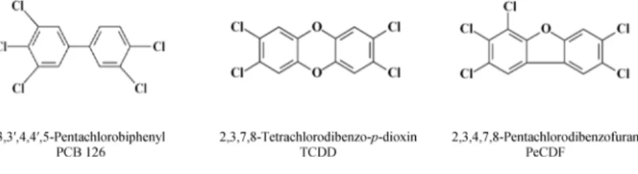
Several PHAHs were chosen by the National Toxicology Program (NTP) as model compounds including TCDD, PCB
126, and 2,3,4,7,8-pentachlorodibenzofuran (PeCDF).21−24
PCB 126 is a non-ortho-substituted PCB with a TEF value of
0.1. As the most potent DLC in the environment, PCB 126
accounts for 40−90% of the toxic potency of dioxin-like
PCBs.22PeCDF, with a TEF value of 0.5, represents the most
potent PCDF present in human tissues.23 The structures of
these compounds are shown in Figure 2. An important
assumption for the TEF methodology is that the toxicity of a mixture of DLCs is dose additive based on the TEF value of the individual components. This study has evaluated this assumption from the standpoint of the number of induced DNA oxidation products and their relationship to toxicity and carcinogenicity of PHAHs.
In this study, we collaborated with the NTP to understand
the DNA oxidation product profile in hepatic DNA of female
Sprague−Dawley rats that were exposed to TCDD and the
tertiary mixture of TCDD, PCB 126, and PeCDF (Figure 2) for
14, 31, and 53 weeks. Nine DNA oxidation lesions (7-OEG; 8-oxo-dGuo; 1,N6-εdAdo; 1,N2-εdGuo; N2,3-εG; M1dGuo;
AcrdGuo; CrdGuo; HNEdGuo) were measured in hepatic
DNA isolated from female Sprague−Dawley rats. Since each
product has distinct metabolic pathways in vivo, our assessment of a battery of DNA oxidation lesions provides extensive information on DNA oxidation damage. This knowledge enables us to better estimate the toxicity of PHAHs and
improve the scientific basis of human risk assessment of
PHAHs in the environment.
■
EXPERIMENTAL PROCEDURESChemicals. Nucleic acid purification grade lysis buffer, protein precipitation solution, and proteinase K were purchased from Gentra Systems (Minneapolis, MN). HPLC-grade water and methanol were from Thermo Fisher Scientific Company (Raleigh, NC).15N
5
-8-Oxo-dGuo,15N
5-dGuo, and 13C10-dGuo were purchased from Cambridge
Isotope Laboratories (Andover, MA, USA). Other chemical reagents were from Sigma-Aldrich Chemical Co. (St. Louis, MO).15N5-1,N6
-εdAdo standard was synthesized as described by Ham et al.40 1,N2
-εdGuo and 13C
10-1,N2-εdGuo were synthesized as reported by
Kusmierek et al.41 MDA modified 15N
5 and14N5 DNA were made
using the method in Jeong’s study.42 AcrdGuo, CrdGuo, and
HNEdGuo standards and their15N5labeled internal standards were
synthesized according to previous studies.43−457-OEG,15N 5-7-OEG,
andN2,3-εG were synthesized as described previously by Mutlu et
al.9,46
Animal Exposure and DNA Isolation. Rat liver tissues were provided by Battelle Laboratories (Columbus, OH) and State University of New York at Buffalo, which conducted the studies under NIEHS contract (N01-ES-75411).21−24 Female Sprague− Dawley rats were exposed to either TCDD alone or the tertiary mixture by gavage 5 days per week for 14, 31, and 53 weeks. The doses used for TCDD were 0 and 100 ng/kg/day for 14, 31, 53 weeks; 0, 22, 46, and 100 ng/kg/day for 53 weeks. The TEQ doses used for the tertiary mixture were 0 and 100 ng/kg/day for 14, 31, 53 weeks; 0, 22, 46, and 100 ng/kg/day for 53 weeks. Further explanation of the TEQ doses can be found in the NTP technical report on the toxicology and carcinogenesis studies of a mixture of 2,3,7,8-tetrachlorodibenzo-p-dioxin (TCDD) (Cas No. 1746−01−6), 2,3,4,7,8-pentachloro-dibenzofuran (PeCDF) (Cas No. 57117−31−4), and 3,3′,4,4′ ,5-pentachlorobiphenyl (PCB 126) (Cas No. 57465−28−8) in female Harlan Sprague−Dawley rats (gavage studies).24 Liver tissues were collected from 4−8 female rats per group/day after thefinal exposure and stored frozen at −80 °C. DNA was isolated as described previously.46
8-Oxo-dGuo and 1,N6-εdAdo Assay.The assay was performed
as previously described by Pang et al.7with minor modifications. A 100
μg sample of DNA in NaOAc buffer I (sodium acetate 30 mM, 0.2 mM ZnCl2, pH 5.6) was incubated with nuclease P1 (5μg) at 37°C
for 1 h. Immediately after incubation, DNA solutions were spiked with 2,2,6,6-tetramethyl-1-piperidinyloxy (TEMPO, 5μL, 1.5 M),15N
5
-8-oxo-dGuo (500 fmol), and15N
5-1,N6-εdAdo (20 fmol) followed by
addition of NaOAc buffer II (sodium acetate 30 mM, pH 8.1), alkaline phosphatase (20 units), and phosphodiesterase (0.012 units) then incubated at 37°C for an additional hour. Enzymes and undigested DNA were removed by Microcon-10filtration (11500 rpm, 4°C, 50 min), and thefiltrate was concentrated using a SpeedVac.
Samples were enriched for 8-oxo-dGuo and 1,N6-εdAdo using an
Agilent 1200 HPLC system equipped with a Atlantis T3 column (5
μm, 4.6 mm×150 mm).47 The nucleosides were monitored at 264 nm. The column was eluted at aflow rate of 1 mL/min at 30°C with a 5−80% MeOH gradient in 10 mM ammonium acetate buffer as follows: hold at 5% MeOH for 5 min, 5−10% MeOH over 5 min, 10− 20% MeOH over 10 min, 20−80% over 10 min; re-equilibrate at 5%
for 5 min. 8-oxo-dGuo and 1,N6-εdAdo fractions were collected at 24−
26 min and 33−34 min, respectively.
AcrdGuo, 1,N2-εdGuo, M
1dGuo, CrdGuo, and HNEdGuo
Assay.An assay similar to 8-oxo-dGuo and 1,N6-εdAdo was applied to measure AcrdGuo, 1,N2-εdGuo, M
1dGuo, CrdGuo, and HNEdGuo
with minor modifications. Considering their similar chromatography behaviors, these oxidation products were analyzed simultaneously. A 100μg sample of DNA in NaOAc buffer I was incubated with nuclease P1 (5 μg) at 37 °C for 1 h. Immediately after incubation, DNA solutions were spiked with TEMPO (5μL, 1.5 M), 15N
5−AcrdGuo
(50 fmol), 13C
10-1,N2-εdGuo (100 fmol), MDA modified internal
standard DNA corresponding to 400 fmol 15N
5-M1dGuo, 15N5−
CrdGuo (50 fmol), and 15N
5−HNEdGuo (50 fmol), followed by
addition of NaOAc buffer II, alkaline phosphate (20 units), and phosphodiesterase (0.012 units) then incubated at 37 °C for an additional hour.42 Enzymes and undigested DNA were removed by Microcon-10filtration (11 500 rpm, 4°C, 50 min), and thefiltrate was concentrated using a SpeedVac.
Samples were enriched for AcrdGuo, 1,N2-εdGuo, and M1dGuo by
the same HPLC method as described for 8-oxo-dGuo and 1,N6-εdAdo
with a 100% methanol and 5 mM ammonium formate−0.1% formic acid gradient.47CrdGuo and HNEdGuo were eluted at aflow rate of 0.5 mL/min, with a 35−70% MeOH gradient in 10 mM ammonium acetate buffer over 25 min. Fraction collection times for AcrdGuo, 1,N2-εdGuo, CrdG, and HNEdGuo were 28−30 min, 32−34 min, 15−
18 min, and 34−36 min, respectively. All fractions of the enzyme-digestion, MDA-treated DNA were collected every minute from the HPLC. Following concentration via SpeedVac the fraction containing M1dGuo was determined by using nanoLC−MS/MS.42
N2,3-εG and 7-OEG Assay.N2,3-εG and 7-OEG were analyzed as
described previously by Mutlu et al.9,46
LC−MS/MS Analysis. 8-Oxo-dGuo was analyzed by a Waters Acquity UPLC coupled to a Thermofinnigan TSQ Quantum Ultra triple-quadrupole mass spectrometer in a positive selected reaction mode (SRM) monitoring the signalsm/z284.1→ 168.0 for 8-oxo-dGuo andm/z289.1→173.0 for15N
5-8-oxo-dGuo. Separation was
performed on a T3 HSS column (1.7μm, 2.1 mm×100 mm) with a
flow rate of 200μL/min using gradient (A) 0.1% acetic acid in water and (B) 0.1% acetic acid in methanol. MS settings were as follows: electrospray voltage (3000 V), ion transfer capillary temperature (285
°C), the vaporizer temperature (250 °C), sheath and auxiliary gas pressures (35 and 30 arbitrary units), and collision energy (12 eV).
1,N6-εdAdo, AcrdGuo, 1,N2-εdGuo, M
1dGuo, CrdGuo, and
HNEdGuo were analyzed by nanoAcquity UPLC coupled to a Thermofinnigan TSQ Quantum Ultra triple-quadrupole mass spectrometer in positive SRM monitoring the signalsm/z276.0→ 160.0 for 1,N6-εdAdo,m/z281.0→165.0 for15N5-1,N6-εdAdo,m/z
304.0→188.0 for M1dGuo,m/z309.0→193.0 for15N5-M1dGuo,m/ z292.0→176.0 for 1,N2-εdGuo,m/z302.0→181.0 for13C
10-1,N2
-εdGuo,m/z424.0→308.0 for HNEdGuo,m/z429.0→313.0 for
15N
5-HNEdGuo,m/z338.0→222.0 for CrdGuo,m/z343.0→227.0
for15N
5-CrdGuo,m/z324.0→208.0 for AcrdGuo, andm/z329.0→
213.0 for15N
5-AcrdGuo. Separation was performed on a UPLC BEH
C18 column (1.7μm, 100μm×100 mm) with aflow rate of 1μL/ min using gradient (A) 5 mM ammonium formate in water and (B) 1% formic acid in acetonitrile for 1,N6-εdAdo, or (A) 0.1% formic acid in water and (B) acetonitrile for AcrdGuo, 1,N2-εdGuo, M
1dGuo,
CrdGuo, and HNEdGuo. MS settings were as follows: emitter tip voltage (1500 V), ion transfer capillary temperature (285 °C), and collision energy (12 eV).
Statistical Analysis. Statistical analyses were performed using R (2.11). Considering the limited sample size in certain groups, the nonparametric test was used to assess the differences between control and PHAH-treated rats or various control groups for the number of DNA oxidation products by Wilcox Rank Sum test. Two-sided and one-sidedp-values were considered significant if they were less than 0.05.
■
RESULTS AND DISCUSSIONIn this study, we examined the relationship between exposure to either TCDD or the tertiary mixture of TCDD, PCB 126, and PeCDF and the formation of DNA oxidation products.
Female Sprague−Dawley rats were exposed to 0 and 100 ng/
Table 1. Number of 8-Oxo-dGuo Adducts/106dGuo and 1,N6-εdAdo Adducts/108dAdo Measured in Female Sprague−Dawley
Rat Hepatic DNA Following Exposure to TCDD (100 ng/kg/day) or the Tertiary Mixture of TCDD, PCB 126, and PeCDF (TEQ 100 ng/kg/day) for 14, 31, and 53 Weeks
TCDD tertiary
14 weeks 31 weeks 53 weeks 14 weeks 31 weeks 53 weeks
8-oxo-dGuo add/106dGuo control 2.41±1.28 2.44±0.80 3.20±0.67 2.09±0.75 2.54±0.76 2.81±0.49
exposeda 2.55±0.91 2.72±0.88 3.87±0.47 3.93±1.27c 4.06±1.56c 4.87±0.81b
1,N6-εdAdo add/108dAdo control 1.47±0.65 1.32±0.44 0.91±0.48 1.36±0.32 1.31±0.63 0.93±0.56
exposeda 1.56±0.23 2.76±1.07b 2.13±0.52c 2.75±1.37c 6.55±3.34c 4.13±0.87c
aIndicates the exposures of TCDD, 100 ng/kg/day, and tertiary mixture (TCDD + PCB 126 + PeCDF), TEQ 100 ng/kg/day.bIndicatesp≤0.05 compared to corresponding control groups.cIndicatesp≤0.01 compared to corresponding control groups.
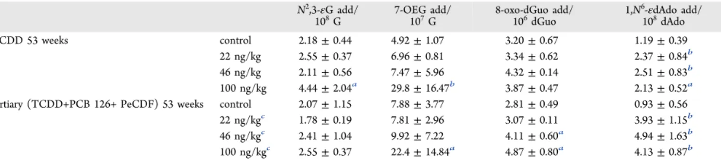
Table 2. Number ofN2,3-εG Adducts/108G, 7-OEG Adducts/107G, 8-Oxo-dGuo Adducts/106dGuo, and 1,N6-εdAdo
Adducts/108dAdo Measured in Female Sprague−Dawley Rat Hepatic DNA Following Exposure to Multiple Concentrations of TCDD or the Tertiary Mixture of TCDD, PCB 126, and PeCDF for 53 Weeks
N2,3-εG add/
108G 7-OEG add/107G 8-oxo-dGuo add/106dGuo 1,N
6-εdAdo add/
108dAdo
TCDD 53 weeks control 2.18±0.44 4.92±1.07 3.20±0.67 1.19±0.39
22 ng/kg 2.55±0.37 6.96±0.81 3.34±0.62 2.37±0.84b
46 ng/kg 2.11±0.56 7.47±5.96 4.32±0.14 2.51±0.83b
100 ng/kg 4.44±2.04a 29.8±16.47b 3.87±0.47 2.13±0.52a
tertiary (TCDD+PCB 126+ PeCDF) 53 weeks control 2.07±1.15 7.88±3.77 2.81±0.49 0.93±0.56
22 ng/kgc 1.78±0.19 7.81±2.96 3.07±0.11 3.93±1.15b
46 ng/kgc 2.41±1.04 9.92±7.22 4.11±0.60a 4.94±1.63b
100 ng/kgc 2.55±0.37 22.4±14.84a 4.87±0.80a 4.13±0.87b
kg/day of TCDD or TEQ doses of 0 and 100 ng/kg/day of the tertiary mixture for 14, 31, and 53 weeks and to 0, 22, 46, and 100 ng/kg/day TCDD or TEQ doses of 0, 22, 46, and 100 ng/ kg/day of the tertiary mixture for 53 weeks.
Measurements of 8-oxo-dGuo and 1,N6-εdAdo formation in
the liver of the female rats exposed to TCDD (0 and 100 ng/ kg/day) or the tertiary mixture (TEQ 0, and 100 ng/kg/day) at 14, 31, and 53 weeks are shown inTable 1. While no significant increase of 8-oxo-dGuo was detected after TCDD exposure for 14, 31, or 53 weeks, 1,N6-εdAdo concentrations for 31 (p =
0.03) and 53 (p= 0.003) weeks were higher in comparison to
their respective control groups. Exposure to the tertiary mixture
for 14 (p = 0.004 for 8-oxo-dGuo, and p = 0.004 for 1,N6
-εdAdo), 31 (p= 0.0001 for 8-oxo-dGuo, and p = 0.0002 for
1,N6-εdA), and 53 weeks (p = 0.02 for 8-oxo-dGuo, andp =
0.0002 for 1,N6-εdAdo) all showed statistically significant
increases in 8-oxo-dGuo and 1,N6-εdAdo. These increases
correspond to a 1.5−1.8-fold increase in 8-oxo-dGuo and a
1.5−5.0-fold increase in 1,N6-εdAdo in the hepatic DNA of
female rats.
The accumulations of N2,3-εG, 7-OEG, 8-oxo-dGuo, and
1,N6-εdAdo were evaluated in the hepatic DNA of the female
Sprague−Dawley Rats exposed to TCDD at 0, 22, 46, 100 ng/
kg/day or the tertiary mixture at TEQ doses of 0, 22, 46, 100
ng/kg/day for 53 weeks (Table 2). No accumulation of
8-oxo-dGuo was observed after TCDD exposure, but a significant
increase of 8-oxo-dGuo was observed following exposure to the
tertiary mixture at TEQ doses of 46 (p= 0.03) and 100 ng/kg/
day (p = 0.02). 1,N6-εdAdo concentrations increased
significantly at all doses (22, 46, 100 mg/kg/day) following
either TCDD (p= 0.008,p= 0.008, andp= 0.02, respectively)
or tertiary (p= 0.003,p= 0.003, andp= 0.002, respectively) exposures.Table 2shows the significant increase in the number of 7-OEG in the liver at the highest dose exposures of both
TCDD (100 ng/kg,p= 0.0098) and the tertiary mixture (TEQ
100 ng/kg,p= 0.014). Endogenous 7-OEG was measured to be
4.92± 1.07 adducts/107G in control samples, while exposure
to 100 ng/kg/day TCDD induced 29.79±16.47 adducts/107G
of OEG. Additionally, a three-fold increase was observed in 7-OEG formation from exposure to the tertiary mixture. No significant increase was observed for N2,3-εG concentration
after exposure to the tertiary mixture, including the highest
TEQ dose group, while a two-fold increase in N2,3-εG was
observed after 100 ng/kg/day TCDD exposure (p= 0.015).
Several other LPO-induced DNA products (1,N2-εdGuo;
M1dGuo; CrdGuo; HNEdGuo; AcrdGuo) were analyzed in the
hepatic DNA of female rats exposed to TCDD (100 ng/kg/ day) and the tertiary mixture (TEQ 100 ng/kg/day) for 53 Table 3. Number of 1,N2-εdGuo Adducts/108dGuo,
M1dGuo adducts/108dGuo, CrdGuo Adducts/108dGuo,
HNEdGuo Adducts/108dGuo, and AcrdGuo Adducts/108
dGuo Measured in Female Sprague−Dawley Rat Hepatic DNA Following Exposure to TCDD (100 ng/kg/day) or the Tertiary Mixture of TCDD, PCB 126, and PeCDF (TEQ 100 ng/kg/day) for 53 Weeks
TCDD add/108dGuo tertiary add/108dGuo
1,N2-εdGuo control 1.61±0.37 2.07±0.43
1,N2-εdGuo exposeda 2.14±0.51 3.80±1.12c
M1dGuo control 4.16±1.02 4.71±2.23
M1dGuo exposeda 6.43±1.68 12.4±6.73b
CrdGuo control 0.24±0.10 0.35±0.07
CrdGuo exposeda 0.24±0.06 0.50±0.12b
HNEdGuo control 1.12±0.05 0.90±0.22
HNEdGuo exposeda 1.18±0.38 1.40±0.38
AcrdGuo control 6.02±1.30 8.26±0.98
AcrdGuo exposeda 7.16±0.84 42.9±24.50b
aIndicates the exposures of TCDD, 100 ng/kg/day, and tertiary
mixture (TCDD + PCB 126 + PeCDF), TEQ 100 ng/kg/day, 53 weeks.bIndicatesp≤0.05 compared to corresponding control groups. cIndicatesp≤0.01 compared to corresponding control groups.
Figure 3. (Top) Cell proliferation in the liver of female Sprague− Dawley rats exposed to TCDD (100 ng/kg/day) or the tertiary mixture of TCDD, PCB 126, and PeCDF (TEQ 100 ng/kg/day) at 14, 31, and 53 weeks.21,24(Bottom) Cell proliferation in the liver of female Sprague−Dawley rats exposed to TCDD (0, 22, 46, and 100 ng/kg/day) or the tertiary mixture of TCDD, PCB 126, and PeCDF (TEQ 0, 22, 46, and 100 ng/kg/day) at 53 weeks.21,24∗, Significantly different (p≤0.05) from the vehicle control group by Shirley’s test.
weeks (Table 3). Concentrations of 1,N2-εdGuo (p= 0.008),
M1dGuo (p= 0.03), CrdGuo (p = 0.05), and AcrdGuo (p =
0.03) were significantly higher in animals exposed to the tertiary mixture, while no significant increase was observed in any of the LPO-induced product concentrations in animals that were exposed to TCDD (100 ng/kg/day) for 53 weeks.
As one of the predominant nongenotoxic pollutants in the environment, the mechanism of PHAH-related carcinogenesis has been studied for decades. Among them, TCDD is the most researched. DNA oxidation products and oxidative stress induced by TCDD have been measured extensively both in vitro and in vivo.18,19,26−31 Hassoun et al. reported the
induction of LPO in the liver of female Sprague−Dawley rats
exposed to TCDD (100 ng/kg/day) for 13 and 30 weeks.27,29
LPO became more pronounced with increasing exposure time, which is similar to our results for 1,N6-εdAdo (Table 1). In our study, no significant increase of 1,N6-εdAdo was observed in
TCDD treated animals at 14-week exposure, but a two-fold
significant increase of this product was detected at 31 weeks
exposure. In the study by Hassoun et al., higher increases of LPO were detected in animals exposed to TCDD for 30 weeks
(6-fold) than 13 weeks (1.5-fold).26Similarly, DNA oxidation
product results found in our study showed statistically
significant increases (7-OEG, p = 0.00008; N2,3-εG, p =
0.0025; 1,N6-εdAdo, p= 0.0016) following 53-week exposure
to TCDD, which are shown inTable 2. Although an increase of
hepatic 8-oxo-dGuo was previously reported to be gender and estrogen dependent in a two-stage carcinogenesis model with
diethylnitrosamine as initiator and TCDD as promotor,18 we
observed no significant induction of 8-oxo-dGuo in the female
rat livers following exposure to TCDD for 14, 31, or 53 weeks (Table 1). These results may be due to differences between the
animal study designs or the analytical assays (LC−MS/MS vs
LC-ECD).18
Induction of superoxide anions, LPO and DNA single-strand breaks following exposure to the tertiary mixture (TEQ 100 ng/kg/day) for 13 and 30 weeks in female rats was also
previously reported by Hassoun et al.26,29 In their study,
exposure for 30 weeks to the tertiary mixture induced more superoxide anions and LPO (8.2-fold, 2.3-fold) than 13 week exposure (6-fold, 1.9-fold), which indicated that production of
these biomarkers is time-dependent.26,29 Consistently, more
DNA oxidation products were induced in liver DNA of our studied animals after exposure to the tertiary mixture for 31
weeks than 14 weeks, as indicated by 1,N6-εdAdo and
8-oxo-dGuo results inTable 1. Significant dose-dependent increases
in the production of superoxide anions, LPO and DNA single-strand breaks in the liver of female rats were also reported in the liver DNA of female rats exposed to the tertiary mixture for
13 weeks and 30 weeks in Hassoun et al.’s studies.26,29
Consistently, we found a significant positive association
between exposure dose and concentrations of 7-OEG (p <
0.05), 8-oxo-dGuo (p= 0.00041), and 1,N6-εdAdo (p< 1 ×
10−5) following exposure to the tertiary mixture for 53 weeks (Table 2).
Similar to other toxic effects of TCDD, DNA oxidation
damage and oxidative stress have been assumed to be connected with the AhR and activation of cytochrome P450
superfamily of enzymes, especially CYP1A1.37,38The
upregu-lation of these enzymes triggers metabolic changes of endogenous compounds in vivo, which may further produce DNA oxidation damage. Since the TEF methodology is based
on the AhR affinity of PHAHs and oxidative stress was reported
in diverse biological systems exposed to PHAHs, the TEF methodology may also be valuable for the evaluation of oxidative stress and oxidative damage induced by mixtures of PHAHs, especially DLCs. However, the DNA oxidation
product findings in this study, and several other studies,
indicate that a simple TEF value cannot be applied to evaluate
oxidative stress and oxidative damage.26,48 Hassoun et al.
showed that following 13-weeks exposure, higher levels of superoxide anions and LPO were induced in the liver of female rats exposed to the tertiary mixture (TEQ 100 ng/kg/day) than
TCDD alone (100 ng/kg/day).26 With similar background
concentrations in control samples, the concentrations of
superoxide anion were ∼1.3 and 0.4 nmol cytochrome c
reduced/mg in the animals treated with the tertiary mixture or TCDD alone, respectively. The concentrations of LPO were
∼3 and 2 nmol 2-thiobarbituric acid substances (TBARS)
formed/mg protein detected in these two groups, respec-tively.26,27Additionally, synergistic effects of TCDD, PeCDF, and PCB126 were indicated in the production of superoxide anion in hepatic tissues, which required only 0.25−0.5% of the doses of the three individual congeners within the tertiary
mixture to produce similar effects for each congener alone.26
The interactions of these three congeners were also seen in the process of superoxide anion production in female rats exposed to the tertiary mixture for 30 weeks.29A significant difference
was observed when comparing the dose−response curves of
superoxide anion production in the hepatic tissues exposed to the tertiary mixture versus TCDD, PeCDF or PCB126 alone,
with larger effects produced by an equivalent dose of the
tertiary mixture than the three congeners alone.
In our study, we compared oxidation DNA product formation induced by either TCDD (100 ng/kg/day) or the tertiary mixture (TEQ 100 ng/kg/day) for 14, 31, and 53
weeks. A significant production of hepatic 8-oxo-dGuo and
1,N6-εdAdo was observed following exposure to the tertiary
mixture at all time points, while TCDD-exposed animals showed a significant increase in 1,N6-εdAdo (31 and 53 weeks) but no significant difference in 8-oxo-dGuo. After 53-week
exposure, significant induction of 1,N2-εdGuo, M
1dGuo,
AcrdGuo, and CrdGuo was observed in the tertiary mixture-treated rat liver DNA; however, none of the products showed
significant changes in TCDD-treated animals. Conversely, a
significant induction of 7-OEG andN2,3-εG was observed in
the liver following 53-week exposure to TCDD, while only 7-OEG was significantly induced in tertiary mixture-exposed animals, as indicated inTable 2.
Complex results were also reported in the toxicogenomic
analysis by Vezina et al.48 and Ovando et al.49 Vezina et al.
examined the gene expression in the hepatic DNA of female
rats exposed to TCDD, PCB 126, and PeCDF for 14 weeks.48
TCDD and PCB126 by Ovando et al.49 A dose-dependent
increase in the number of differentially expressed genes was
observed in animals exposed to PCB 126 for 53 weeks with 30,
300, and 1000 ng/kg/day. While fewer genes were differentially
expressed in animals exposed to PCB 126 for 53 weeks (216)
than 13 weeks (371), many more genes showed differential
expression in animals exposed to TCDD for 53 weeks (299) than 13 weeks (103) with the same TEQ dose. More ROS or
their active metabolite-related detoxification genes were
upregulated or downregulated in the chronic TCDD exposed animals including glutathione-S-transferase (GST), glutathione peroxidase, aldehyde dehydrogenase (Aldh), and aldo-keto reductase (Akr).
DNA oxidation damage in rodents exposed to DLCs is probably induced through upregulation of cytochrome P450 superfamily of enzymes, mediated by AhR-dependent
path-ways.37,38 The dose−response and different time course
induction of CYP1A1 is well characterized in the liver of
female Sprague−Dawley rats following exposure to TCDD and
the tertiary mixture21,24,50CYP1A1 induction occurs in virtually every tissue of the body following exposure to either TCDD or the tertiary mixture.21,24By using the continuous nonlinear Hill model, the induction of hepatic CYP1A1 was compared in female rats following exposure to TCDD, PCB 126, and PeCDF alone or their tertiary mixture with doses or TEQ doses
ranging from 0−100 ng/kg/day for 14, 31, or 53 weeks.50The
estimated parameters indicated that congener-specific dose−
response shapes were significantly different, and the additivity of TEF methodology failed for these individual congeners and their mixture. Six of the 12 equiv time-dose combinations failed to agree between the tertiary mixture and TCDD alone. The same results were found when liver concentrations of TCDD or TEQ dose for the tertiary mixture were used. The maximum
activity of 7-ethoxyresorufin-o-deethylase (EROD) is
substan-tially higher (1.2−3.0-fold) for PeCDF than TCDD with the
same TEQ dose. Toyoshiba et al. observed significant,
nonadditive interaction for EROD activity at 31 and 53 weeks in the tertiary mixture exposed samples but not at 14
weeks.50In summary, although the importance of CYP1A1 in
the induction of oxidative DNA damage was implied previously, the complicated association between DNA oxidation damage and induction of CYP1A1 by DLCs or their mixtures cannot be completely described in our study and still requires further detailed exploration. DNA oxidation products, oxidative stress biomarkers, CYP1A1 induction, and genomic studies consis-tently suggest that the simple TEF methodology cannot be applied to evaluate the diverse patterns of toxic effects induced by DLCs.
Mutation studies have suggested that oxidative stress and
DNA oxidation damage, especially from chronic inflammation,
are associated with carcinogenesis.2,3At present, more than one hundred DNA oxidation products are reported in model studies, but less than 20 of them are measured in cellular
DNA.51 Among them, 8-oxo-dGuo is the most extensively
studied in vivo adduct with G to T transversions as the
dominant mutation pattern.8Many assays have been developed
to detect this product in animal or human tissues,8,52 but
artificial formation during sample preparation has hampered its application as a good biomarker.52,53 1,N6-εdAdo is another
popular biomarker, widely applied to evaluate chronic
inflammation and LPO in animal or human tissues.3 1,N6
-εdAdo, 1,N2-εdGuo, N2,3-εG, M
1dGuo, AcrdGuo, CrdGuo,
and HNEdGuo can also induce distinct mutation spectra,
similar to 8-oxo-dGuo.10−12 However, compared with 1,N6
-εdAdo and 8-oxo-dGuo, the other products examined in our
study are less widely applied in risk assessment of carcinogens. 7-OEG, the predominant product formed by vinyl chloride in rodents, was also recently established as an LPO-induced
product.9 Although it is not a direct promutagenic adduct,
7-OEG can cause AP sites and further induce mutations if they
cannot be repaired before cell replication.54 In addition to
promutagenic properties, 8-oxo-dGuo, 1,N6-εdAdo, 1,N2
-εdGuo,N2,3-εG, M
1dGuo, AcrdGuo, CrdGuo, and HNEdGuo
also have distinct metabolic pathways, as shown in Figure 1.
Furthermore, the primary repair pathways for these products are also different.11,55−58 For small products (8-oxo-dGuo; 1,N6-εdAdo; 1,N2-εdGuo;N2,3-εG), base excision repair (BER)
is the dominant repair pathway, with different glycosylases
involved to repair different products.55−57,59For the medium and bulky products (AcrdGuo; CrdGuo; HNEdGuo;
M1dGuo), nuclear excision repair (NER) is the primary repair
pathway.11,58 Therefore, the simple concentration of a DNA
oxidation product in animals reflects the complex interactions
between environmental and biological systems. The potential
adverse effect of a product is dynamically controlled by its
formation and repair pathways, which could be determined by numerous factors including, but not limited to, exposure dose, exposure time, exposure pathway, chemical metabolism, tissue, age, sex, and species. A corresponding mutation spectrum study is still necessary for further validation of the biological
significance of certain DNA oxidation products in hepatic
toxicity of TCDD and the tertiary mixture exposed animals examined in our study.
Besides the possible indirect genotoxicity of oxidative stress, cytotoxicity, stimulation of cell proliferation, inhibition of apoptosis, and induction of enzymes are all suggested to be
involved in the toxicity of TCDD and its congeners.60−63As
depicted inFigure 3, hepatic cells from female rats showed a
high rate of proliferation following exposure to TCDD alone (100 ng/kg/day) or the tertiary mixture (TEQ 100 ng/kg/day) for 53 weeks, which is consistent with our DNA oxidation
product induction results in Tables 2 and 3. No significant
enhancement of cell proliferation was observed in 14-week TCDD or tertiary mixture exposed animals, but it was observed in 31- and 53-week TCDD and tertiary mixture exposed
animals.22,24 Low-dose exposure of TCDD or the tertiary
mixture for 53 weeks failed to induce significant production of cell proliferation, which agreed with the observation of dose-dependent accumulation of DNA oxidation products following 53-week exposure. Dose- and time-dependent induction of CYP1A1 and CYP1A2 was detected in the livers of female rats exposed to TCDD or the tertiary mixture, which is also in
accordance with our results of DNA oxidation products.21,24
Additionally, increased incidences and severities of hepatocyte inflammation were observed at 31 and 53 weeks in TCDD or the tertiary mixture exposed animals. All these factors are potential players, which can be combined to induce the dose-dependent incidence of hepatocellular adenoma and chol-angiocarcinoma in the 2-year cancer bioassay by the NTP.21,24 Recent advances in scientific understanding of cancer biology support the view that environmental chemicals can act through multiple toxicity pathways, modes, or mechanisms of action to induce cancer.64,65 However, evaluating the relative weight of each possible important contributor to cancer induction is far more complicated than simply identifying them. The role DNA
in female rats may be a good case in point. Although the 2-year tumor incidence data (Figure 4) support the application of the TEF approach in the risk assessment of DLCs based on the
generation of similar dose−response curves in response to
TCDD and the tertiary mixture exposed female rats,66the TEF
approach cannot be applied to evaluate the DNA oxidation products in this study, CYP1A1 or CYP1A2 induction, or oxidative biomarkers.26,27,29,50 Such inconsistency further indicates the complexity of the formation of DNA oxidation damage and carcinogenesis of DLCs in the liver of female rats.
■
AUTHOR INFORMATIONCorresponding Author
*E-mail: [email protected]. Phone: 919-966-6139.
ORCID
Hadley J. Hartwell:0000-0003-4453-3266
Author Contributions
¶These authors contributed equally to this work.
Funding
This work was supported by the NIEHS Superfund Basic Research Program P42-ES05948 and the NIEHS Center for Environmental Health and Susceptibility P30 ES 10126 and in part by the intramural research program of the NIEHS. Notes
The authors declare no competingfinancial interest.
■
ACKNOWLEDGMENTSWe thank Dr. Valeriy Afonin for the technical assistance in DNA isolation.
■
ABBREVIATIONS1,N2-εdGuo, 1,N2-etheno-2′-deoxyguanosine; 1,N6-εdAdo,
1,N6-etheno-2′-deoxyadenosine; 7-OEG,
7-(2-oxoethly)-guanine; 8-oxo-dGuo, 8-oxo-7,8-dihydro-2′-deoxyguanosine;
AcrdGuo, acrolein derived dGuo adducts; AhR, aryl hydro-carbon receptor; AlkB, alpha-ketoglutarate-dependent dioxyge-nase; ANPG, alkyl-N-purine-DNA glycosylase; CrdGuo, crotonaldehyde derived dGuo adducts; DLCs, dioxin-like compounds; HNEdG, 4-hydroxynonenal derived dG adducts;
LPO, lipid peroxidation; M1dGuo, malondialdehyde derived
dGuo adducts; MDA, malondialdehyde; N2,3-εG,
N2,3ethenoguanine; PCB 126, 3,3′,4,4′,5-pentachlorobiphenyl;
PeCDF, 2,3,4,7,8-pentachlorodibenzofuran; PHAHs, polyhalo-genated aromatic hydrocarbons; RNS, reactive nitrogen species; ROS, reactive oxygen species; SRM, selected reaction mode; TCDD, 2,3,7,8-tetrachlorodibenzo-p-dioxin; TEF, toxic equivalency factor; TEMPO, 2,2,6,6-tetramethyl-1-piperidiny-loxy; TEQ, toxic equivalent quotient
■
REFERENCES(1) Cadet, J., Loft, S., Olinski, R., Evans, M. D., Bialkowski, K., Richard Wagner, J., Dedon, P. C., Møller, P., Greenberg, M. M., and Cooke, M. S. (2012) Biologically relevant oxidants and terminology, classification and nomenclature of oxidatively generated damage to nucleobases and 2-deoxyribose in nucleic acids.Free Radical Res. 46, 367−381.
(2) Rasmussen, J. L. (2006) Oxidative damage to DNA and its repair, inOxidative Stress, Disease, and Cancer (Singh, K. K., Ed.), Imperial College Press.
(3) Nair, U., Bartsch, H., and Nair, J. (2007) Lipid peroxidation-induced DNA damage in cancer-prone inflammatory diseases: a review of published adduct types and levels in humans.Free Radical Biol. Med. 43, 1109−1120.
(4) Zhang, S., Villalta, P. W., Wang, M., and Hecht, S. S. (2007) Detection and quantitation of acrolein-derived 1,N2
-propanodeox-yguanosine adducts in human lung by liquid chromatography-electrospray ionization-tandem mass spectrometry.Chem. Res. Toxicol. 20, 565−571.
(5) Chung, F. L., Zhang, L., Ocando, J. E., and Nath, R. G. (1999) Role of 1,N2-propanodeoxyguanosine adducts as endogenous DNA
lesions in rodents and humans.IARC Sci. Publ., 45−54.
(6) Liu, X., Lovell, M. A., and Lynn, B. C. (2006) Detection and quantification of endogenous cyclic DNA adducts derived from trans-4-hydroxy-2-nonenal in human brain tissue by isotope dilution capillary liquid chromatography nanoelectrospray tandem mass spectrometry.Chem. Res. Toxicol. 19, 710−718.
(7) Pang, B., Zhou, X., Yu, H., Dong, M., Taghizadeh, K., Wishnok, J. S., Tannenbaum, S. R., and Dedon, P. C. (2007) Lipid peroxidation dominates the chemistry of DNA adduct formation in a mouse model of inflammation.Carcinogenesis 28, 1807−1813.
(8) Valavanidis, A., Vlachogianni, T., and Fiotakis, C. (2009) 8-hydroxy-2′ -deoxyguanosine (8-OHdG): A critical biomarker of oxidative stress and carcinogenesis.J. Environ. Sci. Health C Environ. Carcinog. Ecotoxicol. Rev. 27, 120−139.
(9) Mutlu, E., Jeong, Y. C., Collins, L. B., Ham, A. J., Upton, P. B., Hatch, G., Winsett, D., Evansky, P., and Swenberg, J. A. (2012) A new LC−MS/MS method for the quantification of endogenous and vinyl chloride-induced 7-(2-Oxoethyl)guanine in Sprague−Dawley rats.
Chem. Res. Toxicol. 25, 391−399.
(10) Niedernhofer, L. J., Daniels, J. S., Rouzer, C. A., Greene, R. E., and Marnett, L. J. (2003) Malondialdehyde, a product of lipid peroxidation, is mutagenic in human cells.J. Biol. Chem. 278, 31426− 31433.
(11) Minko, I. G., Kozekov, I. D., Harris, T. M., Rizzo, C. J., Lloyd, R. S., and Stone, M. P. (2009) Chemistry and biology of DNA containing 1,N2-deoxyguanosine adducts of the alpha,beta-unsaturated aldehydes
acrolein, crotonaldehyde, and 4-hydroxynonenal.Chem. Res. Toxicol. 22, 759−778.
(12) Moriya, M., Zhang, W., Johnson, F., and Grollman, A. P. (1994) Mutagenic potency of exocyclic DNA adducts: marked differences betweenEscherichia coliand simian kidney cells.Proc. Natl. Acad. Sci. U. S. A. 91, 11899−11903.
(13) Munnia, A., Bonassi, S., Verna, A., Quaglia, R., Pelucco, D., Ceppi, M., Neri, M., Buratti, M., Taioli, E., Garte, S., and Peluso, M. (2006) Bronchial malondialdehyde DNA adducts, tobacco smoking, and lung cancer.Free Radical Biol. Med. 41, 1499−1505.
(14) Mangerich, A., Knutson, C. G., Parry, N. M., Muthupalani, S., Ye, W., Prestwich, E., Cui, L., McFaline, J. L., Mobley, M., Ge, Z., Taghizadeh, K., Wishnok, J. S., Wogan, G. N., Fox, J. G., Tannenbaum, S. R., and Dedon, P. C. (2012) Infection-induced colitis in mice causes dynamic and tissue-specific changes in stress response and DNA damage leading to colon cancer.Proc. Natl. Acad. Sci. U. S. A. 109, E1820−1829.
(15) Melnick, R. L., Kohn, M. C., and Portier, C. J. (1996) Implications for risk assessment of suggested nongenotoxic mecha-nisms of chemical carcinogenesis.Environ. Health Perspect. 104(Suppl 1), 123−134.
(16) Jarabek, A. M., Pottenger, L. H., Andrews, L. S., Casciano, D., Embry, M. R., Kim, J. H., Preston, R. J., Reddy, M. V., Schoeny, R., Shuker, D., Skare, J., Swenberg, J., Williams, G. M., and Zeiger, E. (2009) Creating context for the use of DNA adduct data in cancer risk assessment: I. Data organization.Crit. Rev. Toxicol. 39, 659−678.
(17) Spencer, W. A., Lehmler, H. J., Robertson, L. W., and Gupta, R. C. (2009) Oxidative DNA adducts after Cu2+-mediated activation of
dihydroxy PCBs: role of reactive oxygen species. Free Radical Biol. Med. 46, 1346−1352.
(19) Tritscher, A. M., Seacat, A. M., Yager, J. D., Groopman, J. D., Miller, B. D., Bell, D., Sutter, T. R., and Lucier, G. W. (1996) Increased oxidative DNA damage in livers of 2,3,7,8-tetrachlorodibenzo-p-dioxin treated intact but not ovariectomized rats.Cancer Lett. 98, 219−225.
(20) Jeong, Y. C., Walker, N. J., Burgin, D. E., Kissling, G., Gupta, M., Kupper, L., Birnbaum, L. S., and Swenberg, J. A. (2008) Accumulation of M1dG DNA adducts after chronic exposure to PCBs, but not from acute exposure to polychlorinated aromatic hydrocarbons.Free Radical Biol. Med. 45, 585−591.
(21) NTP. (2006) in NTP Technical Report on the Toxicology and Carcinogenesis Studies of 3,3′,4,4′,5-Pentachlorobiphenyl (PCB 126) (CAS No. 57465−28−8) in Female Harlan Sprague−Dawley Rats (Gavage Studies), U.S. Department of Health and Human Services, Washington, DC.
(22) NTP. (2006) NTP Technical Report on the Toxicology and Carcinogenesis Studies of 2,3,7,8-Tetrachlorodibenzo-p-dioxin (TCDD) (CAS No. 1746−01−6) in Female Harlan Sprague−Dawley Rats (Gavage Studies), pp 4−232, U.S. Department of Health and Human Services, Washington, DC.
(23) NTP. (2006) NTP Technical Report on the Toxicology and Carcinogenesis Studies of 2,3,4,7,8-Pentachlorodibenzofuran (PeCDF) (CAS No. 57117−31−4) in Female Harlan Sprague−Dawley Rats (Gavage Studies), pp 1−198, U.S. Department of Health and Human Services, Washington, DC.
(24) NTP. (2006) NTP Technical Report on the Toxicology and Carcinogenesis Studies of a Mixture of 2,3,7,8-Tetrachlorodibenzo-p-dioxin (TCDD) (Cas No. 1746−01−6), 2,3,4,7,8-Pentachlorodibenzofuran (PeCDF) (Cas No. 57117−31−4), and 3,3′,4,4′,5-Pentachlorobiphenyl (PCB 126) (Cas No. 57465−28−8) in Female Harlan Sprague−Dawley Rats (Gavage Studies), pp 1−180, U.S. Department of Health and Human Services, Washington, DC.
(25) Van den Berg, M., Birnbaum, L. S., Denison, M., De Vito, M., Farland, W., Feeley, M., Fiedler, H., Hakansson, H., Hanberg, A., Haws, L., Rose, M., Safe, S., Schrenk, D., Tohyama, C., Tritscher, A., Tuomisto, J., Tysklind, M., Walker, N., and Peterson, R. E. (2006) The 2005 World Health Organization reevaluation of human and mammalian toxic equivalency factors for dioxins and dioxin-like compounds.Toxicol. Sci. 93, 223−241.
(26) Hassoun, E. A., Li, F., Abushaban, A., and Stohs, S. J. (2001) Production of superoxide anion, lipid peroxidation and DNA damage in the hepatic and brain tissues of rats after subchronic exposure to mixtures of TCDD and its congeners.J. Appl. Toxicol. 21, 211−219.
(27) Hassoun, E. A., Li, F., Abushaban, A., and Stohs, S. J. (2000) The relative abilities of TCDD and its congeners to induce oxidative stress in the hepatic and brain tissues of rats after subchronic exposure.
Toxicology 145, 103−113.
(28) Hassoun, E. A., and Periandri-Steinberg, S. (2010) Assessment of the roles of antioxidant enzymes and glutathione in 3,3′,4,4′ ,5-Pentachlorobiphenyl (PCB 126)-induced oxidative stress in the brain tissues of rats after subchronic exposure.Toxicol. Environ. Chem. 92, 301.
(29) Hassoun, E. A., Wang, H., Abushaban, A., and Stohs, S. J. (2002) Induction of oxidative stress in the tissues of rats after chronic exposure to TCDD, 2,3,4,7,8-pentachlorodibenzofuran, and 3,3′,4,4′ ,5-pentachlorobiphenyl.J. Toxicol. Environ. Health, Part A 65, 825−842. (30) Stohs, S. (1990) Oxidative stress induced by 2,3,7,8-tetrachlorodibenzo-p-dioxin (TCDD).Free Radical Biol. Med. 9, 79− 90.
(31) Stohs, S. J., Hassan, M. Q., and Murray, W. J. (1983) Lipid peroxidation as a possible cause of TCDD toxicity.Biochem. Biophys. Res. Commun. 111, 854−859.
(32) Dreiem, A., Rykken, S., Lehmler, H. J., Robertson, L. W., and Fonnum, F. (2009) Hydroxylated polychlorinated biphenyls increase reactive oxygen species formation and induce cell death in cultured cerebellar granule cells.Toxicol. Appl. Pharmacol. 240, 306−313.
(33) Fadhel, Z., Lu, Z., Robertson, L. W., and Glauert, H. P. (2002) Effect of 3,3′,4,4′-tetrachlorobiphenyl and 2,2′,4,4′,5,5′ -hexachlorobi-phenyl on the induction of hepatic lipid peroxidation and cytochrome P-450 associated enzyme activities in rats.Toxicology 175, 15−25.
(34) Glauert, H. P., Tharappel, J. C., Lu, Z., Stemm, D., Banerjee, S., Chan, L. S., Lee, E. Y., Lehmler, H. J., Robertson, L. W., and Spear, B. T. (2008) Role of oxidative stress in the promoting activities of pcbs.
Environ. Toxicol. Pharmacol. 25, 247−250.
(35) Hennig, B., Meerarani, P., Slim, R., Toborek, M., Daugherty, A., Silverstone, A. E., and Robertson, L. W. (2002) Proinflammatory properties of coplanar PCBs: in vitro and in vivo evidence.Toxicol. Appl. Pharmacol. 181, 174−183.
(36) Hori, M., Kondo, H., Ariyoshi, N., Yamada, H., Hiratsuka, A., Watabe, T., and Oguri, K. (1997) Changes in the hepatic glutathione peroxidase redox system produced by coplanar polychlorinated biphenyls in Ah-responsive and -less-responsive strains of mice: mechanism and implications for toxicity.Environ. Toxicol. Pharmacol. 3, 267−275.
(37) Park, J. Y., Shigenaga, M. K., and Ames, B. N. (1996) Induction of cytochrome P4501A1 by 2,3,7,8-tetrachlorodibenzo-p-dioxin or indolo(3,2-b)carbazole is associated with oxidative DNA damage.Proc. Natl. Acad. Sci. U. S. A. 93, 2322−2327.
(38) Reichard, J. F., Dalton, T. P., Shertzer, H. G., and Puga, A. (2005) Induction of oxidative stress responses by dioxin and other ligands of the aryl hydrocarbon receptor.Dose-Response 3, 306−331.
(39) Twaroski, T. P., O’Brien, M. L., Larmonier, N., Glauert, H. P., and Robertson, L. W. (2001) Polychlorinated biphenyl-induced effects on metabolic enzymes, AP-1 binding, vitamin E, and oxidative stress in the rat liver.Toxicol. Appl. Pharmacol. 171, 85−93.
(40) Ham, A. J., Engelward, B. P., Koc, H., Sangaiah, R., Meira, L. B., Samson, L. D., and Swenberg, J. A. (2004) New immunoaffinity-LC− MS/MS methodology reveals that Aag null mice are deficient in their ability to clear 1,N6-etheno-deoxyadenosine DNA lesions from lung and liver in vivo.DNA Repair 3, 257−265.
(41) Kusmierek, J. T., and Singer, B. (1992) 1,N2
-ethenodeox-yguanosine: properties and formation in chloroacetaldehyde-treated polynucleotides and DNA.Chem. Res. Toxicol. 5, 634−638.
(42) Jeong, Y. C., Sangaiah, R., Nakamura, J., Pachkowski, B. F., Ranasinghe, A., Gold, A., Ball, L. M., and Swenberg, J. A. (2005) Analysis of M1G-dR in DNA by aldehyde reactive probe labeling and liquid chromatography tandem mass spectrometry.Chem. Res. Toxicol. 18, 51−60.
(43) Wang, M., McIntee, E. J., Cheng, G., Shi, Y., Villalta, P. W., and Hecht, S. S. (2000) Identification of paraldol-deoxyguanosine adducts in DNA reacted with crotonaldehyde.Chem. Res. Toxicol. 13, 1065− 1074.
(44) Chung, F. L., Young, R., and Hecht, S. S. (1984) Formation of cyclic 1,N2-propanodeoxyguanosine adducts in DNA upon reaction
with acrolein or crotonaldehyde.Cancer Res. 44, 990−995.
(45) Douki, T., Odin, F., Caillat, S., Favier, A., and Cadet, J. (2004) Predominance of the 1,N2-propano 2′-deoxyguanosine adduct among
4-hydroxy-2-nonenal-induced DNA lesions.Free Radical Biol. Med. 37, 62−70.
(46) Mutlu, E., Collins, L. B., Stout, M. D., Upton, P. B., Daye, L. R., Winsett, D., Hatch, G., Evansky, P., and Swenberg, J. A. (2010) Development and application of an LC−MS/MS method for the detection of the vinyl chloride-induced DNA adduct N2
,3-ethenoguanine in tissues of adult and weanling rats following exposure to [13C
2]-VC.Chem. Res. Toxicol. 23, 1485−1491.
(47) Taghizadeh, K., McFaline, J. L., Pang, B., Sullivan, M., Dong, M., Plummer, E., and Dedon, P. C. (2008) Quantification of DNA damage products resulting from deamination, oxidation and reaction with products of lipid peroxidation by liquid chromatography isotope dilution tandem mass spectrometry.Nat. Protoc. 3, 1287−1298.
(48) Vezina, C. M., Walker, N. J., and Olson, J. R. (2004) Subchronic exposure to TCDD, PeCDF, PCB126, and PCB153: effect on hepatic gene expression.Environ. Health Perspect. 112, 1636−1644.
(49) Ovando, B. J., Ellison, C. A., Vezina, C. M., and Olson, J. R. (2010) Toxicogenomic analysis of exposure to TCDD, PCB126 and PCB153: identification of genomic biomarkers of exposure to AhR ligands.BMC Genomics 11, 583.
cytochromes P450 CYP1A1 and CYP1A2 enzyme activity by dioxin-like compounds.Toxicol. Appl. Pharmacol. 194, 156−168.
(51) Cadet, J., and Wagner, J. R. (2013) DNA Base Damage by Reactive Oxygen Species, Oxidizing Agents, and UV Radiation.Cold Spring Harbor Perspect. Biol. 5, a012559.
(52) ESCODD (2003) Measurement of DNA oxidation in human cells by chromatographic and enzymic methods.Free Radical Biol. Med. 34, 1089−1099.
(53) Cadet, J., Douki, T., and Ravanat, J.-L. (2011) Measurement of oxidatively generated base damage in cellular DNA. Mutat. Res., Fundam. Mol. Mech. Mutagen. 711, 3−12.
(54) Barbin, A., Laib, R. J., and Bartsch, H. (1985) Lack of miscoding properties of 7-(2-oxoethyl)guanine, the major vinyl chloride-DNA adduct.Cancer Res. 45, 2440−2444.
(55) Maynard, S., Schurman, S. H., Harboe, C., de Souza-Pinto, N. C., and Bohr, V. A. (2008) Base excision repair of oxidative DNA damage and association with cancer and aging.Carcinogenesis 30, 2− 10.
(56) Slupphaug, G., Kavli, B., and Krokan, H. E. (2003) The interacting pathways for prevention and repair of oxidative DNA damage.Mutat. Res., Fundam. Mol. Mech. Mutagen. 531, 231−251.
(57) Ringvoll, J., Moen, M. N., Nordstrand, L. M., Meira, L. B., Pang, B., Bekkelund, A., Dedon, P. C., Bjelland, S., Samson, L. D., Falnes, P. O., and Klungland, A. (2008) AlkB homologue 2-mediated repair of ethenoadenine lesions in mammalian DNA. Cancer Res. 68, 4142− 4149.
(58) Janowska, B., Komisarski, M., Prorok, P., Sokolowska, B., Kusmierek, J., Janion, C., and Tudek, B. (2009) Nucleotide excision repair and recombination are engaged in repair of trans-4-hydroxy-2-nonenal adducts to DNA bases in Escherichia coli.Int. J. Biol. Sci. 5, 611−620.
(59) Tudek, B., and Speina, E. (2012) Oxidatively damaged DNA and its repair in colon carcinogenesis.Mutat. Res., Fundam. Mol. Mech. Mutagen. 736, 82−92.
(60) Safe, S., and Hutzinger, O. (1984) Polychlorinated biphenyls (PCBs) and polybrominated biphenyls (PBBs): biochemistry, toxicology, and mechanism of action.Crit. Rev. Toxicol. 13, 319−395. (61) Safe, S. H. (1986) Comparative toxicology and mechanism of action of polychlorinated dibenzo-p-dioxins and dibenzofurans.Annu. Rev. Pharmacol. Toxicol. 26, 371−399.
(62) Whysner, J., and Williams, G. M. (1996) 2,3,7,8-Tetrachlor-odibenzo-p-dioxin mechanistic data and risk assessment: gene regulation, cytotoxicity, enhanced cell proliferation, and tumor promotion.Pharmacol. Ther. 71, 193−223.
(63) Huff, J., Lucier, G., and Tritscher, A. (1994) Carcinogenicity of TCDD: experimental, mechanistic, and epidemiologic evidence.Annu. Rev. Pharmacol. Toxicol. 34, 343−372.
(64) Hernandez, L. G., van Steeg, H., Luijten, M., and van Benthem, J. (2009) Mechanisms of non-genotoxic carcinogens and importance of a weight of evidence approach.Mutat. Res., Rev. Mutat. Res. 682, 94− 109.
(65) Guyton, K. Z., Kyle, A. D., Aubrecht, J., Cogliano, V. J., Eastmond, D. A., Jackson, M., Keshava, N., Sandy, M. S., Sonawane, B., Zhang, L., Waters, M. D., and Smith, M. T. (2009) Improving prediction of chemical carcinogenicity by considering multiple mechanisms and applying toxicogenomic approaches. Mutat. Res., Rev. Mutat. Res. 681, 230−240.
(66) Walker, N. J., Crockett, P. W., Nyska, A., Brix, A. E., Jokinen, M. P., Sells, D. M., Hailey, J. R., Easterling, M., Haseman, J. K., Yin, M., Wyde, M. E., Bucher, J. R., and Portier, C. J. (2004) Dose-additive carcinogenicity of a defined mixture of “dioxin-like compounds.