Structural Implications of RNA Dimerization
in a Gamma Retrovirus
By
Christopher S. Badorrek
A dissertation submitted to the faculty of the University of North Carolina at
Chapel Hill in partial fulfillment of the requirements for the degree of Doctor of
Philosophy in the Department of Chemistry.
Chapel Hill
2005
Approved by
Advisor: Kevin M. Weeks
Reader: Linda Spremulli
© 2005
iii
ABSTRACT
Christopher S. Badorrek
Structural Implications of RNA Dimerization in a Gamma Retrovirus (Under the direction of Kevin M. Weeks)
v
ACKNOWLEDGEMENT
TABLE OF CONTENTS
List of Tables………..………...…x
List of Figures……….……..…xi
Abbreviations……….………...xiii
Chapter 1 Introduction………1
1.1. Introduction………..…...2
1.1.1. Retroviral Life Cycle….………..2
1.1.2. Retroviral RNA Dimerization in the DLS……….……..………....4
1.1.3. Mapping Structural Consequences of Retroviral RNA Dimerization in the MIDAS……….…7
1.1.4. An RNA Switch Enforces Stringent Retroviral Genomic RNA Dimerization..………..….12
1.1.5. Summary of Chapters………....13
1.1.6. Conclusion………...14
1.2. References………...………...…………..….15
Chapter 2 RNA Flexibility in the Dimerization Domain of a Gamma Retrovirus………..…...18
2.1. Introduction……….……..……19
2.2. Results……….………..…21
2.2.1. Rigorous Definition of a MiDAS………..………..…...21
vii
2.2.4. A Minimal Dimerization Domain………..……28
2.2.5. MiDAS Monomeric Structure Analyzed by RNA SHAPE Chemistry………..………..30
2.2.6. PAL2 is Unstructured in the MiDAS Monomer………..………..35
2.3. Discussion ……….………..……….38
2.4. Methods and Materials……….……….…………..……..44
2.4.1. Retroviral RNA Transcripts.………..……..……..44
2.4.2. Competitive Dimerization Assay…………..………...……44
2.4.3. Time-resolved Dimerization Assay………..…….45
2.4.4. SHAPE Analysis of Monomer MiDAS..………...……45
2.4.5. Primer Extension………..……..46
2.5. References……….………..……..47
Chapter 3 Quantitative Structure of a Retroviral RNA Dimer…...………..…...….50
3.1. Introduction………..……...51
3.2. Results………..…...…..54
3.2.1. SHAPE Analysis of the Retroviral Dimer..………..……...…..54
3.2.2. Secondary Structure Model for the MiDAS Domain in The Final Dimer State..………...………..…...57
3.2.3. Long-range Interactions in The MiDAS Final Dimer………...….60
3.2.4. Tertiary Structure in the MiDAS Dimer.….………..……62
3.3. Discussion………...………..……64
3.4. Methods and Materials…..………..……..68
3.4.1. Retroviral RNA Transcripts………..….68
3.4.3. 205-BABE………..………69
3.4.4. Site-directed Hydroxyl Radical Cleavage………..……....69
3.4.5. Solvent-based Hydroxyl Radical Cleavage………..…...70
3.4.6. Modeling and Refinement of Three-Dimensional Models………..…..70
3.5. References………...……..……71
Chapter 4 An RNA Switch Enforces Stringent Retroviral Genomic RNA Dimerization...…...73
4.1. Introduction………..….…74
4.2. Results……….………..…76
4.2.1. The SL1-SL2 Domain Undergoes a Conformational Switch Upon Dimerization………..……..….76
4.2.2. The SL1-SL2 Domain is an Autonomous Dimerization Motif……….……….………..……..79
4.2.3. Architecture of SL1-SL2 Domain...………..…….84
4.2.4. Tertiary Structure in the SL1-SL2 Domain………..….88
4.3. Discussion………...…………..……90
4.4. Methods and Materials..……….…………..…….95
4.4.1. Retroviral RNA Constructs………..…..95
4.4.2. SHAPE Analysis of MiDAS and SL1-SL2 RNA………...95
4.4.3. Concentration-dependent Dimerization of SL1-SL2………...…96
4.4.4. 310-BABE and 336-ITE RNAs………..……...96
4.4.5. Site-directed Hydroxyl Radical Cleavage………..……97
4.4.6. Solvent-based Hydroxyl Radical Cleavage………..……….97
ix
LIST OF TABLES
Table 2.1 Dimerization activity of 3’ and 5’ truncation mutants………….………..…..26 Table 4.1 Binding affinities (nM) for SL1-SL2 interactions in the
xi
LIST OF FIGURES
Figure 1.1. Retroviral life cycle……….……….……..…3 Figure 1.2. Schematic of MuSV unspliced genomic RNA framework…………..………..5 Figure 1.3. Conventional RNA secondary structure of a representative
portion of the dimer linkage sequence (DLS)……….………….……..6 Figure 1.4. Exploratory Pb2+ cleavage probing data superimposed on the
conventional secondary structure of the minimal dimerization
active sequence (MiDAS, nt 205-374) in monomer form…………...……8 Figure 1.5. Mechanism for RNA SHAPE chemistry……...………..……10 Figure 1.6. Secondary structural models for monomer and final dimer
MiDAS states………..….11
Figure 2.1. 5’Untranslated region of MuSV……….……...22 Figure 2.2. Competitive dimerization assay for stringent definition of
RNA structures essential for dimerization……….………..…23 Figure 2.3. The minimal dimerization active sequence for MuSV
defined by competitive dimerization………..………...…………..……25 Figure 2.4. Comparison of RNA Dimerization between two viral RNA
constructs (nts 205-374 and 276-374)………..……29 Figure 2.5. SHAPE analysis of the MuSV MiDAS RNA and of the PALSTB
and ∆289-300 mutants……….……..…..32 Figure 2.6. Secondary structure model of the monomeric MuSV MiDAS RNA……..…34 Figure 2.7. Quantitative difference maps for the effects of the PALSTB and
∆289-300 mutations on MiDAS structure………..……...36 Figure 2.8. Proposed secondary structures for MuLV and HaSV
Figure 3.3. Secondary structure model for MiDAS RNA in final dimer state……...…....58 Figure 3.4. Site-directed hydroxyl radical cleavage of the final dimer………..……61 Figure 3.5. Protection from solvent-based hydroxyl radical cleavages in
the final dimer………..…………63 Figure 3.6. Comparison of hydroxyl radical protection for the isolated
SL1-SL2 domain (Chapter 4) and for this domain in the context
of the intact MiDAS RNA in the final dimer state..………..……..……66 Figure 3.7. Architecture of a gamma retroviral RNA dimer………..67 Figure 4.1. Conformational switch in the SL1-SL2 domain during retroviral
RNA dimerization, defined by RNA SHAPE chemistry…..…....………..….75 Figure 4.2. SHAPE analysis of the MiDAS RNA in starting monomer-like
and final dimer conformations and of a simplified SL1-SL2
domain RNA in the final dimer state………...78 Figure 4.3. Structure of the loop-loop interaction formed between
GACG sequences ………….……….…..80 Figure 4.4. Dimerization specificity of the SL1-SL2 domain in monomer-like
versus final dimer conformations ………...81 Figure 4.5. Architecture of the SL1-SL2 interaction in the final dimer
conformation mapped by site-directed hydroxyl radical
footprinting………..………..…..85 Figure 4.6. Global architecture of the SL1-SL2 interaction in the monomer-like
conformation mapped by Fe(II)-BABE mediated site-directed
hydroxyl radical footprinting….……….……….87 Figure 4.7. Solvent-based hydroxyl radical footprinting of the SL1-SL2
domain in the final dimer conformation………..……89 Figure 4.8. Refined model of the SL1-SL2 domain in the final dimer state
based on secondary structure and long-range site-directed cleavage
xiii
LIST OF ABBREVIATIONS
α-[32P]-ATP ATP with 32P at alpha position
A adenine
Å angstrom (10-10 meters)
ATP adenosine triphosphate
BABE bromoacetamidobenzyl-EDTA
BME 2-mercaptoethanol
oC degrees centigrade
C cytidine
Ci curie
cm centimeter
CTP cytidine triphosphate
∆ delta (region deleted)
DMSO dimethylsulfoxide
DNA deoxyribonucleic acid
ds double stranded
DTT dithiothreitol
EDTA ethylenediaminetetraacetic acid γ-[32P]-ATP ATP with 32P at gamma position g gram
G guanosine
GMP guanosine monophosphate
GTP guanosine triphosphate
HEPES 4-(2-hydroxy-ethyl)-1-piperazine-ethanesulfonic acid
H20 water
hr hour
kcal kilocalorie
Kd equilibrium binding constant
ITE isothiocyanobenzyl-EDTA
µg microgram
µl microliter
µM micromolar
µmol micromole
Mg2+ magnesium ion
MgCl2 magnesium chloride
mL milliliter
mM millimolar
mol mole
NaCl sodium chloride
NaOH sodium hydroxide
nt nucleotide
PCR polymerase chain reaction
PNK polynucleotide kinase
RNA ribonucleic acid
xv
T thymidine
Taq Thermus aquaticus
TBE 89 mM tris-borate, 2mM EDTA TE 10 mM Tris (pH 7.5), 1mM EDTA Tris tris(hydroxymethyl)aminomethane
U uridine
UTP uridine triphosphate
V voltage
Chapter 1
2 1.1Introduction
1.1.1 Retroviral Life Cycle. During the early phases of the viral replication cycle, a
mature viral particle introduces its genomic components, consisting of two sense-strand RNAs, into a host cell. The RNA strands are then reverse transcribed into viral DNA and integrated into the host genome where the virus can remain in a dormant state for long periods. The host is later stimulated to transcribe the virally encoded RNA. There are two distinct fates for the viral RNAs. They may be either spliced into mRNA coding for viral proteins or remain unspliced to function as new retroviral genomic RNA. Interestingly, two genomic RNA strands become linked at their 5' ends to form an RNA dimer1 (Fig. 1.1, highlighted in blue box) that has a significant structural role in various stages of the viral cycle, including recombination during reverse transcription2-4 and RNA packaging2,5-7. In viral RNA packaging, only the RNA dimer structure (not the monomer!) is recognized by the nucleocapsid domain of the viral gag protein8,9. Thus, RNA dimerization appears to be a primary mediator used in selectively packaging only the viral genome into nascent viral particles (Fig. 1.1). Notably, the viral RNA dimer is thought to be packaged in an “immature” initial conformational state. Then, in the newly released virion, many viral proteins, like the gag, are digested and the RNA dimer folds into a final “mature” conformation10.
4
conserved among retroviruses. Thus, understanding the structural basis for viral genomic RNA dimerization can yield novel opportunities for generating anti-viral therapies and enhancing vector function.
The Moloney murine sarcoma virus (MuSV), a gamma retrovirus, was chosen as a model since it contains key features conserved among retroviruses. The MuSV sequence is almost identical to that of the well-studied Moloney murine leukemia virus (MuLV), a retrovirus that can heterodimerize11 with the human immunodeficiency virus (HIV), indicating a possible structural similarity. The MuSV retroviral genomic framework is comprised of 2 non-coding regions: 5’ and 3’ untranslated regions (UTR) and 3 coding regions: gag, pol, and v-mos (Fig. 1.2). When the viral genomic RNA is spliced, the three coding regions are translated into the viral proteins required for the viral life cycle. However, if the RNA is not spliced, a 454 nt dimer linkage sequence (DLS) located in the 5’UTR (Fig. 1.2) interacts with another DLS of a second viral genomic RNA strand to form a dimeric complex. The mechanism that directs the genomic RNA to be spliced or remain in the unspliced genomic state is currently unresolved.
1.1.2 Retroviral RNA Dimerization in the DLS. At physiological temperatures, the
6
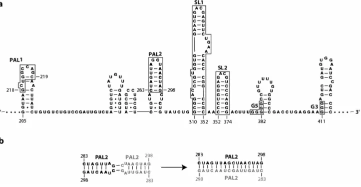
Figure 1.3. Conventional RNA secondary structure for a representative portion of the dimer linkage sequence. (a) Regions thought to be involved with RNA dimerization are highlighted with boxes. (b) Conventional PAL2 loop-loop interaction and intermolecular extended duplex formation.
Previous in vitro chemical mapping studies14 were used to propose what is now the conventional secondary structure for the DLS. The conventional RNA secondary structure of a representative portion for MuSV DLS is shown in Figure 1.3a. Multiple regions within the DLS have been implicated in dimerization (see Fig. 1.3a, highlighted in boxes). PAL115,16 and PAL217,18 are self-complementary sequences that are proposed to interact with PAL1 and PAL2 sequences of a second RNA and form intermolecular extended duplexes (Fig 1.3b). SL1 and SL2 contain well conserved19 GACG tetraloops that form loop-loop interactions20 and, along with PAL2, are proposed to form a structure recognized by the nucleocapsid domain of the gag precursor protein21. G5 and G3 are purine rich sequences that can potentially form G-quartet motifs to stabilize the dimer structure22,23.
Various regions throughout the DLS have been implicated in dimerization, but their specific involvement in the process was still unclear. Thus, my first goal was to resolve the minimal RNA sequence in the DLS that mediated retroviral RNA dimerization. In Chapter 2, I define a 170-nucleotide minimal dimerization active sequence (MiDAS, nts 205-374) for MuSV by stringent competitive dimerization with the full length DLS at 60 oC in near-physiological ionic conditions.
1.1.3 Mapping Structural Consequences of Retroviral RNA Dimerization in the
8
binding sites in RNA26 making it difficult to distinguish between a single-stranded region or divalent metal binding site. Therefore, no final conclusions were made, but this study hinted at the existence of a much more flexible RNA than previously proposed for the dimerization domain. Thus, I sought another chemical and/or enzymatic technique to corroborate these results.
At this time, Edward Merino, Ph.D., Kevin Wilkinson, and Jennifer Coughlan in the Weeks laboratory had been developing a novel chemical mapping method for probing RNA structure at single nucleotide resolution using quantitative Selective 2'-Hydroxyl Acylation analyzed by Primer Extension (SHAPE)27. The reactivity of N-methylisatoic anhydride (NMIA) at the 2’-hydroxyl position of the RNA backbone is correlated directly to the local nucleotide flexibility. That is, NMIA reactivity is high in flexible (single-stranded) positions of the RNA and low in constrained (base-paired) positions (Fig. 1.5). Moreover, by targeting the 2’ hydroxyl position of the ribose moiety in the backbone, this chemistry is not biased to any specific nucleotide as seen in many traditional chemical and/or enzymatic probing technologies. Thus, throughout this thesis, I applied this methodology to understand the secondary structural features of both the monomer and final dimer MiDAS conformations (Chapter 2 and Chapter 3).
10
12
through the interconversion of multiple structures, all of which may have distinct, yet unresolved, dimerization activities.
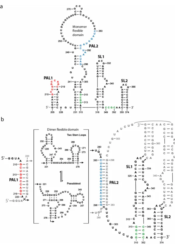
As discussed in Chapter 3, portions of the initial flexible domain become structured, while previously base-paired elements become incorporated into a new flexible domain in the final dimer state (Fig. 1.6b, nts 220-281). Much like the original, this new flexible domain also interconverts between structures and can form a two stem loop motif or even a pseudoknot. As expected, the self-complementary PAL1 and PAL2 sequences form extended duplexes with PAL1 and PAL2 sequences from the second RNA (Fig. 1.6b, colored red and blue, respectively) during dimerization. Interestingly, as the anchoring helix (nts 231-241, 305-315) breaks, the SL1 extends by four base-pairs (Figure 1.6b, colored green). Thus, the SL1-SL2 domain undergoes an RNA conformational switch.
1.1.4 An RNA switch enforces stringent retroviral genomic RNA dimerization.
PAL1 and PAL2, must mediate the selective interactions that are essential for viral RNA recognition.
In Chapter 4, I show that the SL1-SL2 domain undergoes a conformational RNA switch and extends the base of SL1 (Fig. 1.6b, green) upon dimerization. Therefore, this domain has two different conformations: an initial monomer-like and a final dimer. Both of these stem loop structures can form cross-strand loop-loop interactions20 with the SL1-SL2 domain of the second RNA. Strikingly, the initial monomer-like structure (see SL1-SL2 domain in Fig. 1.6a) forms highly selective cross-strand loop-loop interactions with the second viral RNA. But, in the final dimer structure (see Fig. 1.6b), these same interactions convert to a higher affinity, but less selective form.
Thus, viral RNA recognition in MuSV appears to be selectively regulated by a two-step mechanism that advantageously uses two conformations of the SL1-SL2 domain. In the initial stages of dimerization, the virus uses the stringent selectivity of the monomer-like structure to identify another viral RNA and form an interaction. Then, immediately upon recognition and initial dimerization, the SL1-SL2 domain switches to a higher affinity structure that would stabilize the interaction and prevent loss of the viral RNA strand.
1.1.5 Summary of Chapters. In Chapters 2 and 3, I apply two ideas that can be used
14
RNAstructure32. In Chapters 3 and 4, I then show that retroviral RNA dimerization should no longer be regarded as a collection of linked stem-loop and helical structures, but instead as a complex RNA folding problem. Site-directed and solvent-based hydroxyl radical footprinting data indicate that the final dimer structure folds into a compact three-dimensional shape.
1.1.6 Conclusion. Retroviral dimerization is a well conserved event among
retroviruses and thus is not only a potential target for disruption in anti-viral therapies, but is monumental for understanding the retroviral life cycle. However, when I first initiated this project, the RNA elements involved and the structural consequences associated with retroviral dimer formation were unresolved. Thus, in this thesis, I have created a generalizeable approach for characterizing the dimerization domain for any retroviral RNA model. Then, I further show that after resolving the dimerization domain, high throughput RNA SHAPE chemistry can be employed to authoritatively interrogate and define the structural features associated with both monomer and dimer states of the virus. My structural analysis experiments suggest that existing secondary structure models that have led the gamma retroviral field for the past ten years require careful reinterpretation. Thus, I have laid a new solid foundation that researchers can use to create useful theoretical proposals in regards to retroviral RNA dimerization and structure.
1.2References
1. Murti, K.G., Bondurant, M., and Tereba, A. Secondary structural features in the 70S RNAs of Moloney murine leukemia and Rous sarcoma viruses as observed by electron microscopy. J. Virol. 37, 411-419 (1981).
2. Paillart, J., Berthoux, L., Ottmann, M., Darlix, J., Marquet, R., Ehresmann, B., and Ehresmann, C. A dual role of the putative RNA dimerization initiation site of human immunodeficiency virus type 1 in genomic RNA packaging and proviral DNA synthesis. J. Virol. 70, 8348-8354 (1996).
3. Mikkelsen, J.G., Lund, A.H., Duch, M., and Pedersen, F.S. Recombination in the 5' leader of murine leukemia virus is accurate and influenced by sequence identity with a strong bias toward the kissing-loop dimerization region. J. Virol. 72, 6967-6978 (1998).
4. Mikkelsen, J.G., Lund, A.H., Duch, M., and Pedersen, F.S. Mutations of the kissing-loop dimerization sequence influence the site specificity of murine leukemia virus recombination in vivo. J. Virol. 74, 600-610 (2000).
5. Darlix, J.L., Lapadat-Tapolski, M., de Rocquigny, H., and Roques, B.P. First glimpses at structure-function relationships of the nucleocapsid protein of retroviruses. J. Mol. Biol. 254, 523-537 (1995).
6. Laughrea, M., Jette, L., Mak, J., Kleiman, L., Liang, C., and Wainberg, M. Mutations in the kissing-loop hairpin of human immunodeficiency virus type 1 reduce viral infectivity as well as genomic RNA packaging and proviral DNA synthesis. J. Virol. 71, 3397-3406 (1997).
7. Hibbert, C.S., Mirro, J., and Rein, A. mRNA molecules containing murine leukemia virus packaging signals are encapsidated as dimers. J. Virol. 78, 10927-10938 (2004). 8. Russell, R.S., Liang, C., & Wainberg, M.A. Is HIV-1 RNA dimerization a
prerequisite for packaging? Yes, no, probably? Retrovirology 1, 23 (2004).
9. D'Souza, V. & Summers, M.F. How retroviruses select their genomes. Nature Rev. Microbiol. 3, 643-655 (2005).
10. Fu, W., & Rein, A. Maturation of dimeric viral RNA of moloney murine leukemia virus. J. Virol. 67, 5443-5449 (1993).
16
12. Prats, A.C., Roy, C., Wang, P. A., Erard, M., Housset, V., Gabus, C., Paoletti, C., and Darlix, J. L. Cis elements and trans-acting factors involved in dimer formation of murine leukemia virus RNA. J. Virol. 64, 774-783 (1990).
13. Roy, C., Tounekti, N., Mougel, M., Darlix, J., Paoletti, C., Ehresmann, B., and Paoletti, J. An analytical study of the dimerization of in vitro generated RNA of Moloney murine leukemia virus, Mo-MuLV. Nucl. Acids Res. 18, 7287-7292 (1990). 14. Tounekti, N., Mougel, M., Roy, C., Marquet, R., Darlix, J., Paoletti, J., Ehresmann,
B., and Ehresmann, C. Effect of dimerization on the conformation of the
encapsidation psi domain of the Moloney murine leukemia virus. J. Mol. Biol. 223, 205-220 (1992).
15. Oroudjev, E.M., Kang, P. C. E., and Kohlstaedt, L. A. An additional dimer linkage structure in Moloney murine leukemia virus RNA. J. Mol. Biol. 291, 603-613 (1999). 16. Ly, H., and Parslow, T. G. Bipartite signal for genomic RNA dimerization in
Moloney murine leukemia virus. J. Virol. 76, 3135-3144 (2002).
17. Girard, P.M., Bonnet-Mathoniere, B., Muriaux, D., & Paoletti, J. A short
autocomplementary sequence in the 5' leader region is responsible for dimerization of MoMuLV genomic RNA. Biochemistry 34, 9785-9794 (1995).
18. Paillart, J., Marquet, R., Skripkin, E., Ehresmann, C., & Ehresmann, B. Dimerization of retroviral genomic RNAs: structural and functional implications. Biochimie 78, 639-653 (1996).
19. Konings, D.A.M., Nash, M.A., Maizel, J.V. & Arlinghaus, R.B. Novel
GACG-hairpin pair motif in the 5' untranslated region of type C retroviruses related to murine leukemia virus. J. Virol. 66, 632-640 (1992).
20. Kim, C., & Tinoco, I. A retroviral RNA kissing complex containing only two GC base pairs. Proc. Natl. Acad. Sci. USA 97, 9396-9401 (2000).
21. D'Souza, V., Melamed, J., Habib, D., Pullen, K., Wallace, K., & Summers, M.F. Identification of a high affinity nucleocapsid protein binding element within the Moloney murine leukemia virus Psi-RNA packaging signal: Implications for genome recognition. J. Mol. Biol. 314, 217-232 (2001).
23. Ly, H., Nierlich, D., Olsen, J., & Kaplan, A. Functional characterization of the dimer linkage structure RNA of Moloney murine sarcoma virus. J. Virol. 74, 9937-9945 (2000).
24. Behlen, L.S., Sampson, J.R., DiRenzo, A.B., and Uhlenbeck, O.C. Lead-catalyzed cleavage of Yeast tRNAPhe mutants. Biochemistry 29, 2515-2523 (1990).
25. Winter, D., Polacek, N., Halama, I., Streicher, B., and Barta, A. Lead-catalysed specific cleavage of ribosomal RNAs. Nucl. Acids Res. 25, 1817-1824 (1997).
26. David, L., Lambert, D., Gendron, P., and Major, F. Leadzyme. Methods Enzymol 341, 518-540 (2001).
27. Merino, E.J., Wilkinson, K.A., Coughlan, J.L. & Weeks, K.M. RNA structure analysis at single nucleotide resolution by selective 2'-hydroxyl acylation and primer extension (SHAPE). J. Am. Chem. Soc. 127, 4223-4231 (2005).
28. Herschlag, D. Implications of ribozyme kinetics for targeting the cleavage of specific RNA molecules in vivo: more isn't always better. Proc. Natl. Acad. Sci. USA 88, 6921-5 (1991).
29. Wang, S., Friedman, A.E. & Kool, E.T. Origins of high sequence selectivity: a stopped-flow kinetics study of DNA/RNA hybridization by duplex- and triplex-forming oligonucleotides. Biochemistry 34, 9774-84 (1995).
30. Fisher, J., & Goff, S.P. Mutational analysis of stem-loops in the RNA packaging signal of the moloney murine leukemia virus. Virology 244, 133-145 (1998). 31. Aagaard, L., Rasmussen, S.V., Mikkelsen, J. G., & Pedersen, F. S. Efficient
replication of full-length murine leukemia viruses modified at the dimer initiation site regions. Virology 318, 360-370 (2004).
Chapter 2
2.1 Introduction
Retroviruses selectively package two sense-strand genomic RNAs. These genomic RNAs are linked together near their 5' ends1 by a precise, but poorly understood, set of non-covalent interactions. The structure of this RNA "dimer" is implicated in multiple stages of the retroviral infectivity cycle, including RNA encapsidation into nascent viral particles2-5 and recombination during reverse transcription3,6,7. Retroviruses are both valuable biotechnology tools, as gene therapy vectors, and are the causative agents of serious diseases, including the acquired immunodeficiency syndromes and several cancers. Understanding the mechanism of retroviral dimerization thus represents an important opportunity for both enhancing vector function and for disrupting the infectivity cycle of pathogenic viruses.
Because the retroviral dimerization sequences are highly conserved8-10, a consensus secondary structure cannot be inferred by phylogenetic covariation analysis, which is the most robust method to determine the secondary structure for a large RNA11-14. Current secondary structure models for retroviral RNA dimerization domains are still provisional and likely only partially encompass the biological function of these RNAs. Determining the biologically relevant structure of the dimerization domain for any retroviral RNA is thus representative of a broad class of problem in biology. This challenge is to understand an RNA secondary structure in enough detail to be able to formulate hypotheses about biological function, even though only one or a few highly similar sequences are known.
20
prediction of even a few helices in a functionally important region makes it difficult or impossible to develop robust biological models.
Among the gamma retroviruses, several sequences have been consistently proposed as important for dimerization of the RNA genome8-10 (summarized in Fig. 2.1a). PAL1, also known as the 204-227 stem loop17, DIS118, or SL-B'5, and PAL2, also known as DIS218, SL-B19 or H120, are currently postulated to form hairpin loops (in green, Fig. 2.1a). PAL1 and PAL2 span self-complementary ('palindromic') sequences and are conventionally proposed to interact via loop-loop interactions with PAL1 and PAL2 sequences from a second RNA and eventually form extended duplexes in the dimer10,17-19. Highly conserved GACG tetraloops9 (Fig. 2.1a) in stem-loops 1 and 2 (SL1 and SL2, also known as SL-C19 or H220 and SL-D19 or H320) have the potential to form stable loop-loop interactions via cross-loop G-C base pairs19,21 and appear to be important for packaging via interactions with the viral Gag protein22.
conformationally flexible, which may facilitate retroviral RNA dimerization by decreasing the energetic cost of disrupting base pairing or other interactions in the monomer prior to forming functional structures specific to the dimer.
2.2 Results
2.2.1 Rigorous Definition of a MiDAS. In vitro studies using synthetic RNA
transcripts have been essential for identifying candidate structures that contribute to dimerization in the gamma retroviruses8,10,17-20,25-29. A significant challenge in interpreting these experiments is that most RNAs containing a stem-loop structure will dimerize if dimerization reactions are performed at sufficiently high RNA concentration or ionic strength conditions30,31. Neither the minimal region required for dimerization nor the structure of the dimerization-active domain in the viral genome monomeric starting state is currently well defined.
22
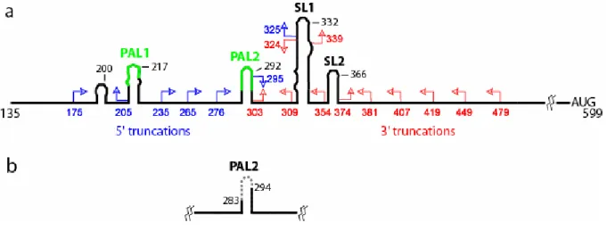
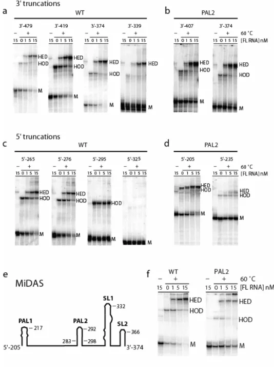
Figure 2.1. 5'-Untranslated region of MuSV. Conserved sequences and conventionally proposed secondary structures are illustrated schematically. The 5' genomic RNA cap is position 1. (a) 5' and 3' truncation mutants are shown in blue and red, respectively. (b)
24
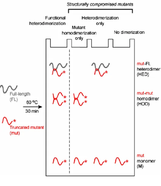
I constructed an extensive series of viral sequences containing systematic truncations from their 5' and 3' ends (blue and red arrows, respectively, in Fig. 2.1a). Competitive dimerization experiments were performed at 60 °C in the presence of the full-length (FL) transcript and visualized by the selective detection of the radiolabeled, truncated RNA variants in non-denaturing gels (Fig. 2.2). Both the radiolabeled mutant-mutant homodimer (HOD) and mutant-full length heterodimer (HED) are visualized directly. Full length RNA homodimers also form but are not radiolabeled and thus are not observed.
To be scored as a structurally competent dimerization active sequence, the mutant RNA (mut) must quantitatively compete with homodimerization by the (unlabeled) full length RNA. RNAs that are only able to homodimerize or only heterodimerize (see middle two lanes in Fig. 2.2) are scored as structurally deficient. Experiments further used a range of full-length RNA concentrations in order to make the experiment structurally stringent (Fig. 2.3).
2.2.2 A Minimal Sequence Active in Dimerization. Truncation mutants are
identified by the 5' or 3' nt at which the mutant sequence terminates (Fig. 2.1a). Competitive dimerization experiments were performed with ~ 1.5 nM radiolabeled, truncated RNA and 1, 5 or 15 nM full length (FL) RNA (Fig. 2.3). Markers for the mobilities of the mutant monomer (M) and for the mutant homodimer (HOD) were obtained by omitting the heating step or addition of FL RNA, respectively.
26
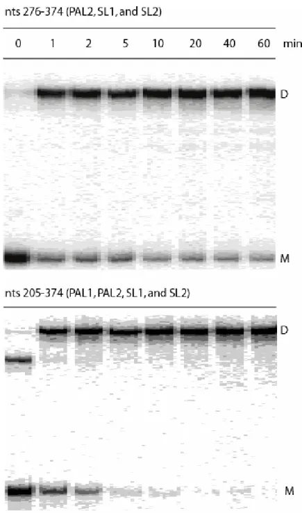
Truncations from the 5' end through position 276 yield RNAs that are fully functional in both homo-and heterodimerization (Fig. 2.3c). In contrast, truncation through position to 295 yields an RNA that homodimerizes well but is essentially incompetent to form heterodimers with the FL RNA (5'-295 panel in Fig. 2.3c). Further truncation through 5'-325 yields an RNA that forms neither homo- nor heterodimers. The 5' boundary for the minimum dimerization active sequence was thus set at position 276.
This minimal dimerization active sequence spans positions 276 to 374, and includes PAL2, SL1, and SL2. Importantly, the behavior of the 5'-295 mutant illustrates the stringency of the competitive dimerization assay. Although this RNA would have been scored as dimerization competent under less stringent conditions, it clearly lacks key elements required to dimerize competitively with the full length RNA.
2.2.3 Deletion of PAL2 Unmasks the Contribution of PAL1. The minimal
dimerization active region, defined by this initial analysis, includes the PAL2 sequence which several groups8,18-20,25-27,32 have proposed plays a role in dimerization. To explore whether any other accessory sequence contributes to dimerization, but is masked by PAL2, I compromised PAL2 by removing nts 283-294 (∆PAL2 mutant, Fig. 2.1b) and re-tested the panel of 5' and 3' truncations by competition with a full-length RNA, also harboring the
∆PAL deletion. All of the 3' truncations in the ∆PAL2 context through nt 374 formed both homo- and heterodimers efficiently (Fig. 2.3b), indicating that the 3' boundary of the minimal dimerization active sequence remains at position 374.
28
the RNA was truncated through 5'-235 in the ∆PAL2 context (5'-235 panel, Fig. 2.3d). In addition, when time resolved dimerization experiments are performed with a construct spanning the PAL2 through SL2 sequences, this RNA dimerizes only to about 80% (at concentrations up to 50 nM). In contrast, RNAs that also include the additional 5' 205-275 sequence dimerize essentially to completion (Fig. 2.3f and compare gels in Fig. 2.4). Thus, I concluded that the role of sequences that include the PAL1 region (at nts 205-217) is partially masked if PAL2 is present and assigned the 5' boundary of the minimal dimerization domain to be position 205 (Fig. 2.3d).
2.2.4 A Minimal Dimerization Domain. I constructed a minimal RNA spanning
MuSV sequences from 205 to 374 and tested the ability of this RNA to function in the competitive dimerization assay, both in the native PAL2 and ∆PAL2 contexts (Fig. 2.3f). Both RNAs form single-conformation monomers, homodimers, and heterodimers identical to those observed for native-like truncations. I infer that nts 205-374 span the minimal dimerization domain for MuSV and, potentially, for most gamma retroviruses.
30
positions 215 to 404 from the Moloney murine leukemia virus is sufficient to increase packaging of a nonviral RNA, in dimeric form, by 50-fold5. To the extent that other viral components like the nucleocapsid protein augment, but do not fundamentally alter, an RNA-centered process, the MiDAS represents a rigorously vetted minimal domain for retroviral dimerization.
2.2.5 MiDAS Monomeric Structure Analyzed by RNA SHAPE Chemistry. The
Weeks laboratory has recently developed a single nucleotide resolution approach to interrogate, quantitatively, the local environment at every nucleotide in an RNA23,24. RNA ribose 2'-hydroxyl groups react with N-methylisatoic anhydride (NMIA) to form the nucleotide 2'-ester as shown in Equation 2.1:
transcriptase-mediated primer extension: the complete experiment involves selective 2'-hydroxyl acylation analyzed by primer extension (SHAPE).
SHAPE experiments were performed in the context of a MiDAS RNA with 30 and 5 nt native sequence extensions, respectively, at the 5' and 3' ends to facilitate analysis of the entire domain by primer extension. I also appended an RNA cassette23 to the 3' end that contains an efficient DNA primer binding site. This 3' cassette folds independently of flanking RNA structures (as described below and in refs. 23,24).
Refolded MiDAS RNA was treated with NMIA under physiological-like conditions (200 mM potassium acetate, 5 mM MgCl2, pH 8, 37 °C); the monomeric state of the RNA was confirmed by native gel analysis. Sites of 2'-O-adduct formation were detected by primer extension and resolved on sequencing gels (Fig. 2.5a). Comparison of reactions performed in the presence of NMIA with reactions omitting the reagent reveals selective formation of 2'-O-adducts at a subset of sites in the RNA (compare + and – NMIA lanes in the MiDAS panel, Fig. 2.5a). Individual band intensities were integrated37 and absolute reactivities were computed for every position in the MiDAS RNA construct.
32
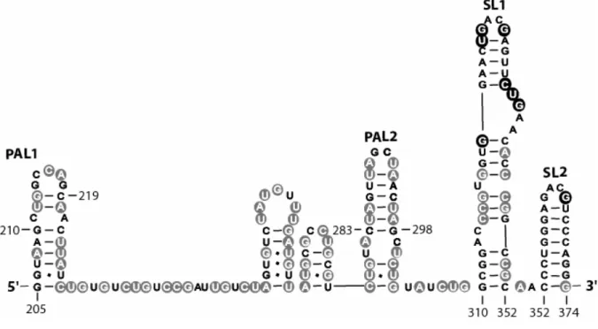
The nucleotide resolution SHAPE experiment provides a large number of constraints that must be accommodated in any secondary structure prediction for the MiDAS RNA. I screened secondary structures for the MiDAS region (residues 205-374) by submitting positions whose calculated reactivity was at least 25% of the strongest observed reactivities (47 nts total) as chemical modification constraints to the RNAStructure 4.11 program15. The quantitative data is shown superimposed on a secondary structure consistent with the entire body of SHAPE reactivity (Fig. 2.6).
Residues with high and moderate reactivity (red and orange, Fig. 2.6) towards NMIA are located in single-stranded loops and connecting structures. Positions with low or undetectable reactivity (blue and black, Fig. 2.6) lie largely in base paired helices. Because SHAPE is sensitive to any interaction, including non-canonical interactions, that constrain a nucleotide23, reactive positions should fall cleanly in flexible RNA structures; whereas, some unreactive nucleotides, that lie in nominally single-stranded regions, may reflect tertiary structure constraints that remain to be defined at this stage of analysis. As expected23,24, the 3' RNA structure cassette (Fig. 2.6, top) has a reactivity pattern exactly consistent with its designed fold, indicating that this appended structure does not interfere with folding of the MiDAS RNA.
34
2.2.6 PAL2 is Unstructured in the MiDAS Monomer. Because the observed
structure is significantly different from conventional models for the dimerization domain, I analyzed the structure of two MiDAS RNAs carrying instructive mutations in PAL2 (Fig. 2.5b). The first mutant, PALSTB (PAL stabilization), was designed to stabilize PAL2 in the
conventional stem-loop structure by increasing the G-C base pair content at flanking helix positions (circled positions, Fig. 2.5b). Inspection of the SHAPE data shows that stabilizing the PAL2 duplex has the desired effect. Nucleotides located in the PAL2 loop are strongly reactive while base paired positions in the stem are now much less reactive than in the native sequence (compare MiDAS and PALSTB lanes, Fig. 2.5a).
The experimental SHAPE reactivity data for the PALSTB mutant was subtracted from that for the native MiDAS RNA to create a quantitative difference map for every position in the PALSTB RNA (Fig. 2.7a). In the difference map, residues that are more reactive or are more constrained in the mutant relative to the native MiDAS sequence are reported as positive and negative amplitudes, respectively (red and blue, Fig. 2.7). If PAL2 already existed as a hairpin in the monomeric native state, stabilizing this stem should have a minimal effect on global MiDAS RNA structure. In strong contrast to this expectation, stabilizing the PAL2 sequence as a stem-loop causes large changes to the SHAPE reactivity in the MiDAS domain.
36
structure that extend almost the entire length of the RNA and up to 80 nts away (see especially nts 252-268 in the flexible domain and nts 374-382 between SL2 and the 3' end of the RNA, Fig. 2.7a). The peaks shown in the difference map are plotted on a scale comparable to that used in Fig. 2.6. Thus, the large positive peaks centered at positions 255, 312, 365 and 380 represent significant enhancements in absolute local nucleotide flexibility in these regions (for example, compare the 255 region in Fig. 2.5 with the difference map in Fig. 2.7a).
38 2.3 Discussion
The model for the minimal dimerization active structure of MuSV makes use of two innovations that are likely to be generalizable to any RNA structure prediction problem. First, I defined a minimal sequence for dimerization using an assay that requires the simplified RNA to functionally compete with a native-like sequence (Fig. 2.2). Second, SHAPE chemistry quantitatively interrogates every nucleotide in an RNA, which means that secondary structure models can be evaluated with much greater confidence than when using traditional chemical and enzymatic reagents.
Although the MiDAS secondary structure (Fig. 2.6) proposed here differs significantly from earlier models, this structure is consistent with the two existing sets of experimental information for dimerization domains in gamma retroviruses. The sequence of the murine leukemia virus (MuLV) is almost identical to that of MuSV. The nucleotide resolution SHAPE information strongly supports the original MuLV model8 for SL2 and the upper portion of SL1. In contrast, SHAPE does not support the earlier proposal that PAL2 forms a stable stem-loop structure. However, superposition of the chemical mapping information for MuLV on our secondary structure for MuSV shows that the prior information is exactly consistent with the current MiDAS proposal (Fig. 2.8). In particular, the PAL2 sequence is strongly reactive towards conventional single-strand-selective chemical reagents8 and thus consistent with the idea that PAL2 lies in a flexible domain.
40
strongly support formation of an internal flexible domain (Fig. 2.8). Many sites in the HaSV domain are cleaved by both single and double strand-selective RNases28 (Fig. 2.8): the HaSV RNA likely contains a flexible domain in which portions of the structure are alternately both paired and flexible in distinct conformations. That the MuLV and HaSV RNAs fold to similar monomeric starting structures provides a structural basis for the observation that these viruses readily heterodimerize28, presumably via PAL1.
I folded the flexible domain, including its anchoring helix (spanning positions 231-315; Fig. 2.6), subject to the requirement that the 27 positions with high and moderate reactivities be single stranded. The lowest energy structure, which is compatible with all of the SHAPE information (Fig. 2.6) has a total calculated15 folding free energy of only –10.5 kcal/mol. This single low energy structure spans 84 nts and thus has a net stability comparable to a simple stem-loop structure containing roughly 3 base pairs. Moreover, although the entire flexible domain from nt 249 through 294 contains no instances in which there are more than two strongly constrained nts in a row (black positions, Fig. 2.6), individual nts vary significantly in their 2'-hydroxyl reactivity.
42
structures in which they are single stranded (always paired and always single stranded are black and white, respectively). Each structure A, B and C is only partially consistent with the SHAPE data. Each of these structures, however, has a calculated folding free energy of approximately –17 kcal/mol and, thus is significantly more stable than the single consensus structure that incorporates all of the flexibility information. In this semi-quantitative analysis, in which the large universe of possible structures is approximated by three low energy structures (Fig. 2.9), there is a very strong correlation between SHAPE reactivity and the extent to which individual nucleotides are constrained, as averaged over these structures.
Models for the genomic RNA retroviral dimerization domain, in which PAL2 forms a stable stem-loop structure (see Figs. 2.1a and 2.5b), have guided the gamma retrovirus field for over a decade. However, the model-independent SHAPE intensity information (Figs. 2.5a and 2.6) emphasize that existing structural models merit careful reinterpretation.
Because PAL2 (and PAL1) sequences are self-complementary, an attractive model for the noncovalent interactions that stabilize the retroviral dimer is for these sequences to form an extended duplex in the dimer10,17-19. Prior models that proposed that PAL2 initially exists as a stable stem-loop recognized that it might be energetically costly to disrupt the extensive pre-existing base pairing in this structure. These models thus generally proposed that dimerization proceeds stepwise, first, via base pairing between nucleotides in the loops of two PAL2 stem-loop structures, followed by helix extension.
44 2.4 Materials and Methods
2.4.1 Retroviral RNA Transcripts. DNA templates for in vitro transcription of the
full length RNA, 5' and 3' truncations, and MiDAS constructs were generated by PCR from the pLNBS26,27 plasmids generously gifted by A. Kaplan. PCR (1 mL; 1 cycle, 95 oC, 5 min, [32 cycles, 95 oC, 30 sec; 55 oC, 30 sec; 72 oC, 1 min], 1 cycle, 72 oC, 10 min) reactions contained 0.2 mM of each dNTP, 1x PCR buffer (0.01 M Tris (pH 7.4), 0.05 M KCl), 0.5 µM each of forward and reverse primer, 2.5 mM MgCl2, 0.1 ng/µl pLNBS template, and 5 µl of Taq polymerase (5 U/µl, Invitrogen). RNA constructs were generated using T7 RNA polymerase-mediated transcription (500 µL; 37 °C, 5 h) containing 80 mM Hepes (pH 7.4), 40 mM dithiothreitol (DTT), 0.01% (v/v) Triton X-100, 2 mM spermidine, 10 mM MgCl2, 2 mM each nucleoside triphosphate, ~ 25 µg of PCR-generated template, 20 U of SUPERase-In (Ambion), and 0.1 mg/mL polymerase. SUPERase-Internally labeled RNAs were synthesized using 20 µCi α-[32P]-ATP and unlabeled ATP at 0.5 mM. RNAs were purified by denaturing gel electrophoresis (5% polyacrylamide, 7 M urea), excised from gel, eluted overnight into 1/2× TBE (45 mM Tris-borate, 1 mM EDTA), and concentrated by ethanol precipitation. RNAs were resuspended in HE [10 mM Hepes (pH 7.5), 1 mM EDTA] and stored at -20 °C.
polyacrylamide in 1x TBE) were pre-run for 15 min prior to sample loading and subsequently run for 2 hr at 20 W. Gel imaging was done on a phosphorimager (Molecular Dynamics) and analyzed using ImageQuant.
2.4.3 Time-resolved Dimerization Assay. [32P]-internally-labeled viral RNA constructs (~1.5 nM) spanning nts 205-374 and 276-374, respectively, were incubated with their same unlabeled RNA (at 50 nM) in 15 µL. Reactions were then heated to 90 oC for 3 min, cooled on ice, and mixed with 5 µL 4× dimerization buffer [25 °C; 200 mM Hepes (pH 7.5), 800 mM potassium acetate (pH 7.5), 20 mM MgCl2], and incubated at 60 oC for 0, 1, 2, 5, 10, 20, 40, and 60 min. Samples were separated at 4 oC as previously discussed above in Competitive Dimerization assay section (5% polyacrylamide in 1x TBE). ImageQuant software was then used to quantify the band intensities and fraction dimer was calculated by dividing intensity of dimer by the total sum of intensities for both monomer and dimer.
2.4.4 SHAPE Analysis of Monomer MiDAS. 2'-Hydroxyl acylation and primer
46
2.4.5 Primer Extension. Two DNA primers were used to analyze the MiDAS RNA
2.5 References
1. Murti, K.G., Bondurant, M., and Tereba, A. Secondary structural features in the 70S RNAs of Moloney murine leukemia and Rous sarcoma viruses as observed by electron microscopy. J. Virol. 37, 411-419 (1981).
2. Darlix, J.L., Lapadat-Tapolski, M., de Rocquigny, H., and Roques, B.P. First glimpses at structure-function relationships of the nucleocapsid protein of retroviruses. J. Mol. Biol. 254, 523-537 (1995).
3. Paillart, J., Berthoux, L., Ottmann, M., Darlix, J., Marquet, R., Ehresmann, B., and Ehresmann, C. A dual role of the putative RNA dimerization initiation site of human immunodeficiency virus type 1 in genomic RNA packaging and proviral DNA synthesis. J. Virol. 70, 8348-8354 (1996).
4. Laughrea, M., Jette, L., Mak, J., Kleiman, L., Liang, C., and Wainberg, M. Mutations in the kissing-loop hairpin of human immunodeficiency virus type 1 reduce viral infectivity as well as genomic RNA packaging and proviral DNA synthesis. J. Virol. 71, 3397-3406 (1997).
5. Hibbert, C.S., Mirro, J., and Rein, A. mRNA molecules containing murine leukemia virus packaging signals are encapsidated as dimers. J. Virol. 78, 10927-10938 (2004). 6. Mikkelsen, J.G., Lund, A.H., Duch, M., and Pedersen, F.S. Recombination in the 5'
leader of murine leukemia virus is accurate and influenced by sequence identity with a strong bias toward the kissing-loop dimerization region. J. Virol. 72, 6967-6978 (1998).
7. Mikkelsen, J.G., Lund, A.H., Duch, M., and Pedersen, F.S. Mutations of the kissing-loop dimerization sequence influence the site specificity of murine leukemia virus recombination in vivo. J. Virol. 74, 600-610 (2000).
8. Tounekti, N., Mougel, M., Roy, C., Marquet, R., Darlix, J., Paoletti, J., Ehresmann, B., and Ehresmann, C. Effect of dimerization on the conformation of the
encapsidation psi domain of the Moloney murine leukemia virus. J. Mol. Biol. 223, 205-220 (1992).
9. Konings, D.A.M., Nash, M.A., Maizel, J.V. & Arlinghaus, R.B. Novel
GACG-hairpin pair motif in the 5' untranslated region of type C retroviruses related to murine leukemia virus. J. Virol. 66, 632-640 (1992).
10. Paillart, J., Marquet, R., Skripkin, E., Ehresmann, C., and Ehresmann, B.
Dimerization of retroviral genomic RNAs: Structural and functional implications. Biochimie 78, 639-653 (1996).
48
12. Frank, D.N., and Pace, N. R. Ribonuclease P: unity and diversity in a tRNA processing ribozyme. Annu. Rev. Biochem. 67, 153-80 (1998).
13. Gutell, R.R., Lee, J. C., and Cannone, J. J. The accuracy of ribosomal RNA comparative structure models. Curr. Opin. Struct. Biol. 12, 301-310 (2002). 14. D'Souza, V., Dey, A., Habib, D., and Summers, M. F. NMR Structure of the
101-nucleotide core encapsidation signal of the Moloney murine leukemia virus. J. Mol. Biol. 337, 427-442 (2004).
15. Mathews, D.H. et al. Incorporating chemical modification constraints into a dynamic programming algorithm for prediction of RNA secondary structure. Proc. Natl. Acad. Sci. USA 101, 7287-7292 (2004).
16. Dowell, R.D. & Eddy, S.R. Evaluation of several lightweight stochastic context-free grammars for RNA secondary structure prediction. BMC Bioinformatics 5, 71 (2004). 17. Oroudjev, E.M., Kang, P. C. E., and Kohlstaedt, L. A. An additional dimer linkage
structure in Moloney murine leukemia virus RNA. J. Mol. Biol. 291, 603-613 (1999). 18. Ly, H., and Parslow, T. G. Bipartite signal for genomic RNA dimerization in
Moloney murine leukemia virus. J. Virol. 76, 3135-3144 (2002).
19. D'Souza, V., Melamed, J., Habib, D., Pullen, K., Wallace, K., and Summers, M. F. Identification of a high affinity nucleocapsid protein binding element within the Moloney murine leukemia virus Ψ-RNA packaging signal: Implications for genome recognition. J. Mol. Biol. 314, 217-232 (2001).
20. De Tapia, M., Metzler, V., Mougel, M., Ehresmann, B., and Ehresmann,C.
Dimerization of the Moloney murine leukemia virus genomic RNA: Redefinition of the role of the palindromic stem-loop H1 (278-303) and new roles for stem-loops H2 (310-352) and H3 (355-374). Biochemistry 37, 6077-6085 (1998).
21. Kim, C., and Tinoco, I. A retroviral RNA kissing complex containing only two GC base pairs. Proc. Natl. Acad. Sci. USA 97, 9396-9401 (2000).
22. Rein, A., Harvin, D. P., Mirro, J., Ernst, S.M., and Gorelick, R. J. Evidence that a central domain of nucleocapsid protein is required for RNA packaging in murine leukemia virus. J. Virol. 68, 6124-6129 (1994).
23. Merino, E.J., Wilkinson, K.A., Coughlan, J.L. & Weeks, K.M. RNA structure analysis at single nucleotide resolution by selective 2'-hydroxyl acylation and primer extension (SHAPE). J. Am. Chem. Soc. 127, 4223-4231 (2005).
25. Girard, P.M., Bonnet-Mathoniere, B., Muriaux, D. & Paoletti, J. A short
autocomplementary sequence in the 5' leader region is responsible for dimerization of MoMuLV genomic RNA. Biochemistry 34, 9785-9794 (1995).
26. Ly, H., Nierlich, D., Olsen, J., and Kaplan, A. Moloney murine sarcoma virus genomic RNAs dimerize via a two-step process: A concentration-dependent kissing-loop interaction is driven by initial contact between consecutive guanosines. J. Virol. 73, 7255-7261 (1999).
27. Ly, H., Nierlich, D., Olsen, J., and Kaplan, A. Functional characterization of the dimer linkage structure RNA of Moloney murine sarcoma virus. J. Virol. 74, 9937-9945 (2000).
28. Rasmussen, S.V., Mikkelsen, J. G., and Pedersen, F. S. Modulation of homo- and heterodimerization of Harvey sarcoma virus RNA by GACG tetraloops and point mutations in palindromic sequences. J. Mol. Biol. 323, 613-628 (2002).
29. Aagaard, L., Rasmussen, S.V., Mikkelsen, J. G., and Pedersen, F. S. Efficient
replication of full-length murine leukemia viruses modified at the dimer initiation site regions. Virology 318, 360-370 (2004).
30. Holbrook, S.R., Cheong, C., Tinoco, I. Jr., and Kim, S. H. Crystal structure of an RNA double helix incorporating a track of non-Watson-Crick base pairs. Nature 353, 579-581 (1991).
31. Wild, K., Weichenrieder, O., Leonard, G. A., and Cusack, S. The 2 Å structure of helix 6 of the human signal recognition particle RNA. Structure Fold Des. 7, 1345-1352 (1999).
32. D'Souza, V., and Summers, M. F. Structural basis for packaging the dimeric genome of Moloney murine leukemia virus. Nature 431, 586-590 (2004).
33. Monie, T.P., Greatorex, J.S., Maynard-Smith, L., Hook, B.D.C., Bishop, N., Beales, L.P., and Lever, A.M.L. Identification and visualization of the dimerization initiation site of the prototype lentivirus, Maedi Visna virus: A potential GACG tetraloop displays structural homology with the α- and γ- retroviruses. Biochemistry 44, 294-302 (2005).
34. Chamberlin, S.I., Merino, E. J., and Weeks, K. M. Catalysis of amide synthesis by RNA phosphodiester and hydroxyl groups. Proc. Natl. Acad. Sci. USA 99, 14688-14693 (2002).
Chapter 3
3.1 Introduction
Retroviruses are well-characterized diploid entities in biology. Retroviruses package exactly two copies of their genomes that are in a dimeric state into each nascent virion1-3. Retroviral RNA genomes dimerize by conserved non-covalent interactions involving sequences near the 5’ end. Expectedly, this dimeric state appears to be a fundamental feature of retroviral biology4-6; thus, it is important to understand the structural details of this domain at high resolution.
I have previously used competitive-dimerization experiments to define a minimal dimerization active sequence (MiDAS)7 for the Moloney murine sarcoma virus (MuSV). This domain, defined in vitro, spans nts 205-374. Strikingly, the MiDAS corresponds very closely to the minimal sequences required to direct packaging of heterologous non-viral RNAs, as dimers, into nascent virions8.
The MiDAS domain includes sequence elements previously proposed to be important for forming the final dimer state including two self-complementary, or palindromic, sequences, PAL1 and PAL2, and two well conserved stem-loop motifs9, SL1 and SL2 (Fig. 3.1a). PAL1 and PAL2 have the potential to form extended heteroduplexes with their
complements in the dimer10-13. As is typical for most RNAs, the MiDAS domain contains well-structured helical elements. In addition, in the monomer state, the MiDAS RNA also includes a large flexible domain that is linked by a stable anchoring helix (nts 231-241, 305-315; Fig. 3.1a).
52
example, conformational changes from an initial stem-loop structure to a heteroduplex will only yield a small number of local structural changes at certain nucleotide positions. Thus, monitoring the monomer-to-dimer conversion at single nucleotide resolution with conventional chemical footprinting methodologies has been difficult. However, the recently developed RNA SHAPE14 chemistry allows local structural analysis at every nucleotide in an RNA to be quantitatively assessed.
54 3.2 Results
3.2.1 SHAPE Analysis of the Retroviral RNA Dimer. The MiDAS RNA forms
well-defined, single conformation monomer and dimer species, as analyzed by non-denaturing gel electrophoresis (Fig. 3.1b). The monomer migrates as a single band and can be converted to the dimer state upon heating at 60 oC for 30 minutes (100 nM RNA; pH 7.5, 200 mM potassium acetate and 5 mM MgCl2). Because the MiDAS RNA in the final dimer state migrates as a single species, it appears that my structural analysis reports on a single conformation without interfering contributions of alternatively folded RNAs.
I analyzed conformational changes that accompanied the monomer-to-dimer transition in the MiDAS domain using RNA SHAPE chemistry. In a SHAPE experiment, 2’-hydroxyl groups at flexible nucleotide positions will preferentially react with N-methylisatoic anhydride (NMIA) to form 2’-O-adducts. Thus, SHAPE chemistry quantitatively monitors local RNA flexibility at single nucleotide resolution.
MiDAS RNAs in the monomer or dimer states (Fig. 3.1b) were treated with NMIA and the sites of 2’-O-adduct formation were detected as stops to primer extension (Fig. 3.2a). Band intensities, corresponding to SHAPE reactivities, were then quantified15 for almost every nucleotide in the MiDAS domain for RNAs in both monomer and dimer states. Absolute reactivities were computed by subtracting background intensities observed in the absence of NMIA (compare (+) and (-) NMIA lanes, Fig. 3.2a; quantitative histograms are shown in Fig. 3.2b).
56
Fig. 3.2b with upper panel in Fig. 2.6 in Chapter 2). This similarity emphasizes the extreme
reproducibility of a SHAPE experiment
Upon forming the final dimer state, SHAPE chemistry reveals large structural changes throughout the MiDAS RNA (compare monomer and dimer experiments in Fig. 3.2a,b). I computed a monomer-to-dimer difference plot by subtracting absolute SHAPE
reactivities in the monomer state from those observed in the final dimer conformation. By this definition, positions that are more reactive in the dimer are positive, while RNA regions that become structured in the dimer are negative (see green and red bars, respectively, in the difference plot, Fig. 3.2b).
SHAPE reactivities in both PAL1 and PAL2 self-complementary sequences (nts 210-219, 283-298) decrease significantly which is consistent with formation of extended heteroduplexes in these regions of the final dimer. In addition, I observe significant conformational changes in the SL1-SL2 region (compare nts 314-315, 319, and 338-343 in upper and middle panel of Fig. 3.2b). This domain has been analyzed and discussed in Chapter 4 and is known to undergo a conformational switch in which SL1 becomes extended by four base-pairs at its base (Badorrek et al., unpublished). Overall, a striking feature of the monomer-to-dimer conformational change is that, with exception of PAL1 and PAL2, local nucleotide flexibility increases in most regions of the dimer, relative to the monomer (see green bars in Fig. 3.2b).
dimer panel, Fig. 3.2b) and is constrained on each side by the PAL1 and PAL2 duplexes. Regions previously with no or lower reactivity in the monomer have now become more reactive and flexible in this domain (see nts 232, 243, 246-247, and 254-258, middle panel, Fig. 3.2b).
3.2.2 Secondary Structure Model for the MiDAS Domain in the Final Dimer
State. The SHAPE information provides high resolution model-independent data about
base-pairing and tertiary interactions in the final dimer. Therefore, I used SHAPE reactivities to constrain RNA secondary structure prediction programs16,17 to obtain specified models of the secondary structure of this domain. I currently constrain my structural predictions using RNAstructure v4.216 by dividing absolute SHAPE reactivities into two classes. Positions with absolute reactivity between 50-100% of the maximum observed are required to be single-stranded (red in Fig. 3.3a). Positions with reactivities in the range of 25-50% of the maximum are required to be either single-stranded or adjacent to an unpaired nucleotide or a G-U base pair as implemented in the RNAstructure algorithm16.
58
predicted10. Instead, the region immediately 5’ to PAL1 (see Fig. 3.1a) does undergo a significant conformational change in which nucleotides 198-207 form a new stem loop containing a GNRA tetraloop at its apex. The SL1-SL2 forms an independent and compact tertiary structure domain (Badorrek et al., unpublished) as discussed in Chapter 4. SHAPE data indicates that the SL1-SL2 domain is linked to the PAL2 and the rest of the MiDAS via a highly flexible junction region (see nts 299-309, Fig. 3.3).
SHAPE reactivities do not support the formation of a single well-defined secondary structure in the region between the PAL1 and PAL2 extended duplexes in the final dimer. The thermodynamic model predicted by RNAstructure16 suggests a loose two stem loop motif (Fig. 3). RNAstructure, though, does not allow pseudoknot formation. I, therefore, searched for alternate secondary structures, consistent with SHAPE constraints, using the HotKnots program17, which is a heuristic algorithm that allows for pseudoknot formations. Interestingly, the experimental constraints for this region indicate a reasonable pseudoknot (Fig. 3.3). Finally, I also note that another alternative structure mediated by cross-strand interactions at nts 268-271 is also consistent with the SHAPE information.
60
3.2.3 Long-range Interactions in the MiDAS Final Dimer. I performed
site-directed hydroxyl radical cleavage18,19 on the MiDAS RNA in the final dimer state. A Fe(II)-EDTA moiety was attached at the 5’ end of the MiDAS RNA via a phosphorothioate-mediated thiol linkage (see insert, Fig. 3.4a). This derivatized RNA was allowed to heterodimerize with the trace concentrations of 3’-[32P]-end labeled MiDAS RNA. Upon addition of hydrogen peroxide and ascorbic acid, hydroxyl radicals are selectively generated in the vicinity of the Fe(II)-EDTA group and will cleave nearby RNA structures. A key feature of this experiment is that, with cleavage observed only in the radiolabeled RNA, I can detect long-range and cross-strand proximal RNA structures.
I calculated the absolute site-directed cleavage intensities by subtracting background observed when using a mock-derivatized RNA lacking the phosphorothioate group (Fig. 3.1a). Cleavages are judged to be strong or moderate if they are 5- or 3- times greater than
the average background, respectively. Cleavages are superimposed on our secondary structure model for the MiDAS domain in the final dimer state (Fig. 3.4b). For clarity, cleavage data is shown on one strand only, but is expected to be symmetrical over the two strands in the dimer.
62
MiDAS domain appears to form a reasonably compact structure in which the PAL1 lies near the PAL2 and SL1-SL2 domain.
3.2.4 Tertiary Structure in the MiDAS Dimer. Site-directed hydroxyl radical
cleavage supports the existence of long-range interactions in the MiDAS dimer such that the 5’ end of the RNA lies near the other conserved elements in the RNA. Thus, I probed for interactions in the MiDAS domain that could sufficiently protect the RNA from cleavages generated by solvent-based or untethered Fe(II)-EDTA reagent. MiDAS RNA was subjected to hydroxyl radical cleavage in conditions that either stabilized the formation of the native final dimer (Fig. 3.1b) or in the absence of mono and divalent ions (denaturing) where the RNA backbone is uniformly accessible to cleavage. A hydroxyl radical protection profile was generated by subtracting integrated band intensities for the RNA in the native final dimer state from those measured under denaturing conditions. Nucleotides that are protected or show enhanced cleavage in the final dimer in relation to the denatured state are indicated as positive and negative amplitudes, respectively (Fig. 3.5a).
64 3.3 Discussion
Using RNA SHAPE chemistry, I design a high resolution secondary structure model of the dimerization domain for the Moloney murine sarcoma virus. This model incorporates many features previously proposed to be important for retroviral genomic RNA dimerization. For example, since the SHAPE method provides robust single nucleotide resolution experimental information, I can now authoritatively define the termini of both the 10mer PAL1 and 16mer PAL2 heteroduplex. Previously, this was a structural detail that was unresolved in the literature.
In addition, SHAPE analysis also emphasizes the existence of flexible domains in retroviral dimerization sequences. Both MiDAS monomeric starting state and final dimer contain well-delimited unstructured, flexible domains that connect structured elements: the anchoring helix (nts 231-241/305-315) and PAL1 and PAL2 heteroduplexes, respectively (Figs. 3.1a, 3.3). Like in the monomer, flexibility in the dimer allows interconversion of multiple structures (see two stem loop and pseudoknot, Fig. 3.3). Thus, RNA flexibility appears to be a prominent feature in retroviral biology that is probably used to facilitate RNA dimerization by reducing the energetic costs for breaking and forming base-pairs during dimerization.
suggests that this duplex is almost completely inaccessible to cleavage and must be buried in the MiDAS dimer. Thus, PAL2 must lie sandwiched between the other RNA elements.
As discussed in Chapter 4, a three-dimensional structure for the isolated SL1-SL2 (Fig. 3.6) domain was experimentally refined. Strikingly, a careful comparison of solvent inaccessible regions in the isolated SL1-SL2 domain versus the SL1-SL2 in the entire MiDAS RNA yields additional strong and moderate hydroxyl radical protections at the base of SL1 (nts 312-315, 351-353) and in the middle of the SL1 (nts 321-323, 343) (see Fig. 3.6). The conservation of the protection pattern for the SL1-SL2 domain in the context of
MiDAS, as compared to its isolated version, suggests that this domain does fold independently as discussed in Chapter 4. Interestingly, the full length MiDAS dimer-specific extra protections lie on one face (side) of the SL1-SL2 RNA region (see asterisks and daggers, respectively; Fig. 3.7). The extensive protection pattern (both sides are protected!) of PAL2 and the additional protection sites (asterisks and daggers, respectively; Fig. 3.7) observed in the SL1-SL2 domain suggest that, after folding, the SL1-SL2 domain forms long-range tertiary structure interactions with PAL2.
66
68 3.4 Materials and Methods
3.4.1 Retroviral RNA Transcripts. DNA templates for in vitro transcription of the
MiDAS RNA construct that contained flanking 5' and 3' extensions of viral sequence of 30 and 5 nucleotides, respectively, were generated by PCR from the pLNBS20,21 plasmid using same conditions previously discussed in Chapter 2. RNA was generated using T7 RNA polymerase-mediated transcription (500 µL; 37 °C, 5 h) containing 80 mM Hepes (pH 7.4), 40 mM dithiothreitol (DTT), 0.01% (v/v) Triton X-100, 2 mM spermidine, 10 mM MgCl2, 2 mM each nucleoside triphosphate, ~ 25 µg of PCR-generated template, 20 U of SUPERase-In (Ambion), and 0.1 mg/mL polymerase. RNA was purified by denaturing gel electrophoresis (5% polyacrylamide, 7 M urea), excised from gel, eluted overnight into 1/2× TBE (45 mM Tris-borate, 1 mM EDTA), and concentrated by ethanol precipitation. RNA was stored in 10 mM Hepes (pH 7.5), 1 mM EDTA at -20 °C.
3.4.2 SHAPE Analysis of MiDAS Monomer and Dimer. NMIA modification,
3.4.3 205-BABE. A 170 nt viral RNA (nts 310-374) was transcribed in vitro in the
presence of 10 mM guanosine monophosphorothioate (GMPS, Biolog Life Science Institute); GMPS is incorporated only at the initiating 5' G during transcription. 50 pmol of this RNA was treated with excess (50 µmol) bromoacetamidobenzyl-EDTA (BABE, Dojindo Labs) and (50 µmol) (NH4)2Fe(SO4)2 in 10 µl 0.15 M potassium phosphate (pH 8.0) at 37 °C for 1.5 h, ethanol precipitated, and resuspended in 10 µl 0.15 M potassium phosphate (pH 8.0).
3.4.4 Site-directed Hydroxyl Radical Cleavage. MiDAS RNA construct was