Received: June 30, 2015; Revised: October 15, 2015; Accepted: January 5, 2016
© The Author 2016. Published by Oxford University Press. All rights reserved. For Permissions, please e-mail: [email protected]. doi:10.1093/jnci/djw004
First published online February 8, 2016 Article
1 of 9
ar
ticle
article
Association of Interferon Regulatory Factor-4
Polymorphism rs12203592 With Divergent
Melanoma Pathways
David C. Gibbs, Irene Orlow, Jennifer I. Bramson, Peter A. Kanetsky,
Li Luo, Anne Kricker, Bruce K. Armstrong, Hoda Anton-Culver,
Stephen B. Gruber, Loraine D. Marrett, Richard P. Gallagher, Roberto Zanetti,
Stefano Rosso, Terence Dwyer, Ajay Sharma, Emily La Pilla, Lynn From,
Klaus J. Busam, Anne E. Cust, David W. Ollila, Colin B. Begg,
Marianne Berwick, Nancy E. Thomas; on behalf of the GEM Study Group
Affiliations of authors: Department of Dermatology, University of North Carolina, Chapel Hill, NC (DCG, NET); Lineberger Comprehensive Cancer Center, University of North Carolina, Chapel Hill, NC (NET, DWO); Department of Epidemiology and Biostatistics, Memorial Sloan Kettering Cancer Center, NY (IO, AS, ELP, KJB, CBB); Department of Surgery, University of North Carolina, Chapel Hill, NC (JIB, DWO); Department of Cancer Epidemiology, H. Lee Moffitt Cancer Center & Research Institute, Tampa, FL (PAK); Department of Internal Medicine, University of New Mexico Cancer Center, University of New Mexico, Albuquerque, NM (LL, MB); Sydney School of Public Health, University of Sydney, Sydney, New South Wales, Australia (AEC, AK, BKA); Department of Epidemiology, University of California, Irvine, CA (HAC); USC Norris Comprehensive Cancer Center, University of Southern California, Los Angeles, CA (SBG); Department of Population Studies and Surveillance, Cancer Care Ontario, Toronto, Ontario, Canada (LDM); Cancer Control Research, British Columbia Cancer Agency, Vancouver, British Columbia, Canada (RPG); Piedmont Cancer Registry, Centre for Epidemiology and Prevention in Oncology in Piedmont, Turin, Italy (RZ, SR); The George Institute for Global Health, Oxford Martin School of Public Health, University of Oxford, Oxford, UK (TD); Department of Pathology, Women’s College Hospital, Toronto, Ontario, Canada (LF).Correspondence to: Nancy E. Thomas, MD PhD, Department of Dermatology, University of North Carolina, 405 Mary Ellen Jones Building, CB#7287, Chapel Hill, NC 27599 (e-mail: [email protected]).
Abstract
Background: Solar elastosis and neval remnants are histologic markers characteristic of divergent melanoma pathways linked to differences in age at onset, host phenotype, and sun exposure. However, the association between these pathway markers and newly identified low-penetrance melanoma susceptibility loci remains unknown.
Methods: In the Genes, Environment and Melanoma (GEM) Study, 2103 Caucasian participants had first primary melanomas that underwent centralized pathology review. For 47 single-nucleotide polymorphisms (SNPs) previously identified as low-penetrant melanoma risk variants, we used multinomial logistic regression to compare melanoma with solar elastosis and melanoma with neval remnants simultaneously to melanoma with neither of these markers, excluding melanomas with both markers. All statistical tests were two-sided.
Results: IRF4 rs12203592 was the only SNP to pass the false discovery threshold in baseline models adjusted for age, sex, and study center. rs12203592*T was associated positively with melanoma with solar elastosis (odds ratio [OR] = 1.47, 95% confidence interval [CI] = 1.18 to 1.82) and inversely with melanoma with neval remnants (OR = 0.65, 95% CI = 0.48 to 0.87)
compared with melanoma with neither marker (Pglobal = 3.78 x 10-08). Adjusting for phenotypic characteristics and total sun
exposure hours did not materially affect rs12203592’s associations. Distinct early- and late-onset age distributions were
ar
ticle
ar
ticle
Conclusions: Our findings suggest a role of IRF4 rs12203592 in pathway-specific risk for melanoma development. We
hypothesize that IRF4 rs12203592 could underlie in part the bimodal age distribution reported for melanoma and linked to
the divergent pathways.
Melanoma incidence has increased in fair-skinned populations worldwide in recent decades (1,2). Understanding melanoma etiology is critical for prevention; however, risk factors may differ by the pathway along which melanoma develops. The divergent pathway hypothesis postulates at least two separate pathways, one related to chronic sun exposure and the other related to the host’s tendency to develop nevi, with melano-mas marked, respectively, by the presence of histologic solar elastosis (SE) and neval remnants (NR) in adjacent tissue (3–5). Predictors of SE and NR adjacent to melanomas include host phenotypic characteristics; however, the association of these pathway markers with recently identified germline variants in melanoma-associated risk loci has yet to be explored (5–7).
The divergent pathway model postulates that melanoma risk associated with UV exposure differs by host characteristics and results in at least two causal pathways linked to differences in age at onset, body site, and histopathologic features (4,5,8). In individuals with low propensity to develop nevi, melanomas seem to require high levels of cumulative sun exposure (4,9). These chronic sun exposure–related melanomas, marked by SE, tend to occur at older ages, in individuals with fewer nevi, and on sun exposed body sites like the head/neck (3,5,9,10). Conversely, melanomas in individuals with a high propensity to develop nevi seem to require lower levels of sun exposure and arise from an existing nevus (4). These nevogenic melano-mas, marked by NR, tend to occur at younger ages, in individuals with higher nevus counts, and on less sun-exposed sites like the trunk (11–15).
Recent genome wide association studies (GWAS) and can-didate pathway studies have identified several low-penetrant genetic variants associated with cutaneous melanoma (16). The majority are in loci associated with pigmentation (eg, TYRP1, TYR, HERC2/OCA2, SLC45A2, and ASIP), nevi (eg, PLA2G6, MTAP,
and NID1), or both (eg, IRF4) (17–24); however, variants in other identified loci (eg, ATM, MX2, PARP1, ARNT, and CASP8) may not be associated with phenotypic risk traits for melanoma (21,25).
Within the large international population-based Genes, Environment, and Melanoma (GEM) Study, we previously inves-tigated 47 single-nucleotide polymorphisms (SNPs) in 21 of these putative low-penetrance melanoma susceptibility loci and found variants in the TERT, TYRP1, MTAP, TYR, NCOA6, and MX2
gene regions and a PARP1 haplotype to be associated with mul-tiple primary melanoma compared with single primary mela-noma (26). The purpose of this study was to determine whether these 47 SNPs influence susceptibility to melanomas arising from the chronic sun exposure or nevogenic pathways by exam-ining their associations with the exclusive presence of SE or NR adjacent to primary melanoma.
Methods
Study Population
The population-based case-only GEM Study enrolled 3579 patients diagnosed between 1998 and 2003 with incident pri-mary melanoma in Australia, Canada, Italy, and the United States (27,28). Of the 2953 (83% of 3567) Caucasian patients with centralized pathology review, 2103 (86% of 2458) were diagnosed
with a first primary melanoma and are included in the analyses reported here. The institutional review boards of participating institutions approved the protocol, and informed consent was obtained from participants.
Hair and eye color, ability to tan, number of back nevi, resi-dential histories, and sun exposure information were collected from phone interviews and self-administered questionnaires as described previously (9,29,30). Number of back nevi is statisti-cally significantly correlated with whole-body nevus density diagrams (31) in GEM, and the suitability of using this variable as a proxy for total body nevus counts has been previously reported (32–34). Total hours of sun exposure from the age of five years until the age at diagnosis were calculated as the sum of reported outdoor sun exposure hours between 9 am and 5 pm on working and nonworking days in warmer and cooler months (based on residential calendars) estimated for each decade of life (29). Average annual sun exposure was calculated as the total exposure hours divided by the participant’s age at diagno-sis minus five years (29).
Three dermatopathologists scored adjacent SE as previously described (9) and adjacent NR as present when benign nevus cells were in the epidermis or dermis immediately adjacent to or below the melanoma cells. Kappa statistics, using the three cat-egories, were 0.60 and 0.64 for scoring NR and SE, respectively, in a test set of 19 H&Es reviewed by the three dermatopathologists.
SNP Selection, Genotyping, and Principal Component Analysis
We selected 47 SNPs from 21 loci based on evidence they were low-penetrant risk variants for melanoma in other studies (26). DNA was collected from buccal swabs. SNPs were genotyped using the MassArray iPLEX chemistry and platform (Agena Bioscience), with quality control measures as described previ-ously (35). Proxy SNPs (r2 > 0.95; 1000 Genomes, CEU population; Proxy SNP; Broad Institute) rs6735656 and rs12278954 were sub-stituted, respectively, for CASP8 rs10931936 and ATM rs1801516 (21)—two SNPs of interest rejected during the assay design steps.
We performed principal component analysis (PCA) of the 47 SNPs with and without MC1R (available to this dataset [36,37]) to detect potential population structure within our data. Using an eigen-value decomposition approach, we transformed and pro-jected our data into lower dimensional spaces by extracting the top principal components (PCs) that explain most of the total variance.
Statistical Analyses
ar
ticle
ar
ticle
inheritance of the minor allele of each SNP and were adjusted for baseline features: age (tertiles), sex, and study center. The false discovery threshold adjusted for multiple comparisons was computed using a resampling method that takes into account the linkage disequilibrium information among SNPs evaluated (38,39).
For the one SNP that passed false discovery (IRF4 rs12203592), different models of inheritance were compared using the Akaike information criterion statistic corrected for the number of model parameters (AICc). We selected the model with the lowest value indicating superior fit of the data (40). Next, the associations of
IRF4 rs12203592 with phenotypic characteristics were estimated using multinomial logistic regression models adjusted for base-line features and limited to patients with no missing phenotypic data. We then estimated the associations of rs12203592, each phenotypic characteristic, and total sun exposure hours with SE+/NR- and SE-/NR+ melanomas compared simultaneously with SE-/NR- melanomas in separate baseline-adjusted models limited to patients with no missing data for rs12203592, pheno-type, or sun exposure hours. The associations of each variable with SE+/NR- and SE-/NR+ melanomas were then estimated in a multivariable model.
Age density distributions were plotted by the presence/ absence of SE/NR and by the IRF4 rs12205932 genotype using R, version 3.2.0 (R package ‘sm’: nonparametric smoothing meth-ods, version 2.2–5.4). All other statistical analyses were per-formed in SAS (SAS Institute, Cary NC) version 9.4. The Wilcoxon rank-sum test was used to compare the median patient age at onset of melanomas with SE+/NR- and SE-/NR+ melanomas, each relative to SE-/NR- melanomas. All tests of statistical sig-nificance were two-sided, with a threshold of .05.
Results
Of the 2103 Caucasian patients in GEM with centrally reviewed incident first primary melanomas, the median age was 55 years and 51.7% were male (Table 1). Data for SE were available for 2048 (97.4% of 2103) and NR for 2090 (99.4% of 2103) of the mela-nomas, with missing data mainly a result of insufficient sur-rounding dermal/stromal tissue to score SE and NR. Data for both SE and NR were available for 2039 (97.0% of 2103) patients, of whom 285 (13.6% of 2103) had both SE and NR present adja-cent to their melanoma and were excluded in the analyses, as our primary goal was to investigate the SE and NR pathways separately. SNP locations, GEM minor allele frequencies (MAF) overall and by study center, MAF from Hapmap CEU or pilot CEU, and RegulomeDB (42)-predicted functional impact are in
Supplementary Table 1 (available online). The SNP associations
were adjusted for center to account for possible MAF differ-ences among the study centers. To further evaluate potential confounding by genetic ancestry, we performed PCA of the 47 SNPs with and without MC1R to detect population structure. Scatterplots for PC1 and PC2 with study center color coded are shown in Supplementary Figure 1 (available online). We observed similar PCA loadings for participants in different study centers. The first three principal components were not statistically significantly associated with either solar elastosis or neval remnants and thus did not show evidence of potential confounding by population structure.
The associations using an additive model of inheritance of the 47 SNPs genotyped with SE+/NR- and SE-/NR+ relative to SE-/NR- melanoma are presented in Figure 1 and Supplementary
Table 2 (available online). IRF4 rs12203592 was the only SNP to
pass the false discovery threshold (P = .002). rs12203592 was
positively associated with SE+/NR- (OR = 1.47, 95% CI = 1.18 to 1.82) and inversely associated with SE-/NR+ (OR = 0.65, 95% CI = 0.48 to 0.87) melanomas (Pglobal = 3.78 x 10-08). Comparing other models of inheritance for rs12203592, we found that the additive model had the lowest AICc value indicating the best fit of the data (data not shown). IRF4 rs12203592*T was statisti-cally significantly associated (P < .05) with having 10 or fewer back nevi, dark hair color, light eye color, and decreased abil-ity to tan in multinomial models adjusted for baseline features
(Supplementary Table 3, available online).
The associations of IRF4 rs12203592, phenotypic characteris-tics, and total sun exposure hours with SE+/NR- and SE-/NR+ rel-ative to SE-/NR- melanoma in baseline and multivariable models limited to participants with complete data are shown in Table 2. rs12203592*T, older age, increased total sun exposure hours, 10 or fewer back nevi, and light eye color were each statistically
Table 1. Clinical characteristics among patients with first primary
melanomas that underwent centralized pathology review in the GEM Study (n = 2103)*
Characteristic No. (%)
Age at diagnosis, y
Median (IQR) 55 (25)
Sex
Male 1087 (51.7)
Female 1016 (48.3)
Solar elastosis
Absent 720 (34.2)
Present 1328 (63.2)
Missing 55 (2.6)
Neval remnants
Absent 1523 (72.4)
Present 567 (27.0)
Missing 13 (0.6)
Solar elastosis/neval remnants
SE-/NR- 444 (21.1)
SE+/NR- 1037 (49.3)
SE-/NR+ 273 (13.0)
SE+/NR+ 285 (13.6)
Missing 64 (3.0)
No. of back nevi
0–10 1186 (56.4)
>10 883 (42.0)
Missing 34 (1.6)
Hair color
Dark hair (dark brown, black) 643 (30.6)
Light hair (light brown, blonde) 1251 (59.5)
Red 186 (8.8)
Missing 23 (1.1)
Eye color
Dark eyes (brown, black) 405 (19.3)
Light eyes (blue, gray, green, hazel) 1678 (79.8)
Missing 20 (1.0)
Ability to tan
Deep/moderate tan 1213 (57.7)
Mild/no tan 840 (39.9)
Missing 50 (2.4)
ar
ticle
ar
ticle
significantly associated (P < .05) with SE+/NR- compared with SE-/NR- melanomas in baseline and multivariable models. Only rs12203592*C was statistically significantly associated (P < .05) with SE-/NR+ melanomas compared with SE-/NR- melanomas
in baseline and multivariable models. Adjusting for phenotypic characteristics and total sun exposure hours did not materi-ally affect rs12203592’s association with SE+/NR- or SE-/NR+ melanomas relative to SE-/NR- melanomas. Re-analyses were
ar ticle ar ticle Ta ble 2. Associations of IRF4 rs12203592, patient char acteristics, and sun e xposur e with histolo
gic solar elastosis and ne
val r
emnants adjacent to first primar
y melanomas in the GEM Stud
y
(n = 1590)* Varia
ble
Histolo
gic markers of
di
ver
gent melanoma pathw
ays
Baseline adjusted†
Full
y adjusted‡
Compar
ed with
SE-/NR-Compar
ed with
SE-/NR-SE-/NR- (n = 400) SE+/NR- (n = 943) SE-/NR+ (n = 247)
SE+/NR-SE-/NR+
SE+/NR-SE-/NR+
OR (95% CI)
P
OR (95% CI)
P
P global
OR (95% CI)
P
OR (95% CI)
P
P global
IRF4 rs12203592 Per T allele 400 943 247
1.43 (1.15 to 1.79)
.001
0.60 (0.44 to 0.82)
.001
<.001
1.30 (1.02 to 1.65)
.03
0.61 (0.44 to 0.85)
.003
<.001
Ag
e at dia
gnosis, y Median (IQR) 48 (22) 60 (24) 47 (20) <.001§ .68§ -<49 214 264 135 1.00 (r efer ent) <.001|| 1.00 (r efer ent) .15|| <.001 1.00 (r efer ent) <.001|| 1.00 (r efer ent) .11|| <.001 50–70 141 385 89
2.80 (2.09 to 3.76)
0.85 (0.60 to 1.22)
2.09 (1.52 to 2.86)
0.82 (0.56 to 1.21)
>70
45
294
23
6.73 (4.55 to 9.97)
0.67 (0.38 to 1.18)
4.10 (2.65 to 6.36)
0.62 (0.34 to 1.14)
Sex Male 180 504 121 1.00 (r efer ent) .69 1.00 (r efer ent) .26 .53 1.00 (r efer ent) .67 1.00 (r efer ent) .43 .51 Female 220 439 126
0.95 (0.73 to 1.23)
0.83 (0.59 to 1.15)
1.06 (0.80 to 1.41)
0.87 (0.61 to 1.24)
Total sun e
xposur e, h <28 418 213 297 124 1.00 (r efer ent) <.001|| 1.00 (r efer ent) .44|| <.001 1.00 (r efer ent) <.001|| 1.00 (r efer ent) .47|| <.001
28 418–50 050
125
309
80
1.43 (1.05 to 1.97)
1.01 (0.75 to 1.61)
1.47 (1.07 to 2.03)
1.08 (0.73 to 1.59)
>50 050
61
337
43
2.36 (1.57 to 3.55)
1.23 (0.72 to 2.08)
2.49 (1.64 to 3.77)
1.22 (0.72 to 2.09)
Number of bac
k moles 0–10 198 591 118 1.00 (r efer ent) .006 1.00 (r efer ent) .62 .004 1.00 (r efer ent) .03 1.00 (r efer ent) .94 .04 >10 202 352 129
0.69 (0.53 to 0.90)
1.09 (0.78 to 1.51)
0.73 (0.56 to 0.96)
1.01 (0.73 to 1.41)
Hair color Dark hair
121 311 61 1.00 (r efer ent) .74 1.00 (r efer ent) .20 .19 1.00 (r efer ent) .58 1.00 (r efer ent) .63 .52 Light hair 245 551 168
0.91 (0.69 to 1.21)
1.38 (0.95 to 2.00)
0.84 (0.61 to 1.16)
1.18 (0.78 to 1.78)
Red
34
81
18
1.05 (0.64 to 1.72)
1.06 (0.55 to 2.05)
0.85 (0.50 to 1.45)
0.95 (0.47 to 1.91)
Ey
e color Dark e
yes 94 182 51 1.00 (r efer ent) .04 1.00 (r efer ent) .53 .13 1.00 (r efer ent) .04 1.00 (r efer ent) .50 .12 Light e yes 306 761 196
1.38 (1.01 to 1.88)
1.14 (0.77 to 1.68)
1.43 (1.02 to 2.00)
1.16 (0.76 to 1.77)
Ability to tan Dee
p/moder ate tan 245 528 155 1.00 (r efer ent) .09 1.00 (r efer ent) .64 .09 1.00 (r efer ent) .17 1.00 (r efer ent) .87 .27 Mild/no tan 155 415 92
1.26 (0.97 to 1.64)
0.92 (0.66 to 1.29)
1.22 (0.92 to 1.62)
0.97 (0.68 to 1.38)
* Excluded w
er
e participants with both solar elastosis and ne
val r
emnants adjacent to their melanoma (n = 285),
non-Caucasians (n = 7),
and participants with one or mor
e missing data point for an
y of the v
aria
bles pr
esented
in the ta
ble
. Multinomial lo
gistic r
egr
ession w
as used to estimate the odds r
atios and 95% confidence interv
als comparing melanomas with solar elastosis but without ne
val r
emnants (SE+/NR-) and melanomas with ne
val r
em-nants but without solar elastosis (SE-/NR+) sim
ultaneousl
y to melanomas without either marker (SE-/NR-).
All statistical tests w
er e tw o-sided. CI = confidence interv al; GEM = Genes, En vir
onment and Melanoma; OR = odds r
atio;
NR = ne
val r
emnants; SE = solar elastosis.
† Se
x is adjusted for a
ge (tertiles) and stud
y center
. Ag
e is adjusted for se
x and stud
y center
. All other v
aria
bles ar
e adjusted for a
ge (tertiles), se x, and stud y center . ‡
Adjusted for a
ge (tertiles),
se
x,
stud
y center
, total sun e
xposur
e hours (tertiles),
and all other v
aria
bles in the ta
ble
.
§
P
v
alues calculated using
W
ilco
xon r
ank-sum test comparing the median a
ge of patients with SE+/NR- vs SE-/NR- melanomas and SE-/NR+ vs SE-/NR- melanomas.
||
Wher
e noted,
linear tr
end w
as tested using the
W
ald statistic when the tertiles of the v
aria
ble w
er
e tr
eated as a single or
dinal v
aria
ble
ar
ticle
ar
ticle
performed of the fully adjusted model, adding either self-reported ancestry, log Breslow, or American Joint Committee on Cancer (AJCC) tumor stage (43), or substituting average annual total sun exposure hours for total exposure hours, but none of these changes materially affected rs12203592’s associations (results not shown).
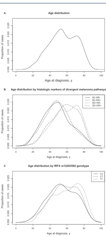
Using all cases, including those with SE+/NR+ tumors, prob-ability density functions revealed a bimodal distribution for age at diagnosis with an early-onset peak around age 50 years and a late-onset peak around age 75 years (Figure 2A). Age density plots stratified by the presence/absence of SE/NR revealed SE+/ NR- melanomas to have a predominantly late-onset distribu-tion, SE-/NR- or SE-/NR+ melanomas to have an early-onset dis-tribution, and SE+/NR+ melanomas to have a distribution falling between the early- and late-onset density peaks (Figure 2B). The median age at onset for SE+/NR- melanomas was statistically significantly higher than SE-/NR- melanomas while the median age at onset of SE-/NR+ and SE-/NR- melanomas did not statisti-cally significantly differ (Table 2).
Patients with the IRF4 rs12203592 (CC) genotype had a predominantly early-onset distribution, peaking around age 45 years, while patients with the (TT) genotype had a predomi-nantly late-onset distribution, peaking around age 75 years. Patients with the rs12203592 (CT) genotype had a bimodal age distribution, with approximately equal density peaks around ages 50 and 70 years (Figure 2C). The age distributions differed by the rs12203592 genotype, even when stratified by Australia and North America (data not shown).
Discussion
IRF4 encodes interferon regulatory factor-4, a transcription factor involved in B- and T-cell development, pigmentation, and poten-tially melanocyte differentiation and proliferation (44–47). IRF4
(also known as MUM1) is predominantly expressed in immune cells but is also present in melanocytes, nevi, and primary mela-nomas (44–47). The variant T allele of rs12203592, in intron 4 of
IRF4, is a functional SNP shown to increase IRF4 promoter ity in Burkitt Lymphoma B-cells and repress promoter activ-ity in human epidermal melanocytes and thus may increase or decrease IRF4 expression in different contexts (45,48,49). In mouse melanocytes, decreased expression of IRF4 as a result of rs12203592*T also led to decreased levels of certain downstream effectors of IRF4, such as the melanin-synthesizing enzyme TYR (45). Also, RegulomeDB (42) predicts that IRF4 rs12203592 is likely to affect binding.
We found that IRF4 rs12203592*T was strongly associated with light eye color, poor tanning ability, dark hair color, and lower nevus counts, consistent with previous epidemiological studies (18,50–52). Decreased TYR expression in melanocytic cells as a result of rs12203592*T might explain, at least in part, its association with certain fair-pigmentary traits. Adjusting for pigmentary traits and nevi, however, did not materially change the odds ratio for the association of rs12203592 with the exclu-sive presence of SE or NR adjacent to melanomas. This suggests that rs12203592 could influence the development of divergent melanoma pathways independent of host phenotypic traits associated with melanoma risk. Experimental studies are needed to elucidate this influence, which may be linked to dif-ferences in expression of IRF4 and its downstream targets, regu-lated in part by the rs12203592 genotype in melanocytes and/or immune system cells.
IRF4 rs12203592’s association with histologic SE and NR adja-cent to melanomas, representing divergent causal pathways,
may also explain why this variant has been inconsistently asso-ciated with melanoma risk in recent epidemiological studies. In a combined analysis of Australian, UK, and Swedish subjects, rs12203592*C was positively associated with melanoma, most statistically significantly with melanoma on the trunk (50). Similarly, in a UK study rs12203592*T was inversely associated
0 20 40 60 80 100
0.000
0.005
0.010
0.015
0.020
0.025
Age at diagnosis, y
Proportion of case
s
Age distribution by histologic markers of divergent melanoma pathways
SE-/NR-SE-/NR+ SE+/NR-SE+/NR+
0 20 40 60 80 100
0.000
0.005
0.010
0.015
0.020
0.025
Age distribution
Age at diagnosis, y
Proportion of case
s
0 20 40 60 80 100
0.000
0.005
0.010
0.015
0.020
0.025
Age at diagnosis, y
Proportion of case
s
Age distribution by IRF4 rs12203592 genotype
CC CT TT A
B
C
ar
ticle
ar
ticle
with melanoma, most statistically significantly with melanoma on the trunk (53). In contrast, rs12203592*T was positively asso-ciated with melanoma risk in two US studies (52,54). In another Australian study, rs12203592*T was inversely associated with melanoma in children/adolescents but was not associated with melanoma in adults (55). Further, rs12203592 was not statisti-cally significantly associated with melanoma in a combined analysis of GenoMEL data with patients from Europe and Israel (21), nor was it associated with multiple compared with single primary melanoma in a recent GEM study (26). The differing associations in these studies may depend upon the proportion of melanomas that arose via the chronic sun exposure vs nevo-genic pathway, given rs12203592’s strong and opposing asso-ciation with the exclusive presence of SE and NR found in the present study.
We are not aware of another study that has reported asso-ciations of IRF4 rs12203592 with SE or NR adjacent to melano-mas. Previous GEM investigations and independent studies have found older age, fewer nevi, and higher self-reported sun expo-sure hours to be associated with SE, with odds ratios in direc-tions consistent with those reported here (3,5,9).
We also found a bimodal age distribution in GEM with dis-tinct early- and late-onset peaks consistent with those reported previously for melanoma and which have been linked to the divergent causal pathways (56,57). This bimodal distribution has been described as displaying an early-onset peak fre-quency for trunk melanomas and a late-onset peak frefre-quency for face/ear melanomas using Surveillance, Epidemiology, and End Results (SEER) data (56). In GEM, melanomas without his-tologic SE displayed an early-onset distribution similar to that found in SEER for trunk melanomas while melanomas without histologic NR but with SE had a late-onset distribution simi-lar to that found in SEER for face/ear melanomas (56). A simi-lar early-onset distribution was also observed in patients with the IRF4 rs12203592 (CC) genotype, while a similar late-onset distribution was observed in patients with the rs12203592 (TT) genotype. This suggests that the rs12203592 genotype could be an inherited characteristic that, in part, underlies the bimodal age distribution of melanomas related to divergent causal path-ways linked to differences in age at onset, body site, and histo-pathologic features. Further work could be done to investigate this possibility.
Major advantages of our study are its large sample size, population-based ascertainment, and centralized pathol-ogy review. The majority of GEM participants (70%) reported Northern European ancestry, and the remaining persons divided by center into Southern European and other groups. While population stratification could have been an issue, the PCA results did not show evidence of a population substruc-ture confounding effect. We adjusted for study center in our analysis, and further adjustment for self-reported ances-try had little effect on the associations in the fully adjusted model. Melanomas with NR have also been reported as hav-ing lower Breslow thickness (13,15), possibly because thicker melanomas overgrow and obscure any NR; however, adding log Breslow or AJCC tumor stage to the fully adjusted multivari-able model did not change the ORs for rs12203592. The effect of age on cumulative sun exposure was of potential concern, but substituting average annual total sun exposure hours for total exposure hours did not materially affect rs12203592’s associations. Another limitation could be bias in the selection of cases for pathology review; however, none of the predic-tor variables used in the fully adjusted multivariable model were associated with missing vs nonmissing pathology review.
Also, we recognize that our SE and NR measures must contain some misclassification as evidenced by the modest agreement between our pathologists, but misclassification typically has the effect of attenuating observed associations. Power may also have been insufficient to detect associations of SNPs with lower MAFs (eg, SNPs in SLC45A2) in GEM. Lastly, the age dis-tributions presented could potentially be affected by age struc-ture and secular trends for sun exposure among the different populations; however, the age distributions among the GEM populations are quite similar, and the age distributions shifted by the rs12203592 genotype, even when stratified by Australia and North America.
IRF4 rs12203592’s independent yet contrasting associations with SE+/NR- and SE-/NR+ melanoma suggest that this inher-ited genetic variant may play a pivotal role underlying suscep-tibility to divergent pathways in melanoma development. We are not aware of a similar strong germline genetic crossover effect for another cancer with an inverse association with one subtype and positive association with another subtype of can-cer. Knowledge of the genetic etiology of divergent melanoma pathways with resultant differences in host characteristics and tumor features among melanoma patients could inform future risk models and prevention efforts for this complex and hetero-geneous disease.
Funding
This work was supported by National Cancer Institute (NCI) grants R01CA112243, R01CA112524, R01CA112243-05S1, R01CA112524-05S2, CA098438, U01CA83180, R33CA10704339, P30CA016086, P30CA008748, and P30CA014089; the National Institute of Environmental Health Sciences (P30ES010126); a University of Sydney Medical Foundation Program grant (Bruce Armstrong); and a Michael Smith Foundation for Health Research Infrastructure Award (Richard Gallagher).
Notes
The study funders had no role in the design of the study; the collection, analysis, or interpretation of the data; the writing of the manuscript; or the decision to submit the manuscript for publication.
No potential conflicts of interest were disclosed.
ar
ticle
ar
ticle
Canada: Loraine D. Marrett, PhD (PI), Elizabeth Theis, MSc (co-Investigator), Lynn From, MD (Dermatopathologist); CPO, Center for Cancer Prevention, Torino, Italy: Roberto Zanetti, MD (PI), Stefano Rosso, MD, MSc (co-PI); University of California, Irvine, CA: Hoda Anton-Culver, PhD (PI), Argyrios Ziogas, PhD (Statistician); University of Michigan, Ann Arbor, MI: Stephen B. Gruber, MD, MPH, PhD (PI, currently at University of Southern California, Los Angeles, CA), Timothy Johnson, MD (Director of Melanoma Program), Duveen Sturgeon, MSN (co-Investiga-tor, joint at USC-University of Michigan); University of North Carolina, Chapel Hill, NC: Nancy E. Thomas, MD, PhD (PI), Robert C. Millikan, PhD (previous PI, deceased), David W. Ollila, MD (co-Investigator), Kathleen Conway, PhD (co-Investiga-tor), Pamela A. Groben, MD (Dermatopathologist), Sharon N. Edmiston, BA (Research Analyst), Honglin Hao (Laboratory Specialist), Eloise Parrish, MSPH (Laboratory Specialist), David C. Gibbs, BS (Research Assistant), Jill S. Frank, MS (Research Assistant), Jennifer I. Bramson (Research Assistant); University of Pennsylvania, Philadelphia, PA: Timothy R. Rebbeck, PhD (PI), Peter A. Kanetsky, MPH, PhD (co-Investigator, currently at H. Lee Moffitt Cancer Center & Research Institute, Tampa, FL); UV data consultants: Julia Lee Taylor, PhD, and Sasha Madronich, PhD, National Centre for Atmospheric Research, Boulder, CO.
References
1. Diepgen T, Mahler V. The epidemiology of skin cancer. Br J Dermatol. 2002;146(s61):1–6.
2. Garbe C, Leiter U. Melanoma epidemiology and trends. Clin Dermatol. 2009;27(1):3–9.
3. Kvaskoff M, Pandeya N, Green AC, et al. Solar elastosis and cutaneous mela-noma: A site-specific analysis. Int J Cancer. 2015;136(12):2900–2911. 4. Whiteman DC, Watt P, Purdie DM, et al. Melanocytic nevi, solar
kera-toses, and divergent pathways to cutaneous melanoma. J Natl Cancer Inst. 2003;95(11):806–812.
5. Lee EY, Williamson R, Watt P, et al. Sun exposure and host phenotype as pre-dictors of cutaneous melanoma associated with neval remnants or dermal elastosis. Int J Cancer. 2006;119(3):636–642.
6. Law MH, MacGregor S, Hayward NK. Melanoma genetics: recent findings take us beyond well-traveled pathways. J Invest Dermatol. 2012;132(7):1763– 1774.
7. Ward KA, Lazovich D, Hordinsky MK. Germline melanoma susceptibil-ity and prognostic genes: a review of the literature. J Am Acad Dermatol. 2012;67(5):1055–1067.
8. Mishima Y. Melanocytic and nevocytic malignant melanomas. Cellular and subcellular differentiation. Cancer. 1967;20(5):632–649.
9. Thomas NE, Kricker A, From L, et al. Associations of cumulative sun expo-sure and phenotypic characteristics with histologic solar elastosis. Cancer Epidemiol Biomarkers Prev. 2010;19(11):2932–2941.
10. English DR, Heenan PJ, Holman CDAJ, et al. Melanoma in Western Australia in 1980–81: incidence and characteristics of histological types. Pathology. 1987;19(4):383–392.
11. Lee EY, Williamson R, Watt P, et al. Sun exposure and host phenotype as pre-dictors of cutaneous melanoma associated with neval remnants or dermal elastosis. Int J Cancer. 2006;119(3):636–642.
12. Cho E, Rosner BA, Colditz GA. Risk factors for melanoma by body site. Cancer Epidemiol Biomarkers Prev. 2005;14(5):1241–1244.
13. Purdue MP, From L, Armstrong BK, et al. Etiologic and other factors predict-ing nevus-associated cutaneous malignant melanoma. Cancer Epidemiol Bio-markers Prev. 2005;14(8):2015–2022.
14. Olsen CM, Zens MS, Stukel TA, et al. Nevus density and melanoma risk in women: a pooled analysis to test the divergent pathway hypothesis. Int J Cancer. 2009;124(4):937–944.
15. Bevona C, Goggins W, Quinn T, et al. Cutaneous melanomas associated with nevi. Arch Dermatol. 2003;139(12):1620–1624.
16. Law MH, Montgomery GW, Brown KM, et al. Meta-analysis combining new and existing data sets confirms that the TERT-CLPTM1L locus influences melanoma risk. J Invest Dermatol. 2012;132(2):485–487.
17. Gudbjartsson DF, Sulem P, Stacey SN, et al. ASIP and TYR pigmentation variants associate with cutaneous melanoma and basal cell carcinoma. Nat Genet. 2008;40(7):886–891.
18. Han J, Kraft P, Nan H, et al. A genome-wide association study identifies novel alleles associated with hair color and skin pigmentation. PLoS Genet. 2008;4(5):e1000074.
19. Bishop DT, Demenais F, Iles MM, et al. Genome-wide association study iden-tifies three loci associated with melanoma risk. Nat Genet. 2009;41(8):920– 925.
20. Nan H, Xu M, Zhang J, et al. Genome-wide association study identifies nido-gen 1 (NID1) as a susceptibility locus to cutaneous nevi and melanoma risk.
Hum Mol Genet. 2011;20(13):2673–2679.
21. Barrett JH, Iles MM, Harland M, et al. Genome-wide association study iden-tifies three new melanoma susceptibility loci. Nat Genet. 2011;43(11):1108– 1113.
22. Amos CI, Wang LE, Lee JE, et al. Genome-wide association study identi-fies novel loci predisposing to cutaneous melanoma. Hum Mol Genet. 2011;20(24):5012–5023.
23. Jannot AS, Meziani R, Bertrand G, et al. Allele variations in the OCA2 gene (pink-eyed-dilution locus) are associated with genetic susceptibility to mel-anoma. Eur J Hum Genet. 2005;13(8):913–920.
24. Fernandez LP, Milne RL, Pita G, et al. SLC45A2: a novel malignant melanoma-associated gene. Hum Mutat. 2008;29(9):1161–1167.
25. Macgregor S, Montgomery GW, Liu JZ, et al. Genome-wide association study identifies a new melanoma susceptibility locus at 1q21.3. Nat Genet. 2011;43(11):1114–1118.
26. Gibbs DC, Orlow I, Kanetsky PA, et al. Inherited genetic variants associated with occurrence of multiple primary melanoma. Cancer Epidemiol Biomarkers Prev. 2015;24(6):992–997.
27. Millikan RC, Hummer A, Begg C, et al. Polymorphisms in nucleotide excision repair genes and risk of multiple primary melanoma: the Genes Environ-ment and Melanoma Study. Carcinogenesis. 2006;27(3):610–618.
28. Begg CB, Hummer AJ, Mujumdar U, et al. A design for cancer case-control studies using only incident cases: experience with the GEM study of mela-noma. Int J Epidemiol. 2006;35(3):756–764.
29. Kricker A, Armstrong BK, Goumas C, et al. Ambient UV, personal sun exposure and risk of multiple primary melanomas. Cancer Causes Control. 2007;18(3):295–304.
30. Thomas NE, Edmiston SN, Alexander A, et al. Number of nevi and early-life ambient UV exposure are associated with BRAF-mutant melanoma. Cancer Epidemiol Biomarkers Prev. 2007;16(5):991–997.
31. Marrett L, King W, Walter S, et al. Use of host factors to identify people at high risk for cutaneous malignant melanoma. CMAJ. 1992;147(4):445. 32. English J, Swerdlow A, MacKie R, et al. Site‐specific melanocytic naevus
counts as predictors of whole body naevi. Br J Dermatol. 1988;118(5):641–644. 33. English DR, Armstrong BK. Melanocytic nevi in children I. Anatomic sites
and demographic and host factors. Am J Epidemiol. 1994;139(4):390–401. 34. Autier P, Boniol M, Severi G, et al. The body site distribution of melanocytic
naevi in 6–7 year old European children. Melanoma Res. 2001;11(2):123–131. 35. Orlow I, Roy P, Reiner AS, et al. Vitamin D receptor polymorphisms in patients
with cutaneous melanoma. Int J Cancer. 2012;130(2):405–418.
36. Kanetsky PA, Rebbeck TR, Hummer AJ, et al. Population-based study of natu-ral variation in the melanocortin-1 receptor gene and melanoma. Cancer Res. 2006;66(18):9330–9337.
37. Taylor NJ, Reiner AS, Begg CB, et al. Inherited variation at MC1R and ASIP and association with melanoma-specific survival. Int J Cancer. 2015;136(11):2659– 2667.
38. Lin DY. An efficient Monte Carlo approach to assessing statistical signifi-cance in genomic studies. Bioinformatics. 2005;21(6):781–787.
39. He Q, Avery CL, Lin DY. A general framework for association tests with multi-variate traits in large-scale genomics studies. Genet Epidemiol. 2013;37(8):759– 767.
40. Burnham KP, Anderson DR. Model selection and multimodel inference: a practical information-theoretic approach. Springer Science & Business Media; 2002. 41. Genomes Project C, Abecasis GR, Auton A, et al. An integrated map of genetic
variation from 1,092 human genomes. Nature. 2012;491(7422):56–65. 42. Boyle AP, Hong EL, Hariharan M, et al. Annotation of functional variation in
personal genomes using RegulomeDB. Genome Res. 2012;22(9):1790–1797. 43. Balch CM, Gershenwald JE, Soong SJ, et al. Final version of 2009 AJCC
mela-noma staging and classification. J Clin Oncol. 2009;27(36):6199–6206. 44. Shaffer AL, Emre NC, Romesser PB, et al. IRF4: Immunity. Malignancy!
Ther-apy? Clin Cancer Res. 2009;15(9):2954–2961.
45. Praetorius C, Grill C, Stacey SN, et al. A polymorphism in IRF4 affects human pigmentation through a tyrosinase-dependent MITF/TFAP2A pathway. Cell. 2013;155(5):1022–1033.
46. Natkunam Y, Warnke RA, Montgomery K, et al. Analysis of MUM1/IRF4 pro-tein expression using tissue microarrays and immunohistochemistry. Mod-ern Pathol. 2001;14(7):686–694.
47. Sundram U, Harvell JD, Rouse RV, et al. Expression of the B-cell prolifera-tion marker MUM1 by melanocytic lesions and comparison with S100, gp100 (HMB45), and MelanA. Mod Pathol. 2003;16(8):802–810.
48. Do TN, Ucisik-Akkaya E, Davis CF, et al. An intronic polymorphism of IRF4 gene influences gene transcription in vitro and shows a risk association with childhood acute lymphoblastic leukemia in males. Biochim Biophys Acta. 2010;1802(2):292–300.
ar
ticle
ar
ticle
50. Duffy DL, Iles MM, Glass D, et al. IRF4 variants have age-specific effects on nevus count and predispose to melanoma. Am J Hum Genet. 2010;87(1):6– 16.
51. Duffy DL, Zhao ZZ, Sturm RA, et al. Multiple pigmentation gene polymor-phisms account for a substantial proportion of risk of cutaneous malignant melanoma. J Invest Dermatol. 2010;130(2):520–528.
52. Zhang M, Song F, Liang L, et al. Genome-wide association studies identify several new loci associated with pigmentation traits and skin cancer risk in European Americans. Hum Mol Genet. 2013:22(14):2948–2959.
53. Newton-Bishop JA, Chang Y-M, Iles MM, et al. Melanocytic nevi, nevus genes, and melanoma risk in a large case-control study in the United Kingdom.
Cancer Epidemiol Biomarkers Prev. 2010;19(8):2043–2054.
54. Han J, Qureshi AA, Nan H, et al. A germline variant in the interferon regulatory factor 4 gene as a novel skin cancer risk locus. Cancer Res. 2011;71(5):1533–1539.
55. Kvaskoff M, Whiteman DC, Zhao ZZ, et al. Polymorphisms in nevus-associ-ated genes MTAP, PLA2G6, and IRF4 and the risk of invasive cutaneous mela-noma. Twin Res Hum Genet. 2011;14(05):422–432.
56. Lachiewicz AM, Berwick M, Wiggins CL, et al. Epidemiologic support for mel-anoma heterogeneity using the surveillance, epidemiology, and end results program. J Invest Dermatol. 2008;128(5):1340.