THE EFFECTS OF EXOGENOUS ORIENTING ON SUSTAINED ENDOGENOUS ATTENTION
Prerna Bholah
A thesis submitted to the faculty of the University of North Carolina at Chapel Hill in partial fulfillment of the requirements of the degree of Masters of Arts in the Department of Psychology (Cognitive).
Chapel Hill 2012
Approved by:
ii ABSTRACT
PRERNA BHOLAH: The Effects of Exogenous Orienting on Sustained Endogenous Attention (Under the direction of Joseph B. Hopfinger)
iii
ACKNOWLEDGEMENTS
iv
TABLE OF CONTENTS
LIST OF ABBREVIATIONS ... vi
Chapter I. INTRODUCTION ... 1
Exogenous Attention ... 2
Endogenous Attention ...5
Event-related potentials and selective attention ...8
Evidence for separable mechanisms ...13
Evidence for interacting mechanisms ...15
Motivation ...18
II. METHOD... 24
Participants ... 24
Materials and Procedure ... 24
Recording and Analysis ... 26
III. RESULTS ... 29
Behavioral Results ... 29
ERP Results ... 31
v
APPENDIX ... 40
REFERENCES ... 53
vi
LIST OF ABBREVIATIONS
EEG Electroencephalogram
ERP Event-related otentials
FEF Frontal eye-fields
fMRI functional Magnetic resonance imaging
IC Endogenously Invalid and exogenously Cued
IOR Inhibition of return
IU Endogenously Invalid and exogenously Uncued
LRP Lateralized readiness potential
SMA Sensory-motor area
SOA Stimulus onset asynchrony
SPL Superior parietal lobule
VC Endogenously Valid and exogenously Cued
CHAPTER 1 INTRODUCTION
Selective attention allows us to focus on relevant stimuli among the flow of incoming information that overloads our visual sense at any given time. Previous and current research show that there are at least two ways of operating one’s focus of attention: involuntary, reflexive or exogenous orienting, generated in response to salient stimuli, and voluntary orienting, an effortful and endogenous orienting of attention, typically elicited by central symbolic cues (Jonides, 1981; Muller & Rabbitt, 1989; Cheal & Lyon, 1991; Posner & Cohen, 1984).
2
type of interaction. Does involuntary capture remain robust across different levels of engagement of sustained voluntary attention or does maintaining voluntary attention strongly override reflexive orienting? Moreover, the current study aims to provide a quantifiable measure of when interactions between the attentional systems take place.
Exogenous attention
Involuntary, exogenous or reflexive attention is said to be at play when an abrupt change in the environment captures attention. For instance, a flash of lightning in the sky will automatically cause one to direct attention to it. This automatic capture could be in response to the appearance or disappearance of an object, a change in luminance or orientation of stimuli around the environment (Jonides, 1981; Yantis & Jonides, 1984; Hopfinger & Maxwell, 2005).
3
location (Posner & Cohen, 1984; Posner et al., 1985; Muller and Rabbitt, 1989; Klein, 2000 for a review).
4
There are studies however that postulate that reflexive orienting is not completely automatic and is dependent on the task demands and the attentional states. Yantis and Jonides (1990) showed that an onset distractor was less effective in capturing attention when attention was directed elsewhere by a cue with the right spatial and temporal attributes. When they manipulated predictabilities, abrupt onsets still captured attention even when the predictability was as high as 75%. This was shown with faster reaction times for onset targets as opposed to no-onset targets when attention was in a diffused state. Only when attention was 100% focused was the effect of abrupt onset completely eliminated (Yantis & Jonides, 1990).
5
Since they used greater eccentricities than previous studies, they argue that the ecological validity of the robust effect of peripheral stimuli in capturing attention could be explained by the need of human beings to avoid potentially dangerous organisms, even when located at the boundaries of our visual field. Based on the different conclusions from the studies described above, the specific conditions under which reflexive orienting can be considered to be automatic, are still open to consideration.
Endogenous Attention
Voluntary or endogenous attention is thought to be goal-driven and under the control of top-down processing (Jonides, 1981). For instance, if an individual is purposefully looking for a red item amongst other colored items, then endogenous orienting mechanisms will be recruited and the processing of irrelevant items will be suppressed. Endogenous orienting is an effortful process that is affected by capacity demands such as memory load and is influenced by predictability (Jonides, 1981). While it is slower to develop than reflexive orienting, it has longer lasting effects.
6
around 300 ms). Participants were therefore more accurate at earlier SOAs with peripheral cues while their performance was slightly improved with central cues at later SOAs. This difference in the time course and effects on accuracy due to reflexive and voluntary orienting confirm that distinct mechanisms are at play in the orienting, engagement and inhibition of attention. Not only are participants faster at orienting with peripheral cues, they also need to decode and engage their attention voluntarily with the central cue (Cheal & Lyon, 1991). In a similar vein, Muller and Rabbitt (1989) manipulated voluntary orienting, using central arrow cues that indicated the appearance of the target letter T, and reflexive orienting through peripheral flashes. For centrally-cued trials, where the target appeared on the same side indicated by the arrow, accuracy showed a steady increase with increasing SOA, especially between 100 and 275 ms. The central cuing condition showed a slower mechanism than the reflexive cuing one with maximal effects appearing at SOAs 400 to 725 ms as opposed 100 to 175 ms for the peripheral cuing condition.
7
network. More specifically, the frontal eye-fields (FEFs), the superior parietal lobule (SPL) and the sensory-motor area (SMA) showed significant activations. Conditions in which voluntary attention was triggered using colored arrowheads, also recruited similar areas. Critically, these arrow-cued shifts showed similar activations in the left and right hemispheres while self-initiated shifts exhibited a hemispheric asymmetry. The right hemisphere was equally activated in trials where attention was shifted to the right or left visual fields, while the left hemisphere showed increased activation when attention was deployed to the contra-lateral side. Their results showed that while a cue is not necessary for voluntary attention to be engaged, the cue might be causing differential activation and processing due to disengagement of attention or inhibition of the centrally-presented cue.
8
(form or color) to focus on at every trial while in the sustained attention condition, they were informed of the feature before each block. Their behavioral results showed faster responses in the sustained attention condition in addition to faster reaction times to the color condition within the sustained attention block (Eimer, 1997). Sustained attention therefore has a non-spatial component that is processed differently than transient attention. This implies that while sustained attention and trial-by-trial cuing share some commonalities, they do not always recruit the same processes. Since reaction time measures do not give enough insight into the neural stages of sustained attention, EEGs have typically been used to investigate sustained and transient attention mechanisms (as discussed further). Voluntary attention is a slowly-developing process under volitional control and could be internally generated or oriented in response to a symbolic cue. Involuntary capture could have specific effects on endogenous attention, whether the latter is deployed in the presence of a cue or sustained in the absence of one. How sustained endogenous attention unfolds in the presence of an involuntary capture and whether neural processing will be affected are questions to be explored in this study.
Event-related potentials and selective attention
9
the P1, N1 and P300 components (Hillyard & Munte, 1984; Luck, Heinze, Mangun & Hillyard, 1990; Dunchan & Donchin, 1978).
The P1 component, occurring around 90-130 ms over posterior cortical areas, is evoked by the presence of visual sensory stimuli and has been shown to be enhanced by both voluntary and reflexive orienting in isolation (Hillyard & Munte, 1984; Luck, Heinze, Mangun & Hillyard, 1990; Mangun & Hillyard, 1991; Eimer, 1994; Hopfinger & Mangun, 1998). The amplitude of the P1 component amplitude is preferentially enhanced at the spatially attended location and this enhancement of the P1 component indicates that selective visual attention is amplifying the flow of visual information (Mangun & Hillyard, 1991; Hillyard, Vogel & Luck, 1998).
10
at long SOAs (566-766ms). Their behavioral results showed a significant facilitation for cued versus uncued locations with no behavioral patterns of inhibition of return. Their ERP results revealed that at short SOAs, the amplitudes of the occipital contra-lateral P1 were enhanced at the cued location. Bottom-up processes therefore have significant effects at this early stage of neural processing (P1 component) even when the type of the task does not require a discrimination. Irrelevant stimuli also modulate the P1 component, which shows that exogenous orienting, irrespective of task relevance, can affect processes early in the neural stream. Hopfinger & Maxwell (2005) showed the enhancement of the P1 component by an irrelevant stimulus when it occurred at the same location as an uninformative visual event.
11
required a discrimination response and was not dependent on whether the design was blocked (testing sustained attention) or hard (Mangun & Hillyard, 1991). In a similar vein, Vogel and Luck (2000) showed that the N1 component was elicited under voluntary attention restrictions during discrimination tasks.
12
response is required (Hopfinger & Maxwell, 2005). This shows that top-down effects are critical in the generation of this late positivity.
The phenomenon of IOR where participants are slower to respond to peripherally cued targets at long SOAs of about 200 ms has also been investigated using ERP measures. One such study looked at the P1, N1 and the lateralized readiness potential (the LRP), which indexes the preparation of motor responses, was carried out by Prime and Ward (2004). They tested letter discrimination after the brightening of an outline box at periphery at an SOA of 290 to 590 ms. Their results showed no onset differences for the LRP and a reduction of the P1 and N1 amplitudes for the cued versus the uncued targets, which suggests that the IOR is at least in part a perceptual process (Prime & Ward, 2004). In the study looking at event-related potentials to non-predictive bottom-up stimuli, the authors found that at long SOAs, the cueing effect at the P1 component was reversed (uncued target locations had greater amplitudes than cued target locations) and there was no difference between the cued and uncued location targets at the P300 level (Hopfinger & Mangun, 1998). While IOR could not be observed behaviorally, this phenomenon could be the underlying reason why the P1 component was larger at the uncued target location than the cued target location (Hopfinger & Mangun, 1998). This dissociation between reaction times and event-related potentials gives us insight on different cognitive processes and their timings and is another reason why having both measures is beneficial in the current study.
13 Evidence for Separable Mechanisms
There exist behavioral and neuroimaging studies in support of the distinct effects and neural networks underlying voluntary and involuntary attention. Prinzmetal, Park and McCool (2005) showed the dissociation between voluntary and involuntary by carrying out a series of behavioral experiments which used reaction times and accuracies as the dependent measure. They used physical stimuli which differed only on the basis of their predictive values to initiate involuntary (25% valid) and voluntary orienting (80% valid). Voluntary and involuntary attention both showed similar reaction time results with valid trials being faster than invalid trials. However, the accuracy results showed a different pattern for voluntary and involuntary attention. While accuracy was better for valid than invalid trials in endogenous attention trials, exogenous trials did not show any improvement in accuracy across valid and invalid trials. This led them to conclude that there were different mechanisms at play for each attentional type.
14
condition revealed an enlarged and delayed P1 and N1 component for cued versus uncued targets. The authors propose that this delay component could be due to cue activity due to disengagement and re-orienting. Critically, smaller cues in the involuntary condition elicited a larger P1 component than larger cues, while the voluntary attention condition evoked a larger P1 to larger cues compared to the smaller cues. This difference in electrophysiological data confirms the independence of voluntary and involuntary attention within the same task.
15 Evidence for Interacting Mechanisms
16
Another study that looked at the neural correlates of endogenous and exogenous orienting when both central and peripheral cues were presented in the same trial, was carried out by Thomsen, Specht, Ersland & Hugdahl (2005). Thomsen et al. (2005) presented both cues simultaneously to test how participants would orient their focus of attention when presented with conflicting information at the same time. Their reaction time data showed an advantage of the exogenous cue when there was a conflict in both types of cues, that is, participants were slower at the location that was exogenously uncued and endogenously valid. While they found activation in the visual cortex, left parietal lobule and left cingulate gyrus when both cues were in conflict, they also found increased activation in the inferior and middle frontal gyrii as well as the precentral gyrus in the exogenously uncued/endogenously valid condition. The increased costs of the exogenous cue could have been due to the timing of the stimuli since it was designed for the optimal time-course of exogenous orienting. Nevertheless, their findings show that both involuntary and voluntary attention have separate modes of operation, with involuntary attention causing interference in the top-down control of voluntary attention.
17
dominated the later phase of the P1 component (120-150 ms). Interactions between endogenous and exogenous attention were observed at the N1 component, where there was a reduced endogenous effect on the N1 amplitude at the exogenously cued location and the onset of this neural mark was delayed by the extended positivity on P1 due to exogenous attention. This is evidence of both modes of orienting having separable effects happening nearly at the same time. While the P300 has previously been shown to be modulated by exogenous orienting (Hopfinger & Mangun, 1998), in this case, endogenous orienting overrode exogenous orienting at this later stage of processing. When Hopfinger & West (2006) tested the effects of endogenous and exogenous orienting separately, they found significant endogenous effects on the P1 and N1 components while exogenous attention in isolation did not affect the early P1 component. The authors suggest that the lack of an exogenous effect on the early P1 could be due to the task and paradigm. While their findings show the endogenous and exogenous orienting systems have separable effects, but they also interact at specific time-points in the processing stream, for instance at the N1 component (Hopfinger & West, 2006).
18
reflexive orienting would affect sustained attention within the visual sensory modality but also across the visual and auditory sensory modality. They found that the reflexive cuing effects were suppressed when attention was engaged elsewhere, which they interpreted as reflexive orienting not being truly automatic especially with increasing task demands.
The above studies have shown that endogenous and exogenous orienting have separable effects, with distinct neural correlates (Natale et al., 2009; Thomsen et al., 2005; Wang et al., 2010). While these two attentional types are thought to be distinct, their effects could interact at various levels of the cognitive stream for optimal processing of information (Hopfinger and West, 2006; Thomsen et al., 2005; Santangelo et al., 2007).
Motivation
19
The way we propose to vary the strength of the endogenous orienting is by manipulating its predictability. Participants will be required to orient their attention to a specific visual field based on the instructions of the experimenter. The target will appear on the side that subjects are instructed to attend 80% of the time (strong endogenous condition) or 65% of the time (weak endogenous condition). Voluntary attention will therefore be maintained by the participants, in the absence of cues. This would eliminate any cue activity that could potentially cloud our interpretation of ERP waveforms since both reflexive and voluntary attention will be elicited during the same trial. The way we propose to vary the strength of exogenous orienting is by manipulating the salience level: bright or dim. This would enable us to explore how capture would affect sustained voluntary attention when its effectiveness is varied. Moreover, deploying and maintaining voluntary attention to one side in the absence of a cue eliminates the possibility of involving other attentional processes such as disengagement from a cue or inhibition of the cue once attention has been dispensed at a peripheral location.
reflexively-20
cuing paradigm. Highly salient peripheral cues would therefore capture attention more robustly than stimuli of low salience.
Typically, in a Posner-cuing paradigm, the central cue has a predictive value which confers greater advantage to orienting to the cued location. Greater cue validity is associated with greater magnitude of endogenous effects and hence, larger top-down effects (Jonides, 1980, 1983; Eriksen and Yeh, 1985; Madden, 1992; Riggio and Kirsner, 1997). Vossel, Thiel & Fink (2006) manipulated central cue validity while recording imaging data. Their study showed greater activation in the right inferior parietal and right frontal cortex when cue validity was decreased from 90% to 60%.
21
22
Sustained and transient attention affect similar ERP components and might be recruiting the same selective attention mechanisms to amplify signals at the attended location. While transient attention may be more difficult to disengage from, non-spatial attentional processes might be more effective under sustained attention conditions. Therefore, while they both recruit voluntary attention mechanisms, they do not operate in the exact same manner and might interact differently in the presence of exogenous cues.
It should be noted that since participants will be paying attention to one side of the visual field across trials, any ERP effect will index sustained voluntary attention to that peripheral location instead of a shift to the periphery. In a study using neglect patients, Malhotra, Coulthard & Husain (2008) showed that the right PPC, involved in orienting voluntary attention, was critical in tasks requiring the use of sustained attention, irrespective of working memory retrieval or higher-order processing.
In the current study, since participants will be maintaining attention to one visual field
across trials, apossible caveat with the blocked-design experiment would be that participants
would not maintain attention at the intended location across trials. To motivate them to sustain their attention throughout the block, monetary compensation was provided to participants who were able to maintain fixation while keeping their accuracy higher than 75%. They will be instructed that paying attention to the instructed side is not only critical to the experimental paradigm but it will also help them improve their accuracy and speed. They were also required to respond to targets at the invalid locations.
23
mainly driven by voluntary attention in the strong endogenous-weak exogenous condition and by reflexive attention in the weak endogenous-strong exogenous condition. We also hypothesize that the levels of engagement of each attentional type will manifest themselves at the neural level. In both the strong endogenous-weak exogenous condition and the weak endogenous-strong exogenous conditions, we predict that reflexive attention will dominate at the earliest processing level and the P1 component will be modulated by involuntary
CHAPTER 2 METHOD Participants
Twenty-nine right-handed participants (ages 18-33, 10 females) were recruited to participate in two sessions of the study. They had no history of neurological disorders and presented normal (20/20) or corrected to normal vision. They were compensated $10/hour for their time and earned up to $25 for maintaining covert fixation, performing with high accuracy (above 75% correct trials) and for showing up to the second session. Nine participants’ data were not usable. Two participants’ data were rejected for not coming back in the second session, four were rejected due to low signal-to-noise ratio, two others were rejected due to noisy mastoid electrodes and one was rejected due to technical problems during recording. The final analysis included data from 20 participants.
Materials and Procedure
25
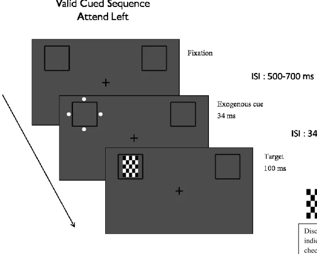
A central fixation cross flanked by two boxed outlines (3.13° x 3.13°) was presented on a grey background. Participants were required to covertly maintain their attention to a given visual field throughout a block of 40 trials, while keeping their eyes fixated on a cross in the middle of a computer screen. At the beginning of each block, participants were instructed to covertly maintain attention to the left or right side of the fixation cross. The side they were sustaining attention to was counter-balanced across blocks. A webcam was used to ensure that they kept their eyes at fixation (Applied Science Laboratories, Bedford, MA). After a jittered ISI of 500-700 ms, four dots (the exogenous cue) appeared around one of the outline boxes for 34 msec. The exogenous cue was non-predictive of where the target would appear and equally likely to flash around one of the two outline boxes. In the strong endogenous-weak exogenous condition, the target appeared on the attended side 80% of the time and the exogenous cue was dim (barely perceivable). In the weak endogenous condition-strong exogenous condition, the target appeared on the attended side 65% of the time and the four dots were bright. A rectangular checkerboard, either with black and white checks only or grayed out in some areas, served as target. This checkerboard target was presented for 100 ms in one of the outline boxes, 34-234 ms after the flashing of the dots. Participants were required to discriminate between the targets that were plain black and white or contained grey in it, using their index and middle fingers to respond on a SAITEC digital gamepad (Figure 1).
26
sustained. Exogenously cued and uncued condition types refer to the exogenously cued location target (target was preceded at the same location by an exogenous cue) and exogenous uncued location targets respectively.
Within each session, all subjects will respond to four different conditions types: (1) endogenously valid and exogenously cued (VC), (2) endogenously valid and exogenously uncued (VU), (3) endogenously invalid and exogenously cued (IC) and (4) endogenously invalid and exogenously uncued (IU) (Figure 2).
Recording and Analysis
Behavioral Data
The software program Presentation (Neurobehavioral Systems, Inc.) was used to generate stimulus displays and to record accuracy and reaction times. Accuracy data was recorded to ensure that participants were actually performing the task correctly. A webcam was used to monitor eye fixation at the central fixation cross and to ensure that participants’ gaze do not drift to the visual field they were covertly sustaining attention to.
ERP Analysis
27
The analysis software BESAResearch 5.3 was used to reject trials containing excessive
CHAPTER 3 RESULTS
Behavioral results
Responses that occurred before 100 ms (anticipations) and longer than 1500 ms were rejected from the behavioral analysis. Overall accuracy was high (mean accuracy rate was 96.86%) and there was no significant difference in accuracy between the right and left target side.
In order to investigate whether the degrees of engagement of endogenous and exogenous attention had an effect on behavioral responses, a repeated measures ANOVA was carried out with factors of condition (strong endogenous-weak exogenous condition vs. weak endogenous-strong endogenous condition), endogenous attention (endogenously valid vs. invalid), exogenous attention (exogenously cued vs. uncued) and target side (right or left). Further t-tests were carried out on interactions across conditions (corrected for multiple comparisons by the Benjamini-Hochberg procedure).
There was no main effect of condition (strong endogenous-weak exogenous condition or weak endogenous-strong exogenous condition) which indicates that participants’
responses did not change with the strength of the sustained voluntary attention or the
29
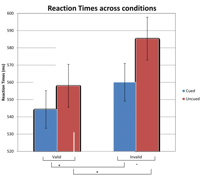
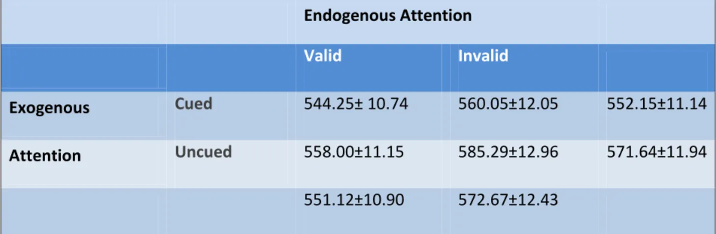
faster than uncued (M = 571.64, SD = 11.94). There was also a significant main effect of endogenous on reaction time, F (1, 19) = 28.804, p <0.001, where participants’ responses were faster at valid locations (M = 551.12, SD = 10.90) than at invalid locations (M = 572.67, SD = 12.43). A significant main effect of target side on reaction times was found, F (1, 19) =4.570, p<0.050, with participants responding faster on the left side (M = 557.59, SD = 12.38) versus the right (M = 566.20, SD = 10.96).
We also found a significant interaction between endogenous attention and exogenous attention, F (1, 19) = 11.104, p =0.004). Planned comparisons revealed that when
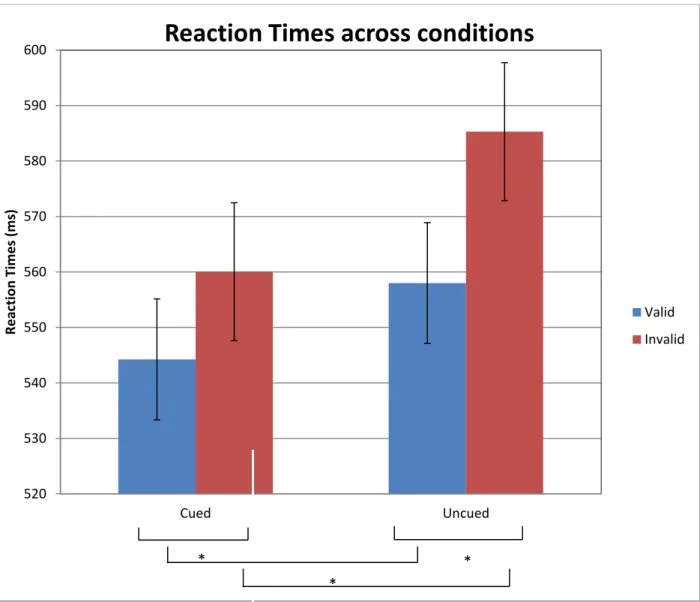
endogenous attention was allocated at the valid location, there was a significant effect of exogenous cueing, t (19) = 6.472, p<0.05, where exogenously cued targets (M = 544.25, SD = 10.74) were responded to significantly faster than uncued targets (M = 558.00, SD = 11.15). There was also a significant effect of exogenous cueing at the endogenously invalid location, t (19) = 8.677, p<0.05, where exogenously cued targets (M = 560.05, SD = 12.05) were responded to significantly faster than uncued targets (M = 585.29, SD = 12.96). Similarly, there was a significant effect of endogenous attention at the exogenously cued location, t (19) = 3.211, p<0.05, where endogenously valid targets (M = 544.25, SD = 10.74) were responded to significantly faster than invalid targets (M = 560.05, SD = 12.05). A
significant effect of endogenous attention was found at the exogenously uncued location, t
30
To ensure that the main effects and interactions that were found were not a result of a general slowing down of the participants in the invalid uncued condition, we looked at the relative percentages of the reaction times. There was a significantly greater validity effect ((Invalid RTs – Valid RTs)/Valid RTs) at the uncued location (4.9 %) than at the cued location (2.9%; t (19) =3.29, p = 0.004). Moreover, there was a significantly greater cueing effect ((Invalid RTs – Valid RTs)/Valid RTs) at the invalid location (4.5 %) than at the valid location (2.5 %; t (19) =3.28, p = 0.004).
ERP results
ERPs to targets
A within-subjects 4x2 repeated-measures analysis of variance (ANOVA) was carried out with factors of condition (strong endogenous-weak exogenous condition vs. weak
endogenous-strong exogenous condition), endogenous attention (endogenously cued vs uncued), exogenous attention (exogenously cued versus uncued), and target side (right versus left) on the mean amplitudes of the P1, N1 and P300 components at specific time-intervals.
P1 component.
Since maximal effects of the P1 component happen on the electrode contralateral to the stimuli, we combined amplitudes from the posterior left electrode for right targets and the posterior right electrode for left targets to get a higher number of trials per condition type. These electrodes were the posterior contralateral P07 and P08 electrodes of the International 10-20 System of electrode placement (Figure 4).
31
There was no main effect of condition for the P1 component (strong endogenous-weak exogenous condition or endogenous-weak endogenous-strong exogenous condition). There was a significant main effect of exogenous cuing on the P1 component (110-140 ms), where exogenously cued targets (3.02 µV) were enhanced more than exogenously uncued targets (1.39 µV; F(1, 19) =10.2, p=0.005). There was no significant main effect of endogenous attention on the P1 component. However, there was a significant interaction of condition with exogenous attention (F (1, 19) =6.09, p=0.023). The cueing effect (amplitudes at uncued location – amplitudes at cued location) in the strong endogenous condition was significantly
greater (1.94 µV) than the cueing effect in the weak endogenous condition (1.32 µV; t (19) =
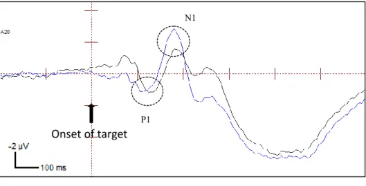
2.47, p = 0.023). A possible explanation for this is that the strong endogenous-weak exogenous condition has stronger effects than the weak endogenous-strong exogenous condition. There was a significant interaction between exogenous and endogenous attention (F (1, 19) = 8.165, p = 0.010). The cueing effect at the invalid location (2.11 µV) was significantly greater than at the valid location (1.15 µV; t (19) = 2.86, p = 0.001). (Please see Figure 5 for P1 mean amplitudes and Figure 6A for ERP waveforms)
32
condition at the cued location only, where the invalid targets had significantly greater amplitudes (3.81 µV) than valid targets (2.55 µV; t(19) = 4.00, p = 0.01). Typically, valid targets have greater amplitudes than invalid targets if endogenous attention is being enhanced at that stage of processing. Since we are finding the opposite only at the exogenously cued location in the strong endogenous condition, reflexive capture is interacting with the endogenous effects in that condition.
N1 component .
Analyses were run on the mean amplitudes of the N1 component in the 160-190 ms time range. There was a main effect of condition with the strong endogenous condition having smaller amplitudes (-2.02 µV) overall, than the weak endogenous condition (-2.57 µV; F (1, 19) =11.7, p=0.003). Surprisingly, there were no significant main effects of endogenous attention (F (1, 19) =0.015, p=0.904) or interactions of endogenous attention with condition (F (1, 19) =1.242, p=0.279) or exogenous cueing (F (1, 19) =2.025, p=0.171).
There was also a main effect of exogenous attention, F (1, 19) = 40.682, p=0.000, with cued targets 1.20 µV) having significantly smaller amplitudes than uncued targets (-3.39 µV). This could be a result of the extended positivity from the P1 component that is causing a latency shift for the cued targets as opposed to the cued targets (Fu et al., 2001).
We also found an interaction between condition and target side, (F (1, 19) = 40.682,
p=0.000) such that the right targets in the strong endogenous condition (-3.07 µV) had
33 P3 component.
There was no main effect of condition on the P300 amplitude (380-440ms), measured at the posterior electrode Cpz of the International 10-20 System of electrode placement, nor significant interaction of condition with endogenous or exogenous attention. There was a significant main effect of exogenous cuing on the P3 component, where exogenously cued targets (7.66 µV) were enhanced more than exogenously uncued targets (6.83 µV; F (1, 19) =10.2, p=0.005). There was a significant main effect of endogenous attention on the P3 component, F (1, 19) =11.49, p=0.003), where invalid locations (6.48 µV) showed smaller amplitudes than valid locations (8.00 µV).
Moreover, there was a significant interaction between exogenous and endogenous attention (F (1, 19) = 7.801, p = 0.012). At the endogenously invalid location, cued targets (6.59 µV) had significantly larger amplitudes than uncued targets (5.90 µV; t (19) = 3.36, p = 0.003). Interestingly, there was no significant effect of exogenous attention at the
endogenously valid location. There was a significant effect of endogenous attention at the cued target locations with valid target amplitudes (7.95 µV) being significantly greater than invalid target amplitudes (6.59 µV; t (19) = 3.14, p = 0.005). Similarly, valid target
CHAPTER 4
DISCUSSION
In this study, we investigated how exogenous attention interacted with sustained endogenous attention in a task where participants covertly maintained attention to a specified visual field while discriminating between a horizontal and vertical target. This study is one of the first to our knowledge to manipulate both sustained endogenous attention, by modifying its predictability across sessions, and exogenous attention, by altering the brightness of the exogenous cue, to investigate how the levels of engagement will affect interactions between the two attentional systems. More specifically, our goal was to investigate whether involuntary capture will still be robust across different levels of engagement of sustained endogenous attention or would sustained attention override reflexive capture when the former is strongly engaged.
35
sustaining attention. They were therefore effectively maintaining covert attention to the side they were instructed to. We also found interactions between the two attentional systems on behavioral and electrophysiological measures. The endogenous validity effect was greater at locations where the exogenous cue did not precede the target and the exogenous cueing effect was greater at locations where voluntary attention was not already deployed as measured by behavior. This implied that reflexive capture was maximal at the location where voluntary attention was not already being sustained regardless of how predictive endogenous attention was of where the target would appear. Similarly, voluntary attention effect was maximal in locations where the exogenous cue did not appear previously, irrespective of the brightness of the cue. Thus, endogenous and exogenous attention still operated distinctly and interacted, across all levels of luminance of the exogenous cue and engagement of voluntary attention.
36
component, which is different from typical validity effects (Mangun & Hillyard, 1991; Hillyard, Vogel & Luck, 1998). Since the endogenous reverse validity effects are observed at the exogenously cued location only, exogenous attention is interacting with early sustained endogenous attention to yield atypical P1 effects. One possible explanation for this could be that exogenous attention dominates at the P1 component, when the target appears at the location where endogenous attention was not already deployed. This effect at that stage of processing could be evidence of the robust and automatic nature of reflexive capture, however for it is hard at this point to further any conclusive explanation. Another explanation for this pattern of results could be that sustained attention might be more sensitive to reflexive capture. The reverse validity effect (greater amplitudes at invalid target location than valid target location) shown at that target-locked P1 stage could be representative of a different strategy being employed when both attentional systems are interacting and further research would be necessary to explore these effects.
The N1 component was modulated by exogenous attention where exogenously uncued targets were enhanced more than exogenously cued targets. This seems to be a result of the extended positivity of the P1 component due to the exogenous attention. Moreover, the effect of N1 amplitudes being greater in the strong endogenous condition also seems to be a consequence of the extended effect of exogenous attention on the P1 component. We did not find any effects of endogenous attention at that stage of processing.
37
38
Based on our results, exogenous attention seemed to dominate at the earliest P1 stage of processing. We also observed interactions between endogenous and exogenous attentional systems when they were concurrently engaged in the strong endogenous-weak exogenous condition. While we did not expect effects of endogenous attention at this early visual level of processing, the interactions could indicate that the effects in the strong endogenous-weak exogenous condition were amplified such that it boosted both the effects of exogenous and endogenous attention. Reflexive capture seemed to be robust enough as to dominate at the N1 component such that endogenous attention possibly could not enhance it anymore. At the P300 component, endogenous attention is the attentional mechanism that wins the competition even though exogenous attention still interacts at the cued locations. This could be a result of the robustness of exogenous attention, or that it was a sustained attention task. While voluntary and involuntary attention are two separable mechanisms that interact, it would seem that under some conditions, reflexive capture would override the endogenous mechanisms unless endogenous attention is fully sustained at that location. To further investigate these interacting mechanisms and whether reflexive capture will occur even when attention needs to completely devoted to a visual field, further research is needed.
39
40 APPENDIX
FIGURE 1: Weak endogenous condition trial sequence (Predictability of 65% and Bright
exogenous cue)
41 FIGURE 2: Condition types in each session.
42
FIGURE 3A. Reaction times as measured across strong endogenous-weak exogenous condition and weak endogenous-strong exogenous condition (endogenous attention plotted on the x-axis).
520 530 540 550 560 570 580 590 600
Valid Invalid
R
e
ac
tion
Ti
m
e
s (m
s)
Reaction Times across conditions
Cued
Uncued
* *
43
FIGURE 3B. Reaction Times as measured across strong endogenous-weak exogenous condition and weak endogenous-strong exogenous condition (exogenous attention plotted on the x-axis).
520 530 540 550 560 570 580 590 600
Cued Uncued
R
e
ac
tion
Ti
m
e
s (m
s)
Reaction Times across conditions
Valid
Invalid
* *
44
FIGURE 4. Topographies of amplitudes for the P1 peak for right and left targets at 125 ms at contra-lateral electrodes.
Topography of peak P1 amplitude to right targets
Topography of peak P1 amplitude to left targets
A20/P07
45 FIGURE 5. P1 component mean amplitudes
0 0.5 1 1.5 2 2.5 3 3.5 4 4.5
Valid Cued Valid Uncued Invalid Cued Invalid Uncued
A
m
p
litu
d
e
s
(µ
V)
Condition Types
46
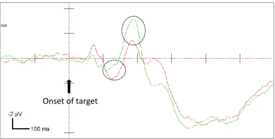
FIGURE 6A. ERPs of exogenously cued and uncued targets at the endogenously valid
location at the contralateral electrode (amplitudes combined across P07 and P08 electrodes). The ERP waveforms shown are collapsed across strong endogenous-weak exogenous condition and weak endogenous-strong exogenous condition.
Onset of target
N1
P1
Valid Cued ValidUncued
47
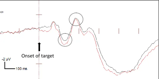
FIGURE 6B. ERPs of exogenously cued and uncued targets at the endogenously invalid
location at the contralateral electrode (amplitudes combined across P07 and P08 electrodes). The ERP waveforms shown are collapsed across strong endogenous-weak exogenous condition and weak endogenous-strong exogenous condition.
Invalid Cued
Invalid Uncued
48
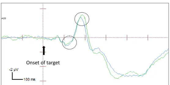
FIGURE 6C. ERPs of endogenously valid and invalid targets at the exogenously cued
location at the contralateral electrode (amplitudes combined across P07 and P08 electrodes). The ERP waveforms shown are collapsed across strong endogenous-weak exogenous condition and weak endogenous-strong exogenous condition.
Onset of target
49
FIGURE 6D. ERPs of endogenously valid and invalid targets at the exogenously uncued
location at the contralateral electrode (amplitudes combined across P07 and P08 electrodes). The ERP waveforms shown are collapsed across strong endogenous-weak exogenous condition and weak endogenous-strong exogenous condition.
Valid Uncued Invalid Uncued
50
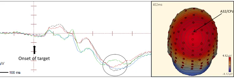
Figure 7. ERP waveforms at the central-posterior electrode CPz showing the P300 component (380-440 ms). The ERPs shown are collapsed across strong endogenous-weak exogenous condition and weak endogenous-strong exogenous condition.
Onset of target
Valid Cued Valid Uncued Invalid Cued
Invalid Uncued
51
TABLE 1. Means and Standard Errors of the mean of behavioral reaction times collapsed across conditions.
Endogenous Attention
Valid Invalid
Exogenous Cued 544.25± 10.74 560.05±12.05 552.15±11.14
Attention Uncued 558.00±11.15 585.29±12.96 571.64±11.94
52
53 REFERENCES
Berger, A., Henik, A., & Rafal, R. (2005). Competition between endogenous and exogenous
orienting of visual attention. Journal of Experimental Psychology, 134, 207-221.
Cheal, M., Lyon, D. R. (1991). Central and peripheral precuing of forced-choice
discrimination. The Quarterly Journal of Experimental Psychology, 43, 859-880.
Di Russo, F. & Spinelli, D. (2002). Effects of sustained voluntary attention on amplitude and latency of steady-state visual evoked potential: a costs and benefits analysis. Clinical Neuropsychology, 113, 1771-1777.
Doallo, S., Lorenzo-Lopez, L., Vizoso, C., Rodriguez, H.S., Amenedo, E., Bara, S., & Cadaveira, F. (2004). The time course of the effects of central and peripheral cues on visual processing: an event-related potentials study. Clinical Neurophysiology, 115, 199-210.
Duncan-Johnson, C. C. & Donchin, E. (1979). The time constant in P300 recording. Psychophysiology, 16, 53-55.
Eimer, M. (1994). An ERP study on visual spatial priming with peripheral onsets. Psychophysiology, 31, 154–63.
Eimer, M. (1996). ERP modulations indicate the selective processing of visual stimuli as a result of transient and sustained spatial attention. Psychophysiology, 33, 13-21. Eimer, M. (1997). Uninformative symbolic cues may bias visual-spatial attention:
behavioral and electrophysiological evidence. Biological Psychology, 46, 67-71.
Fu, S.M., Fan, S.L., Chen, L., & Zhuo, Y. (2001). The attentional effects of peripheral cueing
as revealed by two event-related potential studies. Neurophysiology, 112, 172– 185.
Hillyard, S.A. & Munte, T. F. (1984) Selective attention to color and location: An analysis
with event-related brain potentials. Perception and Psychophysics, 36, 185-198.
Hillyard, S.A., S.J. Luck and G.R. Mangun.(1994) The cuing of attention to visual field
locations: Analysis with ERP recordings. In: H.J. Heinze, T.F. Munte & G.R. Mangun
(Eds.). Cognitive Electrophysiology: Event-Related Brain Potentials in Basic and Clinical Research. Boston: Birkhausen, 1-25.
Hopfinger, J.B., & Mangun, G.R. (1998). Reflexive attention modulates processing of visual stimuli in human extrastriate cortex. Psychological Science, 9, 441-447.
Hopfinger, J. B., Buonocore, M. H., & Mangun, G. R. (2000). The neural mechanisms of
54
Hopfinger, J. B., & Mangun, G. R. (2001). Tracking the influence of reflexive attention on sensory and cognitive processing. Cognitive, Affective, & Behavioral Neuroscience, 1, 56-65.
Hopfinger, J. B., & Maxwell, J. S. (2005). Appearing and disappearing stimuli trigger a reflexive modulation of visual cortical activity. Cognitive Brain Research, 25, 48-56. Hopfinger, J. B., & West, V. M. (2006). Interactions between endogenous and exogenous
attention on cortical visual processing. NeuroImage, 31, 774-789.
Hopfinger, J.B., Camblin, C.C. & Parks, E.L. (2010). Isolating the internal in endogenous attention. Psychophysiology, 47, 739-747.
Jonides, J. (1981). Voluntary versus automatic control over the mind’s eye movement. In: J.B. Long and A.D. Baddeley, Editors, Attentional Performance, vol. IX, Erlbaum Associates, Hillsdale, NJ, 187-203.
Kelley, T.A., Serences, J.T., Giesbrecht, B., & Yantis, S. (2008). Cortical mechanisms for shifting and holding visuospatial attention. Cerebral Cortex, 18, 114-125.
Klein, R.M. (2000). Inhibition of return. Trends in Cognitive Sciences, 4, 138-147. Kraft, A., Pape, N., Hagendorf, H., Schmidt, S., Naito, A., & Brandt, S.A. (2007). What
determines sustained visual attention? The impact of distracter positions, task
difficulty and visual fields compared. Brain Research, 123-135.
Lambert, A., & Hockey, R. (1991). Peripheral visual changes and spatial attention. Acta Psychologica, 76, 149-163.
Luck, S. (2005). An introduction to the event-related potential technique.
Luck, S.J., Heinze, H.J., Mangun, G.R. and Hillyard, S.A. (1990) Visual event-related potentials index focused attention within bilateral stimulus arrays. II: Functional
dissociation of P1and N1 components. Electroencephalography and Clinical
Neurophysiology, 75, 528-542.
Malhotra, P., Coulthard, E.J., & Husain, M. (2008). Role of right posterior parietal cortex in maintaining attention to spatial locations over time. Brain, 132, 645-660.
Posner, M. Cognitive Neuroscience of Attention.
Mangun, G.R. & Hillyard, S. (1991). Modulations of sensory-evoked brain potentials
indicate changes in perceptual processing during visual-spatial priming. Journal of
Experimental Psychology,17, 1057-1074.
Mangun, G.R. & Buck, L.A. (1998). Sustained visual-spatial attention produces costs and
benefits in response time and evoked neural activity. Neuropsychologia, 36,
55
Muller, H.J. & Rabitt, P.M. (1989). Reflexive and voluntary orienting of visual attention:
time course of activation and resistance to interruption. Journal of Experimental
Human Perceptual Performance, 15, 315-330.
Muller, M.M., Mallinowski, P., Gruber, T., & Hillyard, S. (2003). Sustained division of the attentional spotlight. Letters to Nature, 424, 309-312.
Natale E, Marzi CA, Macaluso E. (2009). fMRI correlates of visuo-spatial reorienting
investigates in attention shifting double-cue paradigm. Human Brain Mapp, 30,
2367-2381.
Natale E, Marzi CA, Macaluso E. (2009). Right temporal-parietal junction engagement during spatial reorienting does not depend on strategic attention control.
Neuropsychologia, 48, 1160-1164.
Natale, E., Marzi, C.A., Girelli, M., Pavone, E.F., & Pollmann, S. (2006). ERP and fMRI correlates of endogenous and exogenous focusing of visual-spatial attention. European Journal of Neuroscience, 23, 2511-2521.
Patel, S.H. & Azzam, P.N. (2005). Characterization of N200 and P300: Selected Studies of
the Event-Related Potential. International Journal of Medical Sciences, 2, 147-154.
Posner, M.I., (1980). Orienting of attention. Quat. J. Exper. Psych., 32, 2-25.
Posner, M.I. & Cohen, Y. (1984) Components of visual orienting. Attent. Perform. Erlbaum
Associates, Hillsdale, NJ, X, 531–556.
Prinzmetal, W., McCool, C. & Park, S. (2005). Attention: Reaction time and accuracy reveal different mechanisms. Journal of Experimental Psychology, 134, 73-92.
Prinzmetal, W., Zvinyatskovskiy, A., Gutierrez, P., & Dilem, L. (2009). Voluntary and involuntary attention have different consequences: The effect of perceptual
difficulty. Quarterly Journal of Experimental Psychology, 62, 352–36.
Prime, D. J., & Ward, L. M. (2004). Inhibition of return from stimulus to response. Psychological Science, 15, 272-276.
Proverbio, A.M. & Mangun, G. R. (1994). Electrophysiological and behavioral “costs” and “benefits” during sustained visual-spatial attention. International Journal of
Neuroscience, 79, 221-233.
Robertson, I.H., & Garavan, H. (2004) Vigilant Attention. In M. S. Gazzaniga The Cognitive Neurosciences, 3rd edition. Michael S. Gazzaniga Editor-in-Chief. MIT Press
November 2004, 563-578.
Santangelo, V., Belardinelli, M.O., & Spence, C. (2007). The suppression of reflexive visual
and auditory orienting when attention is otherwise engaged. Journal of Experimental
56
Santangelo, V., & Spence, C. (2008). Is the exogenous orienting of spatial attention truly
automatic? Evidence from unimodal and multisensory studies. Consciousness and
Cognition, 17, 989-1015.
Soltani, M. & Knight, R.T. (2000). Neural Origins of the P300. Critical Review Neurobiology,14, 199-224.
Thomsen, T., Specht,K., Ersland,L.,& Hugdahl,K.(2005) Processing of conflicting cues in an
attention-shift paradigm studied with fMRI. Neuroscience Letters, 380, 138-142.
Van der Lubbe, R.H.J., & Postma, A. (2005). Interruption from irrelevant auditory and visual
onsets even when attention is in a focused state. Experimental Brain Research, 164,
464-471.
Wang, Y., Wu, J., Fu, S., & Luo, Y. (2010). Orienting and focusing in voluntary and involuntary visuospatial attention conditions: an event-related potential study. Journal of Psychophysiology, 24, 198-209.
Yantis, S. & Jonides, J. (1984). Abrupt visual onsets and selective attention: Evidence from
visual search. Journal of Experimental Psychology: Human Perception &
Performance, 10, 601-621.
Yantis, S. & Jonides, J. (1990). Abrupt visual onsets and selective attention: voluntary versus
automatic allocation. Journal of Experimental Psychology, 16, 121-134.
Yantis, S. & Hillstrom, A.P. (1994). Stimulus-driven attentional capture: evidence from
equiluminant visual objects. Journal of Experimental Psychology of Human