R E S E A R C H
Open Access
Novel pathogenic variants and multiple
molecular diagnoses in
neurodevelopmental disorders
Joanne Trinh
1*, Krishna Kumar Kandaswamy
2, Martin Werber
2, Maximilian E. R. Weiss
2, Gabriela Oprea
2,
Shivendra Kishore
2, Katja Lohmann
1†and Arndt Rolfs
2,3†Abstract
Background:
Rare denovo variants represent a significant cause of neurodevelopmental delay and intellectual
disability (ID).
Methods:
Exome sequencing was performed on 4351 patients with global developmental delay, seizures,
microcephaly, macrocephaly, motor delay, delayed speech and language development, or ID according to Human
Phenotype Ontology (HPO) terms. All patients had previously undergone whole exome sequencing as part of
diagnostic genetic testing with a focus on variants in genes implicated in neurodevelopmental disorders up to
January 2017. This resulted in a genetic diagnosis in 1336 of the patients. In this study, we specifically searched for
variants in 14 recently implicated novel neurodevelopmental disorder (NDD) genes.
Results:
We identified 65 rare, protein-changing variants in 11 of these 14 novel candidate genes. Fourteen variants
in
CDK13
,
CHD4
,
KCNQ3
,
KMT5B
,
TCF20
, and
ZBTB18
were scored pathogenic or likely pathogenic. Of note, two of
these patients had a previously identified cause of their disease, and thus, multiple molecular diagnoses were made
including pathogenic/likely pathogenic variants in
FOXG1
and
CDK13
or in
TMEM237
and
KMT5B
.
Conclusions:
Looking for pathogenic variants in newly identified NDD genes enabled us to provide a molecular
diagnosis to 14 patients and their close relatives and caregivers. This underlines the relevance of re-evaluation of
existing exome data on a regular basis to improve the diagnostic yield and serve the needs of our patients.
Keywords:
Neurodevelopmental disorders, De novo variants, Trio exome sequencing
Background
Major congenital malformations, which include
neuro-developmental disorders (NDDs), are present in ~ 2
–
5%
of children [1]. Children with NDD have variable
sever-ity of phenotypic features and different behavioral
abnor-malities. Often times, NDD arises from de-novo variants
in genes important for central nervous system (CNS)
de-velopment [2]. Whole exome sequencing has been
crit-ical and effective in diagnosing patients with NDD.
Thus, treatment for NDD has become more refined
through
molecular
genetic
diagnosis
rather
than
phenotype-driven management of symptoms [3]. Herein,
we find novel pathogenic or likely pathogenic variants in
six recently identified NDD genes, namely
CDK13
,
CHD4
,
KCNQ3
,
KMT5B
,
TCF20
, and
ZBTB18
.
Methods
Patients
From a total of 26,119 house exome data set, we
in-cluded 4351 unrelated NDD patients in this study.
Hu-man Phenotype Ontology (HPO) nomenclature [4] was
applied based on the clinical data provided by referring
physician. In the context of this manuscript, NDD was
defined by HPO terms described in Additional file
1:
Figure S1. Patients had an average age of 7.75 (STD
8.04) years (Additional file
1: Table S1). All patients had
previously undergone whole exome sequencing as part
of their clinical genetic testing, following previously
© The Author(s). 2019Open AccessThis article is distributed under the terms of the Creative Commons Attribution 4.0 International License (http://creativecommons.org/licenses/by/4.0/), which permits unrestricted use, distribution, and reproduction in any medium, provided you give appropriate credit to the original author(s) and the source, provide a link to the Creative Commons license, and indicate if changes were made. The Creative Commons Public Domain Dedication waiver (http://creativecommons.org/publicdomain/zero/1.0/) applies to the data made available in this article, unless otherwise stated.
* Correspondence:[email protected]
reported procedures [5]. These tests focused on NDD
genes established before January 2017. Parents were
available from 2030 patients to test for de novo
occur-rence of variants. Written informed consent was
ob-tained from participants, and this study was approved by
the Ethical Commission of the University of Rostock
(registry no. A2015-0102). All samples were processed in
Centogene
’
s laboratory, which is CAP and CLIA
certi-fied, adhering to the American College of Medical
Gen-etics and Genomics (ACMG) guidelines [6].
Genetic testing
Patient DNA was extracted from EDTA blood or from
dry blood spots in filter cards. WES was performed on
the IonProton (
n
= 911 samples, enrichment with Ion
AmpliSeq Exome RDY Kit (Life Technologies, Carlsbad,
CA, USA)) or Illumina (
n
= 3440 samples, enrichment
with Illumina
’
s NexteraRapid Capture Exome Kit
(Illu-mina, Inc., San Diego, CA, USA)). Sequencing and
bio-informatics were done as previously described [5,
7,
8].
We focused on genes of interest (fourteen recently
nom-inated genes by the DDD study [9]; Additional file
1:
Figure S1), filtered for rare variants (MAF < 0.0001), and
an effect on the encoded protein sequence. Sanger
vali-dations were performed for all indels and variants with
quality Phred score below 300 to rule out false-positive
variants as previously described [5]. Further, we applied
the ACMG criteria to score the pathogenicity of
candi-date variants [6].
Results
Among all 4351 NDD patients, we identified 65
heterozy-gous variant carriers (1.5%), for 65 different rare, protein
sequence-changing variants in 11 out of 14 genes recently
nominated by the DDD study [9] (Additional file
1: Figure
S1 and Table S2). In 11 of 12 carriers for whom parents
were available, the variant was shown to be de novo, and
in one case (
KCNQ3
:p.Arg364Cys) inherited from the
father whereby his affection status is unknown. The
vari-ant
CDK13
: p.His675Arg was found in two affected
sib-lings. For all other patients, no relatives were available for
testing. The 65 variants were either not present or at very
low frequency (< 2.76 × 10
−4frequency) in unaffected
“
in-house
”
exomes or in public databases (ExAC, GnomAD).
Using ACMG recommendations, six of these 65 variants
were scored as pathogenic (
CDK13:
p.Tyr351fs,
CDK13:
p.Gln544*,
CDK13:
p.Asn842Ser,
KMT5B:
p.Pro106fs,
KMT
5B:
p.Ser116fs, and
KCNQ3:
p.Arg230Cys) and eight as
likely pathogenic (
CDK13:
p.Thr500Met,
CDK13:
p.Asn
843Ile,
CDK13:
p.Gly712Arg,
CDK13:
p.Tyr716Cys,
CHD4:
p.Lys634Arg,
KMT5B:
p.Ter394fs,
ZBTB18:
p.Arg436His,
and
TCF20:
p.Pro1147Leu) (Table
1). The remaining 51
variants (78%) were categorized as variants of uncertain
significance (VUS) (Additional file
1: Table S2 and S5;
Fig.
1). This included a de novo splice region variant in
KMT5B
(c.-140+4T>G) which was predicted in silico
(using HumanSplicingFinder and MaxEntScan) to results
in alternative splicing for transcript NM_001300907.1.
However, a fresh sample from this patient was not
avail-able to test for alterations in splicing. Patients
’
clinical
characteristics were compared across
CDK13
and
KMT5B
variant carriers (Additional file
1: Figure S2 and S3).
There were two patients who had previously received
a genetic diagnosis and thus carried an additional
patho-genic variant in a previously established NDD gene
(Additional file
1: Table S3). Thus, these two patients
each carried multiple molecular diagnoses. This included
a patient with a frameshift variant in
FOXG1
(OMIM
number 613454) and a missense change in
CDK13
(OMIM number 603309) who had a complex phenotype
beyond typical Rett-like syndrome presentation
includ-ing MRI abnormalities and visual impairment. This
pa-tient also had delayed motor and language development,
intellectual disability, muscular hypotonia, microcephaly,
ventricular septal defect, failure to thrive and squint
which aligns with the OMIM phenotype of congenital
heart defects, dysmorphic facial features, and intellectual
developmental disorder (CHDFIDD). The onset was at
birth, and her parents were non-consanguineous, and
there were no other affected siblings.
Another patient carried a homozygous c.869+1G>A
variant in
TMEM237
(OMIM number 614424) and a
frameshift variant c.1180_*1delTAAG (p.Ter394fs) in
KMT5B
(OMIM number 617788). This male patient has
been suspected to be affected with Joubert syndrome
which is known to be linked to biallelic
TMEM237
vari-ants, and had defective vision and global developmental
delay. Whether there is an additional contribution of the
likely pathogenic
KMT5B
variant to the phenotype is
dif-ficult to determine, although some features overlap with
the OMIM phenotype of mental retardation.
Discussion
In this study, we identified pathogenic/likely pathogenic
variants in 14 NDD patients in six different, recently
identified genes. Our findings highlight the importance
of reanalyzing and revisiting exome sequencing data to
reclassify variants of uncertain significance by taking
into account novel observations published in the
scien-tific literature. Since the initial study [9], 13 of the 14
in-vestigated genes, with the exception of MSL3, have
independently
been
replicated
[10
–
23]
including
CDK13
,
CHD4
,
KCNQ3
,
KMT5B
,
TCF20
, and
ZBTB18
.
In our sample,
CDK13
(cyclin-dependent kinase 13)
and
KMT5B
(lysine-specific methyltransferase 5B)
amino acid residue asparagine at position 842 in
CDK13
(p.Asn842Ser and p.Asn842Ile). These patients had
de-layed speech and language development, motor delay,
and abnormal facial shape (Additional file
1: Figure S3
and Table S4). The p.Asn842Ser has also been previously
described in the DDD study [9], suggesting that position
842 could be a mutational hot spot.
Notably, there were two patients who carried two
pathogenic/likely pathogenic variants in two different
genes (
n
= 2/65, 3%) each. Of note, this is in the same
range as a recent large-scale study (4.9%) [24], further
underlining the importance to search for genetic causes
with an exome-wide approach not to overlook relevant
genetic diagnoses and also the importance of revisiting
and reanalyzing exomes over time as more and more
new genetic publications surface, even if one genetic
cause has already been identified.
The genetic heterogeneity of NDD with hundreds of
genes in which variants lead to NDD reflects the
complex process of proper brain development. Many
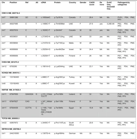
Table 1
List of pathogenic or likely pathogenic variants in this study
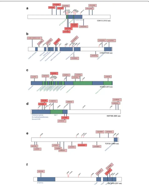
Fig. 1a-fComposite figures of genes with pathogenic or likely pathogenic variants identified in this study:CDK13,CHD4,KCNQ3,KMT5B,TCF20,
andZBTB18(adapted from the“Prevalence and architecture of de novo mutations in developmental disorders”study [9]). Boxes: pink highlighted
of the gene products function in multiple biological
pathways but may result in strikingly different
pheno-types. For example, patients with de novo variants in
CDK13
and
CHD4
may present with overlapping
neu-rodevelopmental features and heart defects; the
func-tion of both genes is different [9,
25,
26]. CHD4 is
part of the SNF2/RAD54 helicase family and is a core
component of the nucleosome remodeling and
his-tone deacetylase repressor complex which is
import-ant for epigenetic regulation of gene transcription. In
contrast, CDK13 forms a complex with cyclin K and
is predicted to have a role in regulating cell cycle but
also transcription. On the other hand, a distinct
phenotype can be seen for variants within the same
gene.
CHD4
somatic variants are also involved in
uterine serous carcinoma, an aggressive endometrial
cancer [27]. This illustrates the high time and spatial
sensitivity of the developing brain/body to genetic
variations.
Many novel NDD genes are involved in epigenetic
mechanisms such as chromatin remodeling, histone
modification, RNA splicing, transcription, and DNA
binding including the two most relevant genes from
our study, i.e.,
CDK13
and
KMT5B.
CDK13 forms a
complex with cyclin K and is predicted to have a role
in regulating cell cycle and transcription. Mutations
can alter complex activity. KMT5B functions as a
his-tone methyltransferases and trimethylate nucleosomal
histone 5 [28]. KMT5B also trimethylates the
onco-gene ERK (extracellular signal-regulated kinases), and
overexpression of KMT5B activates the ERK signaling
pathway [29]. These kinases are important for brain
development, proliferation of cells, and neuronal
mi-gration, and ERK1/2 deficits in mice have shown
im-paired
neurogenesis
[30].
Histone
deacetylase
inhibitors (HDACis) and DNA demethylating drugs
(DNMTis) have been used in cancer therapy trials
[31,
32] and may be emerging drugs in NDD [33].
Conclusions
Our study underlines the relevance of six additional
NDD genes and highlights the significance of multiple
genetic diagnoses in several patients. Our study
accentu-ates the importance of re-evaluating whole exome
se-quencing data in light of new publications enabling
reclassification of previously categorized variants of
un-certain significance.
Additional file
Additional file 1:Table S1.Patient demographics for developmental disorders.Table S2. Variants of unknown significance identified in this study and pathogenicity scoring.Table S3.Individuals with dual molecular diagnoses.Table S4.HPO terms listed for all (likely)
pathogenic mutation carriers.Table S5.Number of rare, protein-changing variants found in the NDD patients.Figure S1.Overview of study: workflow of identification of 14 (likely) pathogenic variants (6 of 14 candidate genes) in 14 of 4351 patients.Figure S2.HPO terms compos-ite for CDK13 pathogenic/likely pathogenic carriers. HPO terms that over-lap in different mutation carriers are highlighted in red.Figure S3.HPO terms composite for KMT5B pathogenic/likely pathogenic variant carriers. HPO terms that overlap in different mutation carriers are highlighted in red. (DOCX 96 kb)
Abbreviations
CDK13:Cyclin-dependent kinase 13; CHD4: Chromodomain-helicase-DNA-binding protein 4; DDD: Deciphering Developmental Disorders; DNA: Deoxyribonucleic acid; EDTA: Ethylenediaminetetraacetic acid; FOXG1: Forkhead box G1; KCNQ3: Potassium voltage-gated channel subfam-ily Q member 3; KMT5B: Lysine methyltransferase 5B;
NDD: Neurodevelopmental disorder; TCF20: Transcription factor 20; TMEM237: Transmembrane protein 237; ZBTB18: Zinc finger and BTB domain containing 18
Authors’contributions
JT contributed to the execution of the research project; designed, executed, reviewed, and critiqued the statistical analysis; and wrote the first draft and reviewed and critiqued the manuscript preparation. KKK contributed to the execution of the research project; executed, reviewed, and critiqued the statistical analysis; and wrote the first draft and reviewed and critiqued the manuscript preparation. MW, MERW, GO, and SK contributed to the execution of the research project; executed, reviewed, and critiqued the statistical analysis; and reviewed and critiqued the manuscript preparation. KL and AR contributed to the conception, organization, and execution of the research project; designed, reviewed, and critiqued the statistical analysis; and wrote the first draft and reviewed and critiqued the manuscript preparation. All authors read and approved the final manuscript.
Funding
JT acknowledges funding from Alexander Von Humboldt, Canadian Institutes of Health Research, and the Joachim Herz Stiftung. We acknowledge financial support by Land Schleswig-Holstein within the funding programme Open Access Publikations fonds.
Availability of data and materials
All data on variants will be available on HGMD.
Ethics approval and consent to participate
Written informed consent was obtained from participants, and this study was approved by the Ethical Commission of the University of Rostock (registry no. A2015-0102). All samples were processed in Centogene’s labora-tory, which is CAP and CLIA certified, adhering to the ACMG guidelines.
Consent for publication
All authors consent for the publication of this work.
Competing interests
The authors declare that they have no competing interests.
Author details
1Institute of Neurogenetics, University of Lübeck, 23538 Lübeck, Germany. 2
Centogene AG, Rostock, Germany.3University of Rostock, 18147 Rostock, Germany.
Received: 18 September 2018 Accepted: 23 May 2019
References
1. Sheridan E, Wright J, Small N, Corry PC, Oddie S, Whibley C, Petherick ES, Malik T, Pawson N, McKinney PA, Parslow RC. Risk factors for congenital anomaly in a multiethnic birth cohort: an analysis of the Born in Bradford study. Lancet. 2013;382:1350–9.
mutations in the context of neurodevelopmental disease. Mol Psychiatry. 2013;18:141–53.
3. Aronson SJ, Rehm HL. Building the foundation for genomics in precision medicine. Nature. 2015;526:336–42.
4. Groza T, Kohler S, Moldenhauer D, Vasilevsky N, Baynam G, Zemojtel T, Schriml LM, Kibbe WA, Schofield PN, Beck T, et al. The human phenotype ontology: semantic unification of common and rare disease. Am J Hum Genet. 2015;97:111–24.
5. Trujillano D, Bertoli-Avella AM, Kumar Kandaswamy K, Weiss ME, Koster J, Marais A, Paknia O, Schroder R, Garcia-Aznar JM, Werber M, et al. Clinical exome sequencing: results from 2819 samples reflecting 1000 families. Eur J Hum Genet. 2017;25:176–82.
6. Richards S, Aziz N, Bale S, Bick D, Das S, Gastier-Foster J, Grody WW, Hegde M, Lyon E, Spector E, et al. Standards and guidelines for the interpretation of sequence variants: a joint consensus recommendation of the American College of Medical Genetics and Genomics and the Association for Molecular Pathology. Genet Med. 2015;17:405–24.
7. Li H, Durbin R. Fast and accurate short read alignment with Burrows-Wheeler transform. Bioinformatics. 2009;25:1754–60.
8. Cingolani P, Platts A, Wang le L, Coon M, Nguyen T, Wang L, Land SJ, Lu X, Ruden DM. A program for annotating and predicting the effects of single nucleotide polymorphisms, SnpEff: SNPs in the genome of Drosophila melanogaster strain w1118; iso-2; iso-3. Fly (Austin). 2012;6:80–92. 9. study DDD. Prevalence and architecture of de novo mutations in
developmental disorders. Nature. 2017;542:433–8.
10. van den Akker WMR, Brummelman I, Martis LM, Timmermans RN, Pfundt R, Kleefstra T, Willemsen MH, Gerkes EH, Herkert JC, van Essen AJ, et al. De novo variants in CDK13 associated with syndromic ID/DD: molecular and clinical delineation of 15 individuals and a further review. Clin Genet. 2018; 93:1000–7.
11. Weiss K, Terhal PA, Cohen L, Bruccoleri M, Irving M, Martinez AF, Rosenfeld JA, Machol K, Yang Y, Liu P, et al. De novo mutations in CHD4, an ATP-dependent chromatin remodeler gene, cause an intellectual disability syndrome with distinctive Dysmorphisms. Am J Hum Genet. 2016;99:934–41.
12. Wang T, Guo H, Xiong B, Stessman HA, Wu H, Coe BP, Turner TN, Liu Y, Zhao W, Hoekzema K, et al. De novo genic mutations among a Chinese autism spectrum disorder cohort. Nat Commun. 2016;7:13316. 13. Ambrosino P, Freri E, Castellotti B, Soldovieri MV, Mosca I, Manocchio L,
Gellera C, Canafoglia L, Franceschetti S, Salis B, et al. Kv7.3 compound heterozygous variants in early onset encephalopathy reveal additive contribution of C-terminal residues to PIP2-dependent K(+) channel gating. Mol Neurobiol. 2018;55:7009–24.
14. Bowling KM, Thompson ML, Amaral MD, Finnila CR, Hiatt SM, Engel KL, Cochran JN, Brothers KB, East KM, Gray DE, et al. Genomic diagnosis for children with intellectual disability and/or developmental delay. Genome Med. 2017;9:43.
15. Moccia A, Srivastava A, Skidmore JM, Bernat JA, Wheeler M, Chong JX, Nickerson D, Bamshad M, Hefner MA, Martin DM, Bielas SL. Genetic analysis of CHARGE syndrome identifies overlapping molecular biology. Genet Med. 2018;20(9):1022–9.
16. Zhao JJ, Halvardson J, Zander CS, Zaghlool A, Georgii-Hemming P, Mansson E, Brandberg G, Savmarker HE, Frykholm C, Kuchinskaya E, et al. Exome sequencing reveals NAA15 and PUF60 as candidate genes associated with intellectual disability. Am J Med Genet B Neuropsychiatr Genet. 2018;177: 10–20.
17. Ververi A, Splitt M, Dean JCS, Brady AF. Phenotypic spectrum associated with de novo mutations in QRICH1 gene. Clin Genet. 2018;93:286–92. 18. Strauss KA, Gonzaga-Jauregui C, Brigatti KW, Williams KB, King AK, Van Hout
C, Robinson DL, Young M, Praveen K, Heaps AD, et al. Genomic diagnostics within a medically underserved population: efficacy and implications. Genet Med. 2018;20:31–41.
19. Stevens SJC, van der Schoot V, Leduc MS, Rinne T, Lalani SR, Weiss MM, van Hagen JM, Lachmeijer AMA, Stockler-Ipsiroglu SG, Lehman A, Brunner HG. De novo mutations in the SET nuclear proto-oncogene, encoding a component of the inhibitor of histone acetyltransferases (INHAT) complex in patients with nonsyndromic intellectual disability. Hum Mutat. 2018;39: 1014–23.
20. Depienne C, Nava C, Keren B, Heide S, Rastetter A, Passemard S, Chantot-Bastaraud S, Moutard ML, Agrawal PB, VanNoy G, et al. Genetic and phenotypic dissection of 1q43q44 microdeletion syndrome and
neurodevelopmental phenotypes associated with mutations in ZBTB18 and HNRNPU. Hum Genet. 2017;136:463–79.
21. Trinh J, Huning I, Budler N, Hingst V, Lohmann K, Gillessen-Kaesbach G. A novel de novo mutation in CSNK2A1: reinforcing the link to
neurodevelopmental abnormalities and dysmorphic features. J Hum Genet. 2017;62:1005–6.
22. Jansen S, Geuer S, Pfundt R, Brough R, Ghongane P, Herkert JC, Marco EJ, Willemsen MH, Kleefstra T, Hannibal M, et al. De novo truncating mutations in the last and penultimate exons of PPM1D cause an intellectual disability syndrome. Am J Hum Genet. 2017;100:650–8.
23. Turner TN, Hormozdiari F, Duyzend MH, McClymont SA, Hook PW, Iossifov I, Raja A, Baker C, Hoekzema K, Stessman HA, et al. Genome sequencing of autism-affected families reveals disruption of putative noncoding regulatory DNA. Am J Hum Genet. 2016;98:58–74.
24. Posey JE, Harel T, Liu P, Rosenfeld JA, James RA, Coban Akdemir ZH, Walkiewicz M, Bi W, Xiao R, Ding Y, et al. Resolution of disease phenotypes resulting from multilocus genomic variation. N Engl J Med. 2017;376:21–31. 25. Sifrim A, Hitz MP, Wilsdon A, Breckpot J, Turki SH, Thienpont B, McRae J,
Fitzgerald TW, Singh T, Swaminathan GJ, et al. Distinct genetic architectures for syndromic and nonsyndromic congenital heart defects identified by exome sequencing. Nat Genet. 2016;48:1060–5.
26. Bostwick BL, McLean S, Posey JE, Streff HE, Gripp KW, Blesson A, Powell-Hamilton N, Tusi J, Stevenson DA, Farrelly E, et al. Phenotypic and molecular characterisation of CDK13-related congenital heart defects, dysmorphic facial features and intellectual developmental disorders. Genome Med. 2017;9:73. 27. Zhao S, Choi M, Overton JD, Bellone S, Roque DM, Cocco E, Guzzo F,
English DP, Varughese J, Gasparrini S, et al. Landscape of somatic single-nucleotide and copy-number mutations in uterine serous carcinoma. Proc Natl Acad Sci U S A. 2013;110:2916–21.
28. Schotta G, Lachner M, Sarma K, Ebert A, Sengupta R, Reuter G, Reinberg D, Jenuwein T. A silencing pathway to induce H3-K9 and H4-K20
trimethylation at constitutive heterochromatin. Genes Dev. 2004;18:1251–62. 29. Vougiouklakis T, Sone K, Saloura V, Cho HS, Suzuki T, Dohmae N, Alachkar H, Nakamura Y, Hamamoto R. SUV420H1 enhances the phosphorylation and transcription of ERK1 in cancer cells. Oncotarget. 2015;6:43162–71. 30. Uriu-Adams JY, Keen CL. Zinc and reproduction: effects of zinc deficiency
on prenatal and early postnatal development. Birth Defects Res B Dev Reprod Toxicol. 2010;89:313–25.
31. Sidhu H, Capalash N. UHRF1: the key regulator of epigenetics and molecular target for cancer therapeutics. Tumour Biol. 2017;39:1010428317692205. 32. Wahid B, Ali A, Rafique S, Idrees M. New insights into the epigenetics of
hepatocellular carcinoma. Biomed Res Int. 2017;2017:1609575. 33. Hauser RM, Henshall DC, Lubin FD. The epigenetics of epilepsy and its
progression. Neuroscientist. 2018;24(2):186–200.https://doi.org/10.1177/ 1073858417705840. Epub 2017 May 4.