R E V I E W
Open Access
MAPKAPK2: the master regulator of
RNA-binding proteins modulates transcript
stability and tumor progression
Sourabh Soni
1,2, Prince Anand
1,2and Yogendra S. Padwad
1,2*Abstract
The p38 mitogen-activated protein kinase (p38MAPK) pathway has been implicated in a variety of pathological conditions including inflammation and metastasis. Post-transcriptional regulation of genes harboring adenine/ uridine-rich elements (AREs) in their 3′-untranslated region (3′-UTR) is controlled by MAPK-activated protein kinase 2 (MAPKAPK2 or MK2), a downstream substrate of the p38MAPK. In response to diverse extracellular stimuli, MK2 influences crucial signaling events, regulates inflammatory cytokines, transcript stability and critical cellular processes. Expression of genes involved in these vital cellular cascades is controlled by subtle interactions in underlying molecular networks and post-transcriptional gene regulation that determines transcript fate in association with RNA-binding proteins (RBPs). Several RBPs associate with the 3′-UTRs of the target transcripts and regulate their expression via modulation of transcript stability. Although MK2 regulates important cellular phenomenon, yet its biological significance in tumor progression has not been well elucidated till date. In this review, we have highlighted in detail the importance of MK2 as the master regulator of RBPs and its role in the regulation of transcript stability, tumor progression, as well as the possibility of use of MK2 as a therapeutic target in tumor management.
Keywords:Mitogen-activated protein kinase-activated protein kinase 2 (MK2), RNA binding proteins (RBPs), Adenine/ uridine-rich elements (AREs), 3′-untranslated region (3′-UTR), Transcript stability, Inhibitors, Therapeutics
Background
A variety of stimuli evokes specific responses in cells via p38 mitogen-activated protein kinase (p38MAPK) signal pathway activation. The stress-activated p38MAPK signal-ing pathway regulates a plethora of cellular processes in particular apoptosis, cell division, cell invasion, and
inflam-matory response [1]. p38MAPK pathway’s downstream
substrate, mitogen-activated protein kinase-activated pro-tein kinase 2 (MAPKAPK2 or MK2) is involved in the post-translational regulation of cytokines as evident in
MK2 knockout (MK2−/−) mice showing attenuated
pro-duction of tumor necrosis factor (TNFα) protein when
compared to wild-type mice. The mRNA levels, however, in wild-type mice were quite similar as compared to
MK2−/− mice, indicating regulation at the translational level which might be impartedviaa MK2 substrate.
In response to stress stimuli, p38MAPK phosphorylates and activates MK2 which further regulates a cascade of biological events and participates in a multitude of pro-cesses like cell apoptosis [2], cell cycle [3], movement [4] and response to oxidative stress [5]. MK2 was discovered as an extracellular signal-regulated kinase (ERK1/2)-acti-vated protein kinase that phosphorylates and inactivates
heat shock protein (Hsp27) [6]. MK2 has been shown to
govern the activation and deactivation of RNA-binding
proteins (RBPs) [7]. These RBPs modulate the gene
ex-pression of mRNAs encoding several proto-oncogenes, cy-tokines, chemokines and pro-inflammatory factors that control cell-cycle progression, proliferation, angiogenesis, metastasis, and cell death [8,9]. Experimental evidence in-dicates that MK2, the prime target of p38MAPK, regulates the stability of essential genes involved in tumor
patho-genesis that harbour adenine/uridine-rich elements
(AREs) in their 3′-untranslated region (3′-UTRs) [8].
* Correspondence:[email protected]
1Pharmacology and Toxicology Laboratory, Food and Nutraceuticals Division, CSIR-Institute of Himalayan Bioresource Technology (CSIR-IHBT), Palampur, Himachal Pradesh, India
2
Academy of Scientific and Innovative Research, Chennai, Tamil Nadu, India
Systemic side effects like hepatic and cardiac toxicity as well as central nervous system disorders caused by the small molecules p38MAPK inhibitors have hindered their translational use. This might be attributed to the fact that p38MAPK regulates more than sixty substrates and therefore its direct inhibitors have failed in their clinical utility due to undesired side effects [10]. This has prompted researchers to look for novel therapeutic targets in downstream regulators of this signaling pathway, prominent among them being MK2. Hence,
insights into the putative role of MK2 in the
post-transcriptional regulation of pathogenesis-linked transcripts have become pertinent. In this review, we have highlighted the importance of MK2 as the master regulator of RBPs and its role in the regulation of tran-script stability and tumor progression. Furthermore, we have discussed the role of MK2 in various cancers and have also deliberated its significance in various cancer processes. The possibility of employing MK2 as a thera-peutic inhibitor has also been reviewed.
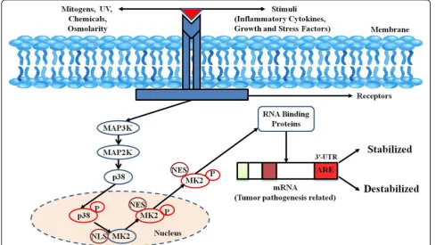
p38MAPK signaling pathway
p38MAPKs are key MAPKs involved in the production of
important inflammatory mediators, including TNFα and
cyclooxygenase-2 (COX-2). Cellular stresses/mitogens
interact in a majorly receptor-mediator manner and help trigger the phosphorylation of a MAPK kinase kinase (MAP3K) specifically which further causes the phosphoryl-ation of its downstream substrate MAPK kinase (MAP2K). After MAP2K phosphorylation, its substrate MAPK is sub-sequently phosphorylated (Fig. 1). Activated MAPKs fur-ther leads to the phosphorylation and activation of several downstream protein kinases, proto-oncogenes, and tran-scription factors [11].
Major kinases in the p38MAPK signaling pathway
MAPK pathways comprises of an array of three ki-nases: Firstly, a MAP3K which is responsible to acti-vates a MAP2K that in turn phosphorylates and activates a MAPK which occurs via a dual phosphor-ylation in the activation motif (Thr-X-Tyr where X could be any amino acid). Mammalian cells are known to express fourteen MAPKs which can be fur-ther segregated into groups based on sequence hom-ology. The classical MAPKs are ERK1 and ERK2 with MAP2Ks, MKK1 or MKK2 activating them. Four
iso-forms of the p38MAPK family are known (p38α,
p38β, p38γ, and p38δ), and these are activated by the
MAP2Ks, MKK3, and MKK6 [12].
Downstream substrates of the p38MAPK signaling pathway
There are numbers of substrates downstream of
p38MAPK signaling pathways. MK2 and MK3 were the first p38MAPK substrates identified [13]. Phosphorylated MK2 or MK3 can activate a variety of substrates, such as small Hsp27 [14], cyclic AMP-responsive element-binding protein (CREB) [15], and tristetraprolin (TTP), a RBP, known to causes mRNA destabilization thus referring at
p38MAPK’s role in mRNA stability [16]. It has been
shown that p38MAPK modulates MK2 expression both transcriptionally and post-transcriptionally in murine cell lines and embryos while it is lost in p38−/−mice [17].
Mitogen-activated protein kinase-activated protein kinase 2
p38MAPK’s downstream substrate responsible for a pleth-ora of signaling cascades in response to numerous extra-cellular stimuli ranging from apoptosis, cell division and differentiation, cell motility to inflammation is a Ser/Tyr protein kinase, MK2 [6]. MK2 acts as an important driver in the signaling pathways triggered in reply to DNA dam-age. A recent report has identified MK2 as protumorigenic with its role been shown in tumor progression [18]. Past reports have elucidated the expression of MK2 in a variety of cell types such as endothelial cells [19], smooth muscle cells [20], and cancers [21].
MK2 substrates
Upon activation MK2 phosphorylates various substrates and leads to regulation of many different biological pro-cesses. The first identified MK2 substrates were Hsp25
and Hsp27 [22]. It has been reported that Hsp27
phos-phorylation by MK2 causes remodeling of actin
cytoskel-eton which leads to cell motility [23]. MK2
increases interleukin (IL)-6 and TNF-α production by
stabilizing their mRNAs or promoting its translation
[24]. MK2 could phosphorylate several important
cancer-related proteins, such as cell division cycle 25 (Cdc25B/C) [25], polo-like kinase 1 (Plk1) [26], tuberin (TSC2), and the ARE-binding proteins (AU-rich element RNA-binding protein 1 (AUF1), human antigen R (HuR), TTP), which are responsible in modulating
tran-script stability of many genes, like TNFα, Cyclin D1,
Plk3, c-Fos, c-Myc, and matrix metalloproteinase
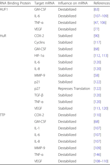
(MMP) affecting cell metabolism, differentiation, and carcinogenesis [27] (Table1). The physiological roles of these substrates are quite different, and each contains a
unique and specific amino acid motif, such as
X-X-Hyd-X-R-X-X-S-X-X (where Hyd is a bulky hydro-phobic residue), essential for efficient MK2-mediated phosphorylation [25, 28]. Recent experimental evidence elucidated that MK2 plays an important role in the maintenance of genomic stability by contributing to the G2/M and the mitotic spindle checkpoints [7].
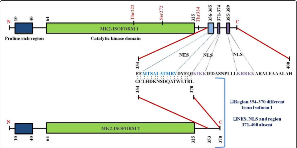
Structure and location of MK2
Human MK2, a 400-residue enzyme, contains in its N-terminus two proline-rich regions followed by the kin-ase and the C-terminal regulatory domain [13]. Except for MK3/4, a very low homology has been shown by the kin-ase domain with other serine/threonine kinkin-ases. On the other hand, no significant homology has been reported in the N-terminal proline-rich and the C-terminal regulatory domain with other non-MAPKAPK proteins. A nuclear export signal (NES) and a bipartite nuclear localization signal (NLS) are located in the C-terminal regulatory
do-main [29] (Fig. 2). Pull-down assays with MK2 and
p38MAPK indicate that the C-terminal region 366–390
represents the p38-docking region [30]. The C-terminal regulatory domain of MK2 (also MK3) contains a func-tional bipartite NLS, 371–374 and 385–389, respectively which is responsible for MK2’s location predominantly in the nuclei of resting cells. Conversely, a functional NES (a Table 1MK2 regulates transcript stability via RBPs
RNA Binding Protein Target mRNA Influence on mRNA References
AUF1 GM-CSF Destabilized [63]
IL-6 Destabilized [107–109] TNF-α Destabilized [47,106]
VEGF Destabilized [77]
HuR COX-2 Stabilized [90]
Cyclins Stabilized [117]
GM-CSF Stabilized [68]
HIF-1α Stabilized [112,113]
IL-6 Stabilized [120]
IL-8 Stabilized [120]
MMP-9 Stabilized [58]
p21 Stabilized [122]
p27 Represses Translation [122]
TGF-β Stabilized [120]
TNF-α Stabilized [120]
VEGF Stabilized [113,120]
TTP COX-2 Destabilized [110]
GM-CSF Destabilized [68]
IL-1 Destabilized [107]
IL-6 Destabilized [107]
IL-8 Destabilized [107]
motif with the sequence 356–365) which is located in the N-terminal region to the NLS is responsible for triggering nuclear export following MK2 activation [10,30] (Fig.2).
Before stimulation, both p38MAPK and MK2 are pre-dominantly located in the nucleus, but they quickly translocate after stimulation to the cytoplasm together
in a phosphorylation-dependent manner [29, 30].
Phosphorylation of MK2 by p38MAPK occurs in the nucleus and involves the interaction between the en-zymatic and catalytic domains of p38MAPK and the
NLS of MK2 [31]. Literature reports revealed that
two kinase domain residues of MK2 (T222 and S272) and one residue located outside the kinase domain
(T334) gets phosphorylated by p38MAPK (Fig. 1).
These phosphorylations have been shown to be re-quired for maximal activation of MK2 in vitro in
mu-tagenesis studies [32]. MK2 activation occurs via the
selective phosphorylation of T222 and T334. Phos-phorylation at T334 abrogates the interaction between kinase and the C-terminal regulatory domain resulting in NES being available for binding to the nuclear re-ceptor as revealed by the crystal structure of MK2
[33]. Once MK2 masks the NLS at the C-terminal
end by phosphorylation, it is rapidly exported to the
cytoplasm via Exportin 1-dependent mechanism to
phosphorylate their downstream cytosolic targets [30]
(Fig. 1).
There are many reports in the literature that confirm the role of MK2 phosphorylation at T222 located in the activation loop, S272 (catalytic domain), and T334 (out-side the catalytic domain within the C-terminal region) in its activation [32]. It has been proposed that an amphi-philicα-helix motif situated within the C-terminus region of MK2 blocks the binding of MK2 with its substrates [34]. There is a hypothesis that suggests the role of T222 and T334 dual phosphorylation in repositioning this α-helix thereby resulting in an enhanced catalytic activity.
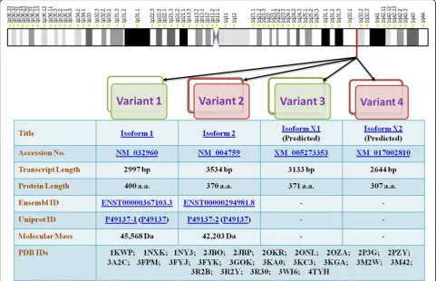
MK2 has been shown to possess different splice variants and protein isoforms (Fig. 3). Sodium dodecyl
sulphate-polyacrylamide gel electrophoresis (SDS-PAGE) [6] and
chromatography [35] led to the description of two differen-tially spliced MK2 isoforms which have comparable migra-tion intensity and which might have arisen as a result of limited proteolysis or post-translational modifications of MK2. The first variant, MK2, contains an NES, NLS and a putative p38-docking domain located near the carboxy terminus [6]. The second shorter variant of MK2 (isoform 2) [13] contains an identical N-terminal kinase domain but
lacks NES, NLS and a p38-docking domain [29, 30] and
bears the substitutive sequence GCLHDKNSDQATWLTRL in place of 354–400 sequence of isoform 1 [10] (Fig.2). Re-cently, automated computational analysis and annotation using gene prediction method have shown that there are two more isoforms of MK2 as detailed in Fig.3.
MK3
The main focus of our review is MK2, but it is still import-ant to discuss MK3 in brief [36]. This kinase has much
lower expression levels as compared to MK2 [37], but
possesses high structural identity and shares approxi-mately similar substrate range with MK2 thus implying al-most identical functional roles in biological systems [38]. The C-terminus of MK3 contains NLS and NES motifs rendering its unphosphorylated form located in the nu-cleus until p38MAPK-dependent phosphorylation induces its translocation to the cytoplasm. Additionally, MK3 could control cytokine biosynthesis in addition to MK2 due to its involvement in post-transcriptional changes in
the ARE-containing mRNAs targeted by MK2 [39].
Fur-thermore, as compared to MK2−/−, the double knockout mice (MK2−/−/MK3−/−) had a higher reduction of
lipo-polysaccharide (LPS)-induced TNFα production [37].
Strikingly, functional dissimilarities among MK2 and MK3 have been portrayed [40].
Copy number variations in MK2
MK2 has been reported to be oncogenic with its
involve-ment shown in growth and invasion of tumors [5].
Hence, genetic variations in MK2 might play a role in susceptibility and prognosis of cancer. Presently, several copy number variations (CNVs) have been shown to be
associated with human diseases including cancers [41,
42]. Studies in the past have reported CNVs causing
MK2 overexpression to influence prognosis of tumors
[43]. Similarly, CNV-30450 which duplicates the MK2
promoter was shown to increase the risk and lead to
poor prognosis of lung cancer [44]. The same group
further assessed the correlation of this CNV to nasopha-ryngeal cancer (NPC) risk [45]. Recently, it was demon-strated that there is a loss of MK2 copy number in non-small cell lung cancer (NSCLC) [46]. These studies have highlighted the need of understanding CNVs and other genomic changes in MK2 as they might act as bio-markers for assessing susceptibility, predicting risk and prognosis of cancers.
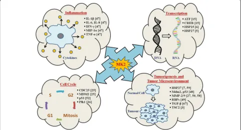
Biological functions of MK2
indicated a crucial role of MK2 in pro-inflammatory
me-diators (TNFα, IL-1β, IL-8, IL-6, and interferon-γ
(IFNγ)) production [47]. MK2 was shown to be essential for up-regulation of cytokine mRNA stability and trans-lation which is LPS-induced and hence, for stimulating cytokine biosynthesis which is integral in inflammatory
responses [47]. Recently, MK2 has been reported to be
intrinsic in control of cell-cycle at CDC25- and
p53-dependent checkpoints [25] (Table 1, Fig. 4).
DNA damage leads to inhibition of CDC25 by CHK1 and CHK2, and it has been reported that MK2
pro-motes G2/M checkpoint during stress response [25].
Further, MK2 was shown to phosphorylate and acti-vate human homolog of mouse double minute 2 (HDM2), thereby causing p53 degradation suggesting the role of MK2 in dampening the p53-mediated
re-sponse to DNA damage/stress [48].
MK2 orchestrates the post-transcriptional regulation of gene expression by modulating the function of RBPs
[49]. It has been demonstrated that MK2 and Hsp27
both modulate cell invasion and MMP-2 activation [50]. Targeting of MK2 could be a more viable option than p38MAPK, due to potentially limited side effects, attrib-uted to limited downstream substrates of MK2 as
com-pared to p38MAPK. Notably, MK2−/− mice are viable
and have a normal phenotype [47]. Hence, much of the
research has been focussed on utilizing MK2 as a mo-lecular target for developing therapeutics for ailments
like alzheimer’s, atherosclerosis, cancer, and rheumatoid arthritis (RA). MK2 modifies the function of RBPs, but MK2’s substrate spectrum is significantly limited than p38MAPK, thereby, MK2 has emerged as an attractive anti-inflammatory and anti-cancer target.
MK2 in cell cycle regulation
Insights into the molecular mechanisms of MK2-medi-ated post-transcriptional regulation indicMK2-medi-ated its
involve-ment in cell-cycle control at the CDC25- and
p53-dependent checkpoints [25, 51]. Reports have
shown that MK2 phosphorylates CDC25B/C at specific sites in ultraviolet (UV)-treated osteosarcoma cells and that MK2−/− causes loss of G2/M checkpoint [25] (Fig.
4). Hence, MK2 can be contemplated as one of the
members of the DNA-damage-checkpoint-kinase family which acts in conjunction with CHK1 and CHK2.
p53 (the tumor-suppressor protein) is also reported to be a p38MAPK cascade target. p53 has been shown to be essential for the regulation of cell-cycle at the G1/S phase and further entry into apoptosis [52]. Strikingly, p53 is a direct substrate of p38MAPK, whereas the p53-interacting ubiquitin ligase, HDM2 which is respon-sible for p53 degradation has been identified as one of
the MK2 targets [25]. HDM2 activation occurs as a
re-sult of its phosphorylation by MK2 which further leads to increased p53 degradation, thereby, resembling the HDM2 activation by the protein kinase B (PKB)/survival
kinase AKT. Hence, it has been hypothesized that MK2
inhibits p53 activity after its stimulation by
p38MAPK-mediated phosphorylation, thereby, contrib-uting to the fine regulation of DNA-damage response. Finally, the p38MAPK/MK2 pathway has been shown to activate signaling leading to G2/M checkpoint arrest and further cell survival post-DNA damage caused due to chemotherapeutics, thus responsible for resistance to treatment regimens. Mouse double minute 2 homolog (Mdm2) acts as a p53 post-transcriptional regulator, functioning by inactivating p53 through augmenting its degradation by the proteasome and repressing its tran-scriptional activity, thereby, down-regulating its protein
levels [53]. MK2 portrays a central role in p53
post-transcriptional regulation, as it has been reported
that Mdm2 phosphorylation occurs via MK2. Further,
MK2−/− cells have elevated p53 levels with reduced
Mdm2 phosphorylation [48]. Reinhardt et al. [54] dem-onstrated that tumors lacking functional p53 can survive the effect of DNA-damage causing chemotherapeutics
via a p38MAPK/MK2-dependent route. Meanwhile, in
p53-overexpressing cells, this pathway was dispensable for survival post-DNA damage. These reports showcase that MK2 follows different mechanisms for the regula-tion of cell survival in response to DNA damage.
Post-transcriptional regulation by MK2 in inflammation
Literature suggests that MK2−/− mice have enhanced re-sistance to endotoxic shock, attributed to impairment in the inflammatory response, in addition to a decreased
production of TNFα and IL-6 cytokines upon
LPS-stimulation [47]. It is quite evident now that MK2 is the prime downstream substrate of p38MAPK, and this signaling cascade regulates the stability and
transla-tion of TNFα and IL-6 mRNAs through AREs
involve-ment in the 3′-UTRs of these transcripts (Fig.4). TTP is
an RBP that has been shown to control TNFα mRNA’s
stability and translation and is a direct substrate of MK2 [30]. TTP’s phosphorylation by MK2 increases its
stabil-ity and binding to 14–3-3 proteins [25] and thereby
stimulates TNFαexpression.
Role of MK2 in actin remodeling
Hsp27 portrays a crucial role in remodeling of actin and cell migration. In its unphosphorylated state, Hsp27 can function as an actin filament cap-binding protein, lead-ing to inhibition of globular actin polymerization into filamentous actin (F-actin). MK2-mediated Hsp27 phos-phorylation [7] blocks capping activity, thus, promoting actin polymerization and remodeling [55] (Fig. 4). Fur-thermore, Hsp27 phosphorylation inhibits its multimeric self-aggregation, causing loss of its chaperone activity
[56]. Association of the MK2/Hsp27 relation in cell
migration and remodeling of actin is also crucial for in-vasion and metastasis of cancer.
Role of MK2 in tumorigenesis and tumor microenvironment
In tumor cells, the emergence of MK2 as an alterna-tive cell-cycle checkpoint, responsible for resistance to apoptosis caused by p53 mutation, has put forward MK2 as an effective target for combination-based
cancer therapies [7]. Depending on stimuli, MK2
reg-ulates phosphorylation, mRNA stability, and expres-sion of various proteins involved in actin remodeling [57], cell migration [58], immune responses [47], cell cycle and apoptosis [7] (Fig. 4).
Role in DSS-induced colitis and colorectal cancer
In colorectal carcinoma, epithelial cell proliferation and apoptosis are the key parameters contributing to tumori-genesis. As discussed earlier, one of the most important
downstream mediators of MK2’s function is Hsp27,
which is phosphorylated by MK2 in response to a variety of stimuli and is strongly associated with cancer progres-sion and metastasis [59]. A recent study on intestinal epithelial cells has shown that MK2 plays a role in pro-gression of colon cancer through downstream activation of Hsp27, which ultimately leads to angiogenesis cyto-kine mediation, cell proliferation, migration, and apop-tosis [60]. This study also emphasized that deletion of MK2 leads to reductions of both tumor size and invasive potential in azoxymethane (AOM)/dextran sodium sul-fate (DSS) induced colon cancer in mice [60]. Surpris-ingly, phosphorylation of Hsp27 is not influenced by MK2 deletion thus indicating that the function of the p38MAPK/MK2/Hsp27 pathway is cell and tissue dependent in colon cancer [60].
Deletion of MK2 in intestinal mesenchymal cells had the most profound effect on tumor multiplicity and size and was found associated with decreased epi-thelial proliferation, increased apoptosis, and
de-creased angiogenesis [60]. Furthermore, induction of
Role in skin cancer
Literature reports demonstrate that MK2 is required for the development of skin tumors. It regulates inflamma-tory response as well as maintains DNA-damaged cells
survival caused by 7,12-dimethylbenz[a]anthracene
(DMBA) during tumor initiation [61]. MK2-deficient
keratinocytes were more prone to carcinogen-induced
apoptosis viaimpaired Mdm2 phosphorylation and
sub-sequently increased p53 stabilization. This suggests an inhibitory role of MK2 in apoptosis induction during tumor promotion. A crucial mediator in response to DNA damage, the p53 protein has been shown to play a pivotal role in apoptosis induction [62].
In a nutshell, MK2 works as a double-edged sword in skin carcinogenesis as it regulates pro-inflammatory
cytokine expression as well as apoptosis via the
p53-signaling pathway. It has been reported the loss of MK2 on the one hand, causes decreased inflammatory response while on the other hand it increased p53
stabilization, thereby increasing the number of
DNA-damaged cells that undergo apoptosis (Fig. 4). In
conclusion, MK2 inhibitors could be potential anticancer agents and be employed to inhibit the early stages dur-ing carcinoma development.
Role in bladder cancer
MK2 and Hsp27 lead to activation of cell invasion and MMP-2 in prostate cancer [50], with past studies show-ing MAPK pathways to be activated durshow-ing growth phase in bladder cancer cells [63]. Further studies have reported that p38MAPK and MK2 regulate the invasion and metastasis of bladder cancer through MMP-2 and MMP-9 mRNA stability modulation [58] (Fig.4).
Up-regulation of MMPs is one of the processes by which p38MAPK promotes cell migration and invasion in tumors. Past reports have shown high MMP-2/9 ac-tivity in HTB9 cells, while in HTB5 cells MMP-9 acac-tivity in a basal state was low. Additionally, it has been ob-served that p38MAPK signaling inhibition reduces MMP-2/9 activity. Hence it could be said that active p38MAPK signaling by modulation of MMP-2/9 activity may regulate the migration/invasion in bladder cancer
[58]. Furthermore, the addition of MMP-2/9 antibody
led to inhibition of tumor invasion, indicating that MMP expression in bladder cancers is directly responsible for it. These reports suggested that p38MAPK pathway could regulate the activity of MMP independent of tissue inhibitor of metalloproteinases (TIMP) regulation. In-deed, it has been observed that a p38MAPK inhibitor and a dominant-negative kinase-inactive mutant of MK2 led to a significant reduction in MMP-2/9 mRNA half-life. Earlier Xu and colleagues [50] had reported the role of MK2 and Hsp27 in prostate cancer cell lines in-vasion. Taken together, it can be concluded that the
invasion of bladder cancer cells is regulated by
p38MAPK-driven MK2 through stabilization of MMP-2/ 9 transcripts [58].
Role in prostate cancer
Transforming growth factor β (TGFβ) is an important
regulator of cell adhesion and motility in a wide range of cell types including prostate and is shown to act in a cell-specific manner [64, 65]. Past studies have shown
that TGFβ-mediated increase in cell invasion in human
prostate cancer is dependent upon p38MAPK activation
[66]. It has also been observed that both Smad3 and
p38MAPK are integral for TGFβ-mediated cell adhesion
in prostate cancer [66]. Recent studies have suggested
that during colorectal cancer progression, TGFβ
pro-motes tumor growth via its involvement in crosstalk
with different pathways like p38MAPK and Wnt [67]. A
study by Xu et al. demonstrated that in human prostate cancer both MK2 and Hsp27 are important for TGFβ-mediated up-regulation of MMP-2 activity and cell invasion which was inhibited by SB203580, a p38MAPK inhibitor [50]. This clearly indicated a direct role of p38MAPK signalling in prostate cancer through a chan-nelized activation of p38MAPK, MK2 and Hsp27 (Fig.4).
MK2 and mRNA stability (interplay between MK2, RBPs and target RNAs)
Adenine/uridine-rich elements (AREs)
Cytokines are essential for cell signaling to facilitate re-sponses to various stimuli necessary for the maintenance of homeostasis and survival. Any malfunction in the cyto-kine signaling network has damaging effects on both the intra-cellular as well as the extracellular environments. An important process in cancer pathogenesis is cytokine and growth factor dysregulation which causes uncontrolled cell growth. Hence, tight regulation of the expression of cytokines at transcriptional and post-transcriptional levels is critical. AREs are conserved sequences located in the 3′-UTR of short-lived transcripts that codes for a multi-tude of proteins responsible for apoptosis, cellular activa-tion, cytokine signaling, and growth. The stability of cytokine mRNAs has been shown to be altered due to the presence of AREs in their 3′-UTRs.
In 1986, conserved AREs were found in the 3′-UTR of
genes encoding short-lived cytokines
(granulocyte-ma-crophage colony-stimulating factor (GM-CSF) and
TNFα) [68]. A lot of information about the role of AREs in the post-transcriptional regulation of many cytokines and growth factors is available [69]. AREs act as binding sites for RBPs that regulate mRNA half-life [70]. Most of the RBPs that bind to AREs target them to exosome thereby promoting rapid deadenylation and degradation
of their substrate mRNAs (e.g., TTP, AUF1) [71].
functionally diverse pool responsible for cellular prolifera-tion, development inflammatory and immune response, RNA metabolism, signaling, and transcription harbour
AU-rich sequences [72, 73]. The recently constructed
human ARE-containing mRNA database encompasses greater than 1000 transcripts [74]. Within the 3′-UTR, the presence of an ARE is a common link in many un-stable mRNAs in mammals which is a part of the regula-tory system responsible for the mRNA degradation or stabilization and is linked to interaction with RBPs [75]. The fate of ARE-containing mRNAs is determined by the integration of functionalities of multiple ARE-binding pro-teins/RBPs [76].
3′-UTR located AREs constitute cis-elements causing
rapid degradation of transcripts encoding many cyto-kines, growth factors, and proto-oncogenes [77]. It has been shown previously and confirmed by findings in
MK2−/− mice that the p38MAPK/MK2 pathway
facili-tates transcript stability of mRNAs that harbour distinct
AREs [8, 24, 78]. In comparison, the stability of
ARE-deficient mRNAs was not affected [79]. The
mo-lecular mechanisms behind the control of ARE-mediated stabilization/decay involve the activity of proteins select-ively interacting with ARE, but their mode of action is not well elucidated [80].
AREs comprises of many large clusters of overlapping AUUUA pentamers repeats and UUAUUUAUU nonamers that are specifically recognized by a variety of different ARE-binding proteins and found in transcripts encoding various cell cycle regulators (p16, p21, p27, cyclins, and
Cdks), cytokines, epidermal growth factor (EGF),
insulin-like growth factor (IGF), proto-oncogenes (c-fos, c-jun, c-myc), TGFβ, and vascular endothelial growth factor (VEGF) [77]. The list has considerably increased as a result of various genome sequencing programs [72]. Continuously active mRNA-decay mechanisms restrict the cytokine ex-pression in resting cells. mRNA stability regulation through AREs is a post-transcriptional control mechanism which al-lows cells under varying environmental conditions to fine-tune the expression of important gene products (reviewed in [81]).
RNA-binding proteins (RBPs)
RBPs are single or double-stranded RNA binding proteins present in cells which participate in the formation of ribonucleoprotein complexes and portray pivotal roles in processes such as cellular functions, transport, and localization. They are responsible for post-transcriptional control of RNAs, such as pre-mRNA splicing, and polya-denylation, as well as mRNA export, turnover, localization, and translation [82]. Apart from regulating mRNA decay, RBPs mediate other post-transcriptional processes like intracellular localization, pre-mRNA processing, transla-tion, and transport (reviewed in [83]). Various reports
have highlighted the function of multiple diverse classes of RBPs in the regulation of mRNA decay and stabilization (reviewed in [84]).
Studies have indicated the role of MK2 in the
modifica-tion of the stability and translamodifica-tion of IL-6 and TNFα
mRNA via activation of RBPs such as TTP, AUF1, and HuR (Table1). These processes of complex post-transcriptional
cytokine synthesis regulation via MK2-mediated RBPs
phosphorylation have been discussed in some excellent re-views [85,86]. A number of proteins having the potential to bind to ARE are known, among them TTP, and AUF1 stimulate target transcript decay by recruiting deadenylases and downstream degradation machinery [87]. By contrast, the embryonic lethal and abnormal vision (ELAV) family member HuR stabilizes its targets by competing with the destabilizing ARE-binding proteins for ARE occupancy (reviewed in [87, 88]). Induction of decay pathways for mRNA allows for the attenuation of cellular cytokines pro-duction through interactions with RBPs [89].
During inflammatory responses, cytokine mRNAs are stabilized via complex interactions with RBPs controlled
by phosphorylation via multiple signaling pathways
in-cluding the MAPKs. Activation of p38MAPK stabilizes
the COX-2 transcriptsviaits effect on AUF-1, HuR, and
TTP [90]. Substantial evidence has highlighted the rele-vance of mRNA stability in the regulation of genes [91]. mRNA fate is regulated by the complex interplay among the cis-acting sequences within mRNA and trans-acting
nuclear and cytoplasmic factors [92]. The mammalian
genome encodes approximately 1000 RBPs which por-tray important roles in mRNA stability, splicing, localization, nuclear export, and translation. RBPs phys-ically interact with mRNA to exert their functionality in a highly sequence-specific manner. AREs are amongst the well-characterized regions that bind RBPs. Different RBPs have been discovered which function by stabilizing, destabilizing or influencing the translation of ARE-con-taining mRNAs (Table 1). A possible hypothesis for role of the p38MAPK cascade is that it stimulates the modifi-cation of RBPs by phosphorylation. RBPs are rightly called as the master regulators of transcript processing and translation with their expression often found to be
aberrant in cancer [93]. In conjunction with
much-studied transcription factors, RBPs have emerged as inte-gral components in tumor development. RBPs along with their mRNAs targets form a complex network of post-transcriptional regulation of gene expression that plays a crucial role in tumorigenesis [94].
Tristetraprolin (TTP)
by post-transcriptional TTP-mediated destabilizing
mechanisms (reviewed in [97]). The role of TTP as a
trans-acting anti-inflammatory RBP first came into light
when TTP−/− mouse showed overexpression of TNFαin
macrophages and developed the pro-inflammatory
phenotype [98]. TTP confers mRNA instability and
deg-radation by binding the conserved ARE in the 3′-UTR
of transcripts [99], which promotes the poly(A) tail
shortening reported in GM-CSF and TNFα [100]. TTP
shows very low constitutive levels and is an early re-sponse gene induced in phagocytes by LPS. It functions as a negative feedback on cytokine mRNAs; hence, mice lacking TTP tend to overproduce cytokines. Contradic-torily, TTP expression is influenced by p38MAPK
signal-ing [16]. TTP becomes hyperphosphorylated, with both
p38MAPK and MK2 having been implicated in this process [16,95].
The p38MAPK pathway regulates the mRNA expres-sion, mRNA decay property and protein expression of
TTP via MK2 [101]. TTP binds the TNFαARE and
de-stabilizes the mRNA [99]. Mice null for TTP develop an inflammatory syndrome because they overexpressed TNFα[98]. MK2 phosphorylates 14–3-3 binding sites at Ser52 and Ser178 in TTP [95], causing the protein to be
sequestered away from TNFα mRNA and prevents it
from recruiting a deadenylase to the bound transcript [102, 103]. These phosphorylations enable complex
for-mation of TTP with multifunction adaptor 14–3-3
pro-teins resulting in ablation of its function as a transcript destabilizing protein [95], hence, allowing efficient
trans-lation via subcellular translocation of the mRNA [51,
104]. Literature suggests that MK2-mediated TTP
phos-phorylation increases the expression of TTP proteinvia
cytoplasmic retention and exclusion from proteasomal
degradation [105]. TTP dephosphorylation causes its
movement from the cytoplasm into the nucleus and causes its degradation [101]. The deletion of AREs in the
3′-UTR of TNFα in mice leads to elevated TNFα
pro-duction and inflammatory disorders [106].
Many studies have shown that TTP overexpression in vitro promoted the decay of mRNAs containing AU-rich
sequences from TNFα [99]. In a p38MAPK-dependent
manner, TTP directs mRNA stability of IL-6 [107]. The
p38MAPK-MK2 axis is responsible for TTP being a mRNA destabilizing factor [100]. Moreover, in head and neck squa-mous cell carcinoma (HNSCC), TTP down-regulation en-hances the stability of mRNAs, promotes IL-6 and VEGF secretion and significantly increases cellular invasion in cancers by increased secretion of IL-6 and MMP-2/9 [108, 109]. Given all these reports, TTP could be considered a
therapeutic target as it can concurrently lead to
down-regulation of multiple cytokines in HNSCC.
It was recently shown that TTP expression is inversely correlated with invasion in HNSCC [108]. In macrophages,
TTP is inactivated by phosphorylation [51]. The mechan-ism by which TTP mediates invasion of HNSCC has been investigated, and it has been shown that the suppression or p38MAPK-mediated phosphorylation of TTP leads to the promotion of invasion due to enhanced secretion of IL-6 and MMP-2/9. TTP promotes mRNAs degradation by binding to AREs in the 3′-UTR [96,107]. In macrophages,
p38MAPK inactivates TTPviaMK2-mediated
phosphoryl-ation at two serine sites [51, 103]. Typical targets of TTP
are mRNAs regulating tumor growth such as TNFα,
COX-2, VEGF, and IL-10 [110] (Table 1). It has been
suggested that decreased TTP expression contributes to cancer-related processes, and reports show that TTP-medi-ated regulation of crucial cancer-relTTP-medi-ated transcripts in breast cancer cell leads to suppression of their invasive potential [111].
Human antigen R (HuR)
HuR, one of the most prominent RBP, is intricately in-volved in tumorigenesis [112], with its overexpression been observed in a number of cancers including brain, breast, colon, gastric, lung, lymphomas, oral, ovarian, pancreatic, prostate, and skin cancers [113]. In normal cells, HuR is generally localized in the nucleus, but in transformed cells, it often translocates to the cytoplasm
[114]. MK2 has been shown to induce the cytoplasmic
accumulation of HuR [114]. MK2 has been shown to
regulate intercellular adhesion molecule-1 (ICAM-1)
and IL-8 expression in acute inflammatory response via
HuR [115]. The sub-cellular localization of HuR is gov-erned by post-translational modifications, and all the HuR modifying enzymes are implicated in cancer
pro-cesses [116]. In the cytoplasm, HuR binds to AREs
lo-cated in 3′-UTR of target mRNAs. HuR is most often
functionally defined as a positive regulator of target mRNAs-stability and translation [112], which generally code for cyclins, favouring cell cycle progression and promoting proliferation of malignant cells [117]. In vivo models suggested a more diverse functional array with multiple complex side-effects [118]. Investigations sug-gested that elevated cytoplasmic localization of HuR cor-responds to high-grade tumor thereby serving as a good prognostic indicator for poor clinical response in many cancers [119].
HuR targets mRNAs which encodes products promot-ing proliferation, increaspromot-ing angiogenesis, inhibitpromot-ing apoptosis, and facilitating invasion and metastasis, viz. COX-2, GM-CSF, IL-6, IL-8, inducible nitric oxide
syn-thase (NOS), TGFβ, TNFα, VEGF, and others [120]
(Table 1). IL-1βactivates the MK2-HuR pathway which
encoding anti-apoptotic factors like B-cell lymphoma 2 (Bcl-2), p21, and Sirtuin 1 (SIRT1) [122]. The mechan-ism behind this feature of HuR is still unclear, but a few studies attribute this to the interplay among HuR and microRNAs [123]. HuR enhances the stability of a set of its target mRNAs by antagonizing their binding to RBPs or microRNAs that destabilizes them [124,125]. Overex-pression of HuR is found in HNSCC, and it leads to in-creases in the stability of COX2 and VEGF mRNAs
[113]. In several cancers (including HNSCC) increased
cytoplasmic HuR localization has been found, which contributes to increased COX-2 expression in metastasis and tumorigenesis [126].
AU-rich element RNA-binding protein 1 (AUF1)
AUF1 is a RBP that regulates the mRNA stability of proto-oncogenes, growth factors, cytokines, and cell cycle regulatory genes. AUF1 generally destabilizes tran-scripts and has been shown to control the stability and
translation of GM-CSF, IL-6, TNF-α, VEGF and many
other ARE-containing mRNAs [127] (Table 1). AUF1
has been reported to be present in a cytosolic fraction and its overexpression in animal models has been shown to be associated with decreased mRNA stability [128]. It has been shown that a p38MAPK-MK2-Hsp27 signaling axis promotes proteasomal degradation of AUF1, further leading to cytokine ARE-mRNAs stabilization [129].
Correlation between MK2-mediated mRNA stabilization and tumorigenesis
An important determinant in modulating gene expres-sion levels is the regulation of mRNA stability.
Numer-ous studies in the past have demonstrated the
importance of mRNA stability-mediated regulation in inflammation and cancer [9, 68, 89]. Modulation of the decay rate of various cytokines, proto-oncogenes, and growth factors, involves AREs in their 3′-UTRs [77]. RBPs tend to fine-tune cellular responses and directly mediate critical inflammatory signals responsible for dis-ease pathogenesis by binding to AREs. It is quite evident that any aberrations in mRNA decay processes can lead to the over-production of certain gene-encoded products that can possibly lead to cancer. Post-transcriptional regulation of gene expression has been shown to be ab-errant in tumors with over-expression of ARE-rich
mRNAs been reported in multiple cancers [130]. RBPs
like HuR have been shown to stabilize VEGF mRNA in various tumors [131]. AREs tend to play a huge role in the post-transcriptional regulation of certain genes in-volved in carcinogenesis [78]. Mechanistic insights on how AREs fine-tune mRNA stability reveal
involve-ment of specific MK2-regulated RBPs [9, 132]. Past
findings have implicated MK2 in mediating tumor
in-vasion via regulating mRNA stability of MMP-2/9 in
bladder cancer [58]. Hence, a better understanding of
MK2-RBP mediated mechanisms will surely enable us to develop novel therapeutics in combating cancer progression [133].
Therapeutic implications of MK2
As therapeutic target?
MK2 modulates the stability and translation of inflam-matory cytokines through phosphorylation of
transacti-vating factors binding to their AREs [51]. Hence, MK2
inhibition could be a target for blocking the production of inflammatory mediators. Traditionally active site in-hibitors of the kinases were employed for therapeutic purposes. But the major issue with this approach is that ATP competitive inhibitors of kinases have been known to be inherently cross-reactive, because of the homology shared by kinase active sites, hence, the development of specific active site kinase inhibitors is difficult.
A more viable approach in the development of selective kinase inhibitors is the search of agents that disrupt the docking among kinases, and their upstream and down-stream signaling partners. MK2 docking domain compris-ing peptide is a potent inhibitor of p38MAPK-dependent phosphorylation of MK2. This might also perturb p38MAPK’s interaction with its upstream activators, like
MKK3 [134]. Thus a more reasonable approach for
inhi-biting this pathway would be the development of inhibi-tors of the docking interactions between p38MAPK and its signaling partners [135].
Inhibitors of MK2: Types, uses and history
Systemic side-effects of p38MAPK inhibitors such as cardiac toxicity, hepatotoxicity and central nervous sys-tem (CNS) disorders have been among foremost hurdles against the developed inhibitors to transform into a suc-cessful drug. This was the main reason behind their fail-ure in phase III clinical trials [136]. To overcome the issue and for effective inhibition of p38MAPK signalling pathway, researchers prompted toward numerous down-stream targets of the pathway such as MK2 [10].
Currently, MK2 is widely considered as a novel disease-modifying anti-rheumatic drug (DMARD) ligand and a promising possible alternative to p38MAPK for the treatment of various inflammatory diseases. Study on the involvement of MK2 in inflammation associated disorders
suggested that health of p38−/− mice suffering from
neurodegenerative disease such as parkinson’s disease, multiple sclerosis and even alzheimer’s disease. So it has been suggested that this linkage could be directly associ-ated with modulation of MK2 activity [10].
Past studies had indicated that targeting MK2 to block its downstream events could be equivalent to direct
in-hibition of upstream p38α (responsible for MK2
activa-tion) of the p38MAPK pathway, with the additional advantage of lacking any p38-dependent side effects [138, 139]. This is the reason that MK2 is currently be-ing considered as a more promisbe-ing target. The inhibi-tors of the MK2 activity could serve as potential therapeutic agents in the treatment of various inflamma-tion and neuro-inflammainflamma-tion associated diseases. The active involvement of MK2 with Hsp27 may also be used to reduce remodelling and migration of cancer cells and metastasis through its abrogation. Furthermore, consid-ering the capability of MK2 to modulate a cell cycle checkpoint, inhibitors of MK2 are also considered as ef-fective tools to evade DNA repair mechanism induced by chemotherapy and thus resulting in increased sensi-tivity of tumor cells to chemotherapy [25,58,60].
Nearly all of the revealed MK2 inhibitors belong to the type I class of inhibitors (ATP competitive MK2 inhibi-tors (which binds to the ATP binding site of the kinase) and therefore compete with intra-cellular ATP molecules to block p38MAPK-mediated phosphorylation and acti-vation of the kinase. Several compounds with in vivo ef-ficacy against MK2 have been already reported by other
researchers also [140]. After discerning various
com-pounds with minimal to modest in vitro activity towards
MK2 [141], researchers have made significant
improve-ments in efficacy and safety as compared to compounds generated earlier. However, low biochemical efficiency (BE) value (generally expressed as the ratio between Ki -the binding affinity of inhibitor molecule to -the target protein and its effective concentration 50 (EC50-cellular activity of the inhibitor) has been one of the major draw-backs of the MK2 inhibitors discovered so far.
Void and lacunas in the area of MK2 inhibitor research
Various studies on the mechanism of action of total marketed drugs demonstrate that around two-thirds of
them have BE values higher than 0.4 [142]. A study by
Swinney et al. [142] reported that BE value higher than 0.4 is an attribute of many approved drugs. If we con-clude strictly, the EC50 values for any successful drug should not be more than 2.5-folds higher than its Ki values. Studies indicated that cellular efficacy reports for MK2 inhibitors in a diseased condition are quite inad-equate in public domain, and indicated BE values of the tested inhibitors are far below the 0.4 threshold, suggest-ing the unlikeness of available MK2 inhibitors to become successful drug candidates [140]. Retaining in mind the
fact that high concentrations of inhibitor compounds are required to ascertain good cellular efficacy in diseased
conditions, their cytotoxicity, non-specificity and
side-effects could be aggravated, thus increasing the prob-ability of attrition. Conversely, compounds not competing with intracellular ATP could remain active at compara-tively lower concentrations and have a greater probability to be optimized to become a drug. Inopportunely, the cur-rently available uncompetitive and non-ATP competitive MK2 inhibitor compounds do not provide any experimen-tal support to this hypothesis, thus, opening the door of possibilities for experimental validation of already avail-able non-competitive MK2 inhibitors.
The higher affinity of inactive MK2 towards intra-cellular ATP has been anticipated as the major deter-minant of lowering the BE values for potential MK2 inhibitors. Consequently, researchers have screened their known inhibitors among the pool of compounds that bind the inactive form of the kinase, have a lesser competition with the high intracellular ATP concentration, and, accordingly, are required at low concentrations to give cellular effects in diseased con-ditions. By looking into all these factors, the import-ance of MK2 in modulating inflammatory conditions, cell cycle process, cytoskeleton remodelling and cell motility, non-ATP-competitive and allosteric inhibitors of MK2 are under continuous investigation as nega-tive regulators or modulators of the p38MAPK/MK2 signalling pathway in various disorders [10].
Current insights into MK2 inhibitors ATP competitive inhibitors
MK2 has been hypothesized as a potent druggable target in inflammatory disorders. The release of 3D structure of MK2 in complex with ADP or other small molecule inhibitors drove the discovery of numerous small mol-ecule ATP-competitive inhibitors (Table 2). Conversely, blocking the MK2 with its ATP binding site in competi-tive mode gave rise to two important challenging issues. Firstly, the similarity of the ATP-binding site of MK2 with other kinases (MK3, MK5, etc.), interferes with the selectivity of inhibitors. Secondly, low BE value of the ATP-competitive inhibitors either due to high affinity of ATP for its binding site on kinase. Finally, solubility in suitable agents and permeability profiles of inhibitors ap-propriate for in vivo administration have been very diffi-cult tasks to be addressed [10,141].
ATP non-competitive inhibitors
In the recent years, due to the inefficiencies associated with ATP-competitive inhibitors, promising inhibitors with non-ATP competitive and ATP-uncompetitive
mechanism of action have been identified (Table 2).
with a binding site in kinase which is different from that of ATP, thus avoiding issues like selectivity with other ki-nases and low BE value. Additional advantage associated with them is effectiveness at low concentration. By def-inition, non-competitive inhibitors are not required to contend with the high ATP concentrations in cells and with high affinity of ATP for the inactive and active forms of MK2, effective lower concentrations of them promises less pronounced side-effects also. Mechanism of action of inhibitor dissimilar to ATP-competitiveness could enhance BE value of potential inhibitors and have better possibilities to be developed as an effective drug candidate against MK2. Thus, inhibitory efficacy of a non-ATP competitive inhibitor is expected to be higher than ATP-competitive inhibitors. Additionally, they could exert higher kinase selectivity profile as a conse-quence of the fact that they do not bind to similar ATP binding sites among related kinases [10].
Studies have shown that a good BE value enables the effi-cacy of a drug at lower concentrations with an increase in the therapeutic index, there is a minimum probability of success in clinical studies in case of ATP-competitive MK2 inhibitors. Mourey et al. [140] demonstrated in vivo efficacy of a selective ATP-competitive MK2 inhibitor PF-3644022 despite its biochemical inefficiency (BE 50.03). This
inhibi-tor has been reported to reduce TNFα production in
in-flammation mice models. Various non-ATP-competitive inhibitors have been reported by Merck [143], and it would be of a topic of great interest to see the progression of this class of compounds in in vivo and clinical studies further (Table2). As of now, the outcomes of MK2 inhibition can only be assumed and solely depend on the analysis of the efficacy of inhibitors of p38MAPK that target MK2 activa-tion. Along these lines, Watterson et al. [144] have recently demonstrated that the anti-neuroinflammatory efficacy of blood-brain-barrier-permeable p38MAPK inhibitors
in the animal model of Alzheimer’s disease correlates
with the inhibition of MK2 activity. Recently,
CDD-450, also called ATI-450 was developed as an unique inhibitor which possesses the property of se-lectively blocking p38MAPK-mediated MK2 activation
while sparing other p38α substrates. ATI-450 has an
efficacy similar to global p38α inhibitors and inhibits
IL-6, IL-1β, and TNF-α production thereby decreasing
inflammation in preclinical models [145].
Conclusions
MK2 activation generates a plethora of different biological effects targeting diverse cellular processes like cell-cycle progression, cytoskeletal architecture, mRNA stability, and protein translationviaregulating the activation and deacti-vation cycles of RBPs [146, 147]. Improved understanding
of MK2’s role in tumor progression could provide new
insight into the enigma behind the post-transcriptional Table 2List of potent ATP competitive and non-competitive
inhibitors of MK2 [151–162]
gene regulation in tumorigenesis. The complex mecha-nisms of post-transcriptional of cytokine regulation via MK2-mediated phosphorylation of RBPs play a pivotal role in tumorigenesis [85,86].
Inhibition of the p38MAPK/MK2 pathway by blocking p38MAPK failed, as none of the inhibitors were found successful in the clinical trials due to the unwanted side effects [10]. Hence, in recent times, MK2 was preferred as a potential candidate for targeted therapies as a p38MAPK alternate to minimize the systemic undesired effects associated with the majority of p38MAPK inhibi-tors. MK2 remains a promising therapeutic target given the importance of the p38/MK2 pathway in processes like cell-cycle, inflammation, and metastasis.
DNA-damage due to chemotherapeutic agents could be repaired by cancer cells by arresting cell cycle pro-gression and escaping apoptosis. It has been shown that MK2 activity is essential for G2/M arrest, hence; it gives an exciting prospective for the utility of MK2 inhibitors as chemo-sensitizers. Importantly, MK2-depleted mice are viable [47], in contrast to Chk1 and p38MAPK−/− mice [148], suggesting that MK2 inhibition could target cancer cells the same way as Chk1 and p38MAPK inhib-itors but with fewer side-effects. Latest reports of MK2 inhibition decreasing production of inflammatory cyto-kines and subsequently leading to reduced tumor vol-umes potentiates its use in therapeutics [149,150].
Pathological roles of MK2 in several diseases has led to a renewed interest in developing drug-like MK2 in-hibitors despite the difficulties encountered in this process. The identification of MK2 inhibitors with suit-able pharmacodynamics and pharmacokinetics is an at-tractive question for medicinal chemists [10]. Scientific advances in the area of molecular oncology have opened novel research directions. Nowadays, numerous research endeavours have been concentrated towards developing
targeted therapies and unveiling novel molecular
markers which could be utilized in predictions of treat-ment outcome or personalized therapies. It is quite evi-dent that further unraveling the molecular tumorigenesis enigma will surely pave the way forward for novel thera-peutics and personalized treatment regimens for the patients.
Abbreviations
3′-UTR:3′-untranslated region; AOM/DSS: Azoxymethane/Dextran sodium sulphate; AREs: Adenine/uridine-rich elements; AUF1: AU-rich element RNA-binding protein 1; BCL-2: B-cell lymphoma 2; BE: Biochemical efficiency; CDC25: Cell division cycle 25; CNV: Copy number variation; COX-2: Cyclooxygenase-2; CREB: Cyclic AMP-responsive element-binding protein; DMARD: Disease-modifying anti-rheumatic drug; DMBA:
7,12-dimethylbenz[a]anthracene; EC50: Effective concentration 50; EGF: Epidermal growth factor; ELAV: Embryonic lethal and abnormal vision; ERK: Extracellular signal-regulated kinase; F-actin: Filamentous actin; GM-CSF: Granulocyte-macrophage colony-stimulating factor; HDM2: Human homolog of mouse double minute 2; HNSCC: Head and neck squamous cell carcinoma; HSP27: Heat shock protein; HuR: Human antigen R; IECs: Intestinal epithelial
cells; IFNγ: Interferon-γ; IGF: Insulin-like growth factor; IL: Interleukin; LPS: Lipopolysaccharide; MAP2K: MAPK kinase; MAP3K: MAPK kinase kinase; MAPKAPK2 or MK2: Mitogen-activated protein kinase-activated protein kinase 2; MDM2: Mouse double minute 2 homolog; MK2−/−: MK2 knockout; MMP: Matrix metalloproteinase; NES: Nuclear export signal; NLS: Nuclear localization signal; NOS: Nitric oxide synthase; NPC: Nasopharyngeal cancer; NSCLC: Non-small cell lung cancer; p38MAPK: p38 mitogen-activated protein kinase; PKB: Protein kinase B; PLK1: Polo-like kinase 1; RA: Rheumatoid arthritis; RBPs: RNA-binding proteins; SDS-PAGE: Sodium dodecyl sulphate-polyacrylamide gel electrophoresis; SIRT1: Sirtuin 1; TGFβ: Transforming growth factorβ; TIMP: Tissue inhibitor of metalloproteinases; TNFα: Tumor necrosis factor; TSC2: Tuberin; TTP: Tristetraprolin; UTR: Untranslated region; UV: Ultraviolet; VEGF: Vascular endothelial growth factor
Acknowledgements
The authors would like to thank the Director, CSIR-IHBT, Palampur, India for his constant support and encouragement. SS is immensely thankful to CSIR and PA to Department of Science and Technology, Government of India for providing Ph.D. fellowship. Both SS and PA acknowledge Academy of Scientific and Innovative Research (AcSIR), Tamil Nadu for Ph.D. registration. The authors are apologetic to many research groups whose work could not be cited due to space constraints.
Funding
The authors acknowledge CSIR for financial support (CSIR-IHBT; Project MLP0204).
Availability of data and materials
Not applicable.
Authors’contributions
SS, PA and YSP conceptualized the manuscript. SS and PA collected the literature, wrote the manuscript and made the figures. YSP edited and made significant revisions to the manuscript. All authors read and approved the final manuscript.
Ethics approval and consent to participate
Not applicable.
Consent for publication
Not applicable.
Competing interests
The authors declare that they have no competing interests.
Publisher’s Note
Springer Nature remains neutral with regard to jurisdictional claims in published maps and institutional affiliations.
Received: 6 December 2018 Accepted: 21 February 2019
References
1. Lee JC, Laydon JT, McDonnell PC, Gallagher TF, Kumar S, Green D, McNulty D, Blumenthal MJ, Keys JR, Strickler JE, McLaughlin MM. A protein kinase involved in the regulation of inflammatory cytokine biosynthesis. Nature. 1994;372:739.
2. Seisenbacher G, Hafen E, Stocker H. MK2-dependent p38b signalling protects Drosophila hindgut enterocytes against JNK-induced apoptosis under chronic stress. PLoS Genet. 2011;7:e1002168.
3. Li Y, Inoki K, Vacrasis P, Guan KL. The p38 and MK2 kinase cascade phosphorylates tuberin, the tuberous sclerosis 2 (TSC2) gene product, and enhances its interaction with 14-3-3. J Biol Chem. 2003;278:13663–71. 4. Wu R, Kausar H, Johnson P, Montoya-Durango DE, Merchant M, Rane MJ.
Hsp27 regulates Akt activation and PMN apoptosis by scaffolding MK2 to Akt signal complex. J Biol Chem. 2007;282:21598–608.
6. Stokoe D, Campbell DG, Nakielny S, Hidaka H, Leevers SJ, Marshall C, Cohen P. MAPKAP kinase-2; a novel protein kinase activated by mitogen-activated protein kinase. EMBO J. 1992;11:3985–94.
7. Gurgis FM, Ziaziaris W, Munoz L. Mitogen-activated protein kinase–activated protein kinase 2 in neuroinflammation, heat shock protein 27
phosphorylation, and cell cycle: role and targeting. Mol Pharmacol. 2014;85: 345–56.
8. Winzen R, Kracht M, Ritter B, Wilhelm A, Chen CY, Shyu AB, Müller M, Gaestel M, Resch K, Holtmann H. The p38 MAP kinase pathway signals for cytokine-induced mRNA stabilization via MAP kinase-activated protein kinase 2 and an AU-rich region-targeted mechanism. EMBO J. 1999;18:4969–80.
9. Pereira B, Billaud M, Almeida R. RNA-binding proteins in cancer: old players and new actors. Trends Cancer. 2017;3:506–28.
10. Fiore M, Forli S, Manetti F. Targeting mitogen-activated protein kinase-activated protein kinase 2 (MAPKAPK2, MK2): medicinal chemistry efforts to lead small molecule inhibitors to clinical trials. J Med Chem. 2015;59:3609–34. 11. Ashwell JD. The many paths to p38 mitogen-activated protein kinase
activation in the immune system. Nat Rev Immunol. 2006;6:532.
12. Arthur JS, Ley SC. Mitogen-activated protein kinases in innate immunity. Nat Rev Immunol. 2013;13:679.
13. Zu YL, Wu FY, Gilchrist A, Ai YX, Labadia ME, Huang CK. The primary structure of a human MAP kinase activated protein kinase 2. Biochem Biophys Res Commun. 1994;200:1118–24.
14. Guay J, Lambert H, Gingras-Breton G, Lavoie JN, Huot J, Landry J. Regulation of actin filament dynamics by p38 map kinase-mediated phosphorylation of heat shock protein 27. J Cell Sci. 1997;110:357–68.
15. Tan YI, Rouse J, Zhang A, Cariati S, Cohen P, Comb MJ. FGF and stress regulate CREB and ATF-1 via a pathway involving p38 MAP kinase and MAPKAP kinase-2. EMBO J. 1996;15:4629–42.
16. Mahtani KR, Brook M, Dean JL, Sully G, Saklatvala J, Clark AR. Mitogen-activated protein kinase p38 controls the expression and posttranslational modification of tristetraprolin, a regulator of tumor necrosis factor alpha mRNA stability. Mol Cell Biol. 2001;21:6461–9.
17. Sudo T, Kawai K, Matsuzaki H, Osada H. p38 mitogen-activated protein kinase plays a key role in regulating MAPKAPK2 expression. Biochem Biophys Res Commun. 2005;337:415–21.
18. Suarez-Lopez L, Sriram G, Kong YW, Morandell S, Merrick KA, Hernandez Y, Haigis KM, Yaffe MB. MK2 contributes to tumor progression by promoting M2 macrophage polarization and tumor angiogenesis. Proc Natl Acad Sci U S A. 2018;115:4236–44.
19. Dalle-Donne I, Rossi R, Milzani A, Di Simplicio P, Colombo R. The actin cytoskeleton response to oxidants: from small heat shock protein phosphorylation to changes in the redox state of actin itself. Free Radic Biol Med. 2001;31:1624–32.
20. Hedges JC, Dechert MA, Yamboliev IA, Martin JL, Hickey E, Weber LA, Gerthoffer WT. A role for p38MAPK/HSP27 pathway in smooth muscle cell migration.J Biol Chem. 1999;274:24211–9.
21. Ray AL, Berggren KL, Restrepo Cruz S, Gan GN, Beswick EJ. Inhibition of MK2 suppresses IL-1β, IL-6, and TNF-α-dependent colorectal cancer growth. Int J Cancer. 2018;142:1702–11.
22. Stokoe D, Engel K, Campbell DG, Cohen P, Gaestel M. Identification of MAPKAP kinase 2 as a major enzyme responsible for the phosphorylation of the small mammalian heat shock proteins. FEBS Lett. 1992;313:307–13. 23. Menon MB, Ronkina N, Schwermann J, Kotlyarov A, Gaestel M.
Fluorescence-based quantitative scratch wound healing assay
demonstrating the role of MAPKAPK-2/3 in fibroblast migration. Cell Motil Cytoskeleton. 2009;66:1041–7.
24. Neininger A, Kontoyiannis D, Kotlyarov A, Winzen R, Eckert R, Volk HD, Holtmann H, Kollias G, Gaestel M. MK2 targets AU-rich elements and regulates biosynthesis of tumor necrosis factor and interleukin-6 independently at different post-transcriptional levels. J Biol Chem. 2002;277: 3065–8.
25. Manke IA, Nguyen A, Lim D, Stewart MQ, Elia AE, Yaffe MB. MAPKAP kinase-2 is a cell cycle checkpoint kinase that regulates the Gkinase-2/M transition and S phase progression in response to UV irradiation. Mol Cell. 2005;17:37–48. 26. Tang J, Yang X, Liu X. Phosphorylation of Plk1 at Ser326 regulates its
functions during mitotic progression. Oncogene. 2008;27:6635.
27. Ronkina N, Menon MB, Schwermann J, Tiedje C, Hitti E, Kotlyarov A, Gaestel M. MAPKAP kinases MK2 and MK3 in inflammation: complex regulation of TNF biosynthesis via expression and phosphorylation of tristetraprolin. Biochem Pharmacol. 2010;80:1915–20.
28. Stokoe D, Caudwell B, Cohen PT, Cohen P. The substrate specificity and structure of mitogen-activated protein (MAP) kinase-activated protein kinase-2. Biochem J. 1993;296:843–9.
29. Engel K, Kotlyarov A, Gaestel M. Leptomycin B-sensitive nuclear export of MAPKAP kinase 2 is regulated by phosphorylation. EMBO J. 1998;17:3363–71. 30. Ben-Levy R, Hooper S, Wilson R, Paterson HF, Marshall CJ. Nuclear export of
the stress-activated protein kinase p38 mediated by its substrate MAPKAP kinase-2. Curr Biol. 1998;8:1049–57.
31. Tanoue T, Maeda R, Adachi M, Nishida E. Identification of a docking groove on ERK and p38 MAP kinases that regulates the specificity of docking interactions. EMBO J. 2001;20:466–79.
32. Ben-Levy R, Leighton IA, Doza YN, Attwood P, Morrice N, Marshall CJ, Cohen P. Identification of novel phosphorylation sites required for activation of MAPKAP kinase-2. EMBO J. 1995;14:5920–30.
33. Meng W, Swenson LL, Fitzgibbon MJ, Hayakawa K, ter Haar E, Behrens AE, Fulghum JR, Lippke JA. Structure of mitogen-activated protein kinase-activated protein (MAPKAP) kinase 2 suggests a bifunctional switch that couples kinase activation with nuclear export. J Biol Chem. 2002;277:37401–5.
34. Engel K, Schultz H, Martin F, Kotlyarov A, Plath K, Hahn M, Heinemann U, Gaestel M. Constitutive activation of mitogen-activated protein kinase-activated protein kinase 2 by mutation of phosphorylation sites and an A-helix motif.J Biol Chem. 1995;270:27213–21.
35. Chevalier D, Allen BG. Two distinct forms of MAPKAP kinase-2 in adult cardiac ventricular myocytes. Biochemistry. 2000;39:6145–56. 36. McLaughlin MM, Kumar S, McDonnell PC, Van Horn S, Lee JC, Livi GP,
Young PR. Identification of mitogen-activated protein (MAP) kinase-activated protein kinase-3, a novel substrate of CSBP p38 MAP kinase. J Biol Chem. 1996;271:8488–92.
37. Ronkina N, Kotlyarov A, Dittrich-Breiholz O, Kracht M, Hitti E, Milarski K, Askew R, Marusic S, Lin LL, Gaestel M, Telliez JB. The mitogen-activated protein kinase (MAPK)-activated protein kinases MK2 and MK3 cooperate in stimulation of tumor necrosis factor biosynthesis and stabilization of p38 MAPK. Mol Cell Biol. 2007;27:170–81.
38. Cheng R, Felicetti B, Palan S, Toogood-Johnson I, Scheich C, Barker J, Whittaker M, Hesterkamp T. High-resolution crystal structure of human Mapkap kinase 3 in complex with a high affinity ligand. Protein Sci. 2010;19:168–73. 39. Ronkina N, Kotlyarov A, Gaestel M. MK2 and MK3–a pair of isoenzymes.
Front Biosci. 2008;13:5511–21.
40. Ehlting C, Ronkina N, Böhmer O, Albrecht U, Bode KA, Lang KS, Kotlyarov A, Radzioch D, Gaestel M, Häussinger D, Bode JG. Distinct functions of mitogen-activated protein kinase-activated protein (MAPKAP) kinases MK2 and MK3: MK2 mediates lipopolysaccharide-induced signal transducers and activators of transcription 3 (STAT3) activation by preventing negative regulatory effects of MK3. J Biol Chem. 2011;286:24113–24.
41. McCarroll SA, Altshuler DM. Copy-number variation and association studies of human disease. Nat Genet. 2007;39:S37.
42. Shlien A, Malkin D. Copy number variations and cancer susceptibility. Curr Opin Oncol. 2010;22:55–63.
43. Birner P, Beer A, Vinatzer U, Stary S, Höftberger R, Nirtl N, Wrba F, Streubel B, Schoppmann SF. MAPKAP kinase 2 overexpression influences prognosis in gastrointestinal stromal tumors and associates with copy number variations on chromosome 1 and expression of p38 MAP kinase and ETV1. Clin Cancer Res. 2012;18:1879–87.
44. Liu B, Yang L, Huang B, Cheng M, Wang H, Li Y, Huang D, Zheng J, Li Q, Zhang X, Ji W. A functional copy-number variation in MAPKAPK2 predicts risk and prognosis of lung cancer. Am J Hum Genet. 2012;91:384–90. 45. Yang L, Liu B, Qiu F, Huang B, Li Y, Huang D, Yang R, Yang X, Deng J, Jiang
Q, Zhou Y. The effect of functional MAPKAPK2 copy number variation CNV-30450 on elevating nasopharyngeal carcinoma risk is modulated by EBV infection. Carcinogenesis. 2013;35:46–52.
46. Erdem JS, Skaug V, Haugen A, Zienolddiny S. Loss of MKK3 and MK2 copy numbers in non-small cell lung cancer. J Cancer. 2016;7:512.
47. Kotlyarov A, Neininger A, Schubert C, Eckert R, Birchmeier C, Volk HD, Gaestel M. MAPKAP kinase 2 is essential for LPS-induced TNF-αbiosynthesis. Nature Cell Biol. 1999;1:94.
48. Weber HO, Ludwig RL, Morrison D, Kotlyarov A, Gaestel M, Vousden KH. HDM2 phosphorylation by MAPKAP kinase 2. Oncogene. 2005;24:1965–72. 49. Morandell S, Reinhardt HC, Cannell IG, Kim JS, Ruf DM, Mitra T, Couvillon
![Table 2 List of potent ATP competitive and non-competitiveinhibitors of MK2 [151–162]](https://thumb-us.123doks.com/thumbv2/123dok_us/9018238.1894653/13.595.49.291.110.713/table-list-potent-atp-competitive-non-competitiveinhibitors-mk.webp)
