R E S E A R C H
Open Access
ALX4, an epigenetically down regulated
tumor suppressor, inhibits breast cancer
progression by interfering Wnt/
β
-catenin
pathway
Juntang Yang
†, Fei Han
†, Wenbin Liu, Hongqiang Chen, Xianglin Hao, Xiao Jiang, Li Yin, Yongsheng Huang,
Jia Cao, Huidong Zhang and Jinyi Liu
*Abstract
Background:ALX4 is a paired-like homedomain transcription factor mainly expressed in the mesenchymal compartment of variety of developing tissues, but its functions, regulation mechanisms and clinical values in breast cancer remains unclear.
Methods:The expression of ALX4 in breast cancer cell lines and patients’tissues were detected by RT-PCR, qPCR and western blot. Furthermore TCGA database was applied to confirm these results. MSP and BSP methods were used to assess the methylation of ALX4 promoter region. In vitro proliferation, metastasis and in vivo nude mice model were used to evaluate the anti-tumor effect of ALX4 on breast cancer cell lines. Luciferase reporter assay, western blot and TCGA database were used to investigate the tumor suppression mechanisms of ALX4. TMA of 142 breast patients was generated to evaluate the clinical significance of ALX4.
Results:Expression analysis revealed that ALX4 expression is down regulated in breast cancer cell lines and tissues. MSP study showed that the promoter region of ALX4 was hyper-methylated 100% (3/3) in breast cancer cell lines and 69.44% (75/108) in primary breast tumors tissues while 0% (0/8) in normal breast tissues. 5-aza-dc
de-methylation treatment restored ALX4 expression in breast cancer cell lines. Functional studies showed that ectopic expression of ALX4 in breast cancer cells inhibited cell proliferation, metastasis in vitro and in vivo. Mechanism study found that ALX4 exerted its anti-tumor function by suppressing the Wnt/β-catenin pathway through promoting the phosphorylation degradation ofβ-catenin in a GSK3βdependent manner. Clinically multivariate analysis showed that ALX4 expression was an independent favorable prognostic factor in breast cancer patients. Conclusions:We reveal for the first time that ALX4 acts as a novel functional tumor suppressor inactivated by DNA methylation and is an independent prognostic factor in breast cancer.
Keywords:ALX4, DNA methylation, Wnt/β-catenin and breast cancer
* Correspondence:[email protected]
†Equal contributors
Institute of Toxicology, College of Preventive Medicine, Third Military Medical University, 30 Gaotanyan Street, Shapingba District, Chongqing 400038, People’s Republic of China
Background
According to the latest survey, breast cancer is the most frequently diagnosed and the leading cause of cancer re-lated death among female worldwide [1, 2]. Despite the dramatic progress achieved in diagnostic and treatment techniques, the prognosis for breast cancer patients has not increased significantly [3, 4]. To improve the survival rate of breast cancer patients, novel strategies especially molecularly targeted therapies are need to be urgently pur-sued, thus better understanding of the key molecular changes in normal cells that lead to malignant tumor cells is of great importance. From the molecular perspective, the tumorigenesis of breast cancer is a multi-step process com-prised of sequential genetic and epigenetic changes [5, 6]. Despite the loss of heterozygosity and acquire of muta-tions, accumulating studies indicate that DNA hyper-methylation is the third most common mechanism of the inactivation of tumor suppressor gene [7, 8].
Recently a series of methylation-silenced tumor sup-pression genes have been reported to be associated with cancer initiation and progression [9, 10]. Thus, the iden-tification of novel functional genes associated with CpG island methylation may help to provide insights into the mechanisms for the inactivation of the tumor suppres-sive pathways involved in breast cancer and identify bet-ter potential targets for the diagnosis and treatment.
ALX4 is a paired-like homedomain transcription factor [11] mainly expressed in the mesenchymal compartment of variety of developing tissues [11–17]. We have previously reported that ALX4 could suppress lung cancer progression by inducing cell apoptosis [18]. Recent studies showed its tumor-promoting or tumor-suppressive roles in HCC and ovarian cancers, indicating its diverse functions among dif-ferent types of cancer [19, 20]. Previous mice model study demonstrated that ALX4 is required for normal mammary gland morphogenesis during mouse puberty and lost expression in stromal and epithelial cells in breast tumors [13, 14], suggesting it may be involved in the precancerous lesions of breast cancer. In the present study we detected the expression and promoter methylation status of ALX4 in breast cancer cell lines, normal human breast tissues and primary human breast tumor tissues. Furthermore the bio-logical function, molecular mechanisms and clinical signifi-cance of ALX4 were investigated in breast signifi-cancer.
Materials and methods Cell lines and patient sample
The human breast cancer cell lines MCF-7, MB-MDA-231 and T-47D were obtained from the Cell Bank of the Chinese Academy of Science (Shanghai, China) respect-ively. All cells were cultured in the corresponding medium (recommended by the suppliers) supplemented with 10% fetal bovine serum, and maintained at 37 °C incubator with 5% CO2. A total of 108 primary paraffin embedded
breast cancer tissue and 8 normal breast tissues of patients who had mammary anaplasty were collected from the Southwest Hospital affiliated to the Third Military Medical University. All experiments and procedures were ap-proved by the Clinical Research Ethics Committee of the Third Military Medical University.
Reverse-transcription polymerase chain reaction (RT-PCR) and real-time qRT-PCR analysis
Total RNA was extracted from cells and tissues with Trizol (Invitrogen, Carlsbad, CA, USA) according to the manufac-turer’s protocol. RT-PCR and real-time quantitative PCR analyses were performed as previously described [9]. Primer sequences are listed in Additional file 1: Table S1.
Western blot and SiRNA
WB was performed as previously described [9]. After in-cubation with the secondary antibody, the proteins were detected by chemiluminescence (Millipore Germany). Pri-mary antibodies used in this study were anti-ALX4 (1:1000; Santa Cruz Biotechnology), anti-CTNNB1 (1:700; Santa Cruz Biotechnology), anti-p-CTNNB1 (1800; Santa Cruz Biotechnology), anti-GSK3β(1:800; Santa Cruz Bio-technology), anti-c-Myc (1:1000; Santa Cruz Biotechnol-ogy), anti-CCND1 (1:1000; Santa Cruz BiotechnolBiotechnol-ogy), anti-MMP7 (1:1000; Abcam) and anti-GAPDH (1:2000; Beyotime China). The siRNA targeting human GSK3β was purchased from Riobio (stQ0004712–1).
Methylation analysis of ALX4 CpG islands by methylation-specific PCR (MSP) and bisulfite genomic sequencing (BGS) DNA samples were modified using the EZ DNA Methylation-Gold Kit (Zymo Research, Orange, CA, USA). The MSP and BGS were performed as previously reported [9, 18]. Primers are listed in Additional file 1: Table S1.
De-methylation experiments
Cells were treated with 5-aza-2-deoxycytidine (Sigma) as previously described [9].
MTS and EdU assay
Briefly, 5000 cells per well were seeded in 96-well plates and transfected with pIRES2-EGFP-ALX4, ALX4-miRNA and control vectors respectively. The absorbance was determined one day after cells were plated to confirm the identical cell number of each group. Cell viability was evaluated by with Cell Proliferation Reagent MTS (Promega) according to the manufacturer’s instructions.
Flow cytometry assay
USA). A total of 30,000 cells were sorted by Accuri-C6 (BD Biosciences, Franklin Lakes, NJ, USA) and cell cycle profiles were analyzed using the Flowjo software.
Tissue microarray (TMA) generation
A total of 142 primary Breast cancer patients who had undergone surgical resection with curative intent between 2004 and 2009, were obtained from the Southwest Hospital in Chongqing, China. The clinico-pathologic information was retrieved from the patients’electronic medical records including age, gender, tumor size, histological grade, lymph node (negative or positive) and clinical stage (defined ac-cording to American Joint Committee on Cancer 7th edi-tion) and follow-up information (5–10 years) for overall survival rates. This study was approved by the ethics com-mittee of the Southwest Hospital Affiliated to Third Mili-tary Medical University, and all experiments were carried out in accordance with approved guidelines of Third Mili-tary Medical University. Informed consent was signed by all of the recruited patients. All samples from breast cancer patients were reviewed histologically by hematoxylin and eosin staining. To construct the TMA slides, two cores were taken from each representative tumor tissues (within a distance of 20 mm). The tissues were stained with hematoxylin-eosin and then reviewed histologically by at least two pathologists. Duplicate cylinders from intratu-moral and perituintratu-moral areas were obtained. Finally, the TMAs were constructed (in collaboration with Shanghai Biochip Company Ltd., Shanghai, China).
Immuno-histochemical (IHC) analysis and scoring
IHC staining was performed using the antibody against ALX4 as described previously [21]. The staining was eval-uated for the tumor cells. The immunostaining was con-sidered positive when ≥10% of the tumor cells being immunoreactive. A double-blind method was carried out independently by two investigators without knowing the patients’clinical and pathological features to analyze the IHC results. Three visual fields from different areas of each specimen were chosen randomly for the IHC evalu-ation. ALX4 expression was scored according to staining intensity and the percentage of positive cells as previously described [22]. The percentage of positive cells was scored as follows: 0% -10% (0), 11%–30% (1), 31%–50% (2), 51%– 80% (3) and 81%–100% (4). Staining intensity was scored as follows: no staining (0), week (1), moderate (2), and strong (3). Comprehensive score = staining percentage × intensity. ALX4 expression was classified as follows: ≤4 low expression, >4 high expression according to the me-dian of 142 breast cancer patients.
Generation of stable cell lines
The ALX4 over expression or knock down cell lines were generated as previously described [9, 21]. Briefly
the PIRES2-EGFP-ALX4 vector or the pcDNA6.2-GW/ EmGFP-miR vector was transfected using ViaFect trans-fection reagent. The stably transfected cells were screened with G418 (Calbiochem, La Jolla, CA, USA) or Blasticidin (Invitrogen Preservation). Single clone was obtained by the cylinder method. Several positive clones were confirmed by qPCR and then mixed for conse-quent experiments.
Colony formation assay
MDA-MB-231 and MCF-7 cells were plated in 6-well plates at 2.5 × 10⁵cells per well. For knockdown, MDA-MB-231-ALX4 and MCF-7-ALX4 stably cells were plated in 6-well plates at 3 × 10⁵cells per well. After culturing for 24 h, cells were transfected with ALX4 or vector control and miRNA or negative control, respectively. After 48 h of transfection, cells were collected, diluted 1:3, plated in 6-well plates and selected with 0.8 mg/ml of G418 or 2μg/ ml of Blasticidin for 14 days to establish stable clones in which the plasmids had stably integrated into genomic DNA. Surviving colonies (⩾50 cells per colony) were stained by using crystal violet (Sigma-Aldrich, St Louis, MO, USA) and counted.
Cell migration and invasion assay
For migration assay, cells (4 × 10⁴/ well) were suspended in 300 μL serum-free medium and seeded in the upper transwell chamber (8μm pore size, BD Biosciences). For invasion assay, cells in serum-free medium were placed into the upper chamber of an insert coated with Matri-gel (BD Biosciences). After incubation for 12 h at 37 °C, non-migrated or non-invaded cells on the upper mem-brane were removed by a cotton swab. Cells that had migrated or invaded through the membrane were stained with 0.1% crystal violet and three fields were ran-domly selected for cell number counting.
Animal experiments
Tumor metastasis were observed by H&E staining and qualified by human-specific β2-MG
(beta-2-microglobu-lin) [23] 5 weeks later. All experimental animal procedures were approved by the Institutional Animal Care and Use Committee of Third Military Medical University, China.
TOP/FOP flash reporter assays
The previously reported promoter region for CTNNB1 (−2760/+27) was amplified and cloned into the pGL3 vec-tor (Promega, Madison, WI, USA) [24]. MDA-MB-231 and MCF-7 cells were transfected with the pGL3-CTNNB1/ promoter together with pIRES2-EGFP-ALX4 or control vector and pRTK-Luc (Renilla-TK-luciferase vector, Pro-mega) to normalize the transfection efficiency. 36 h later, the activities of Firefly luciferase and Renilla luciferase were measured using the Dual Luciferase Reporter Assay System (Promega). For TOP and FOP flash assay, MDA-MB-231 and MCF-7 cells were plated into 24-well plates at a concentration of 2.0 × 10⁴ cells per well. Cells were co-transfected with 300 ng of either TOP flash (T-cell factor reporter plasmid) or FOP flash (mutant T-cell factor re-porter plasmid) expression plasmids (Millipore, Temecula, CA, USA), and 300 ng of pIRES2-EGFP-ALX4 or control vector and 30 ng pRL-TK. Luciferase activity was measured in triplicate using the fluorescence microplate reader meas-urement system Varioskan LUX (Thermo Fisher, Waltham, MA, USA) using a Dual-luciferase reporter kit (Promega) as previously described [25].
Analysis of publicly available datasets
TCGA dataset (https://genome-cancer.soe.ucsc.edu/proj/ site/xena) was applied to analysis the relationship between ALX4 expression prognostic outcomes of breast cancer patients. Log in the website and click “launch Xena Browser” and in the first section “Select a Study to Ex-plore”, select “TCGA breast cancer (BRCA)” data set in the case of our study. In the second section“Select Your First Variable”, select “Phenotypic” for the data type and
“sample type”for the Phenotype. In the third section“ Se-lect Your Second Variable”, select “Genomic” for the data type and input the gene name “ALX4” then select Gene expression. When all the parameters are selected we will proceed to the next page and input“Primary tumor”in the filter actions and click“Filter”. Finally click“Kaplan Meier plotter” the relationship between ALX4 expression prog-nostic outcomes of breast cancer patients will be showed.
The TCGA breast cancer RNAseq (IlluminaHiSeq;N= 1124) data was applied to analyze the expression of ALX4 and genes related to Wnt/β-catenin signaling. The heat maps of gene expression were generated using https://gen-ome-cancer.ucsc.edu/proj/site/hgHeatmap/. First click
“add datasets” on the left corner and the select “TCGA breast cancer RNAseq (IlluminaHiSeq; N = 1124)”. By in-putting the gene names the expression heap map will be
generated automatically. For statistically analysis, down load the raw data in“.tgz”form and open it with Excel soft-ware and analyze the data with relative statistical methods.
Statistical analysis
Statistical analyses were performed using the SPSS 16.0 soft-ware (SPSS, Inc., Chicago, IL). Data were expressed as means ± standard deviation (SD). Kaplan-Meier survival plots and the Cox regression methods were used to com-pare the survival outcome between two ALX4 expression groups. The relationship between ALX4 expression and clinical-pathological parameters was analyzed by Chi-square test and Linear-by-Linear Association (2-sided). P-value <0.05 was considered to denote statistical significance.
Results
ALX4 expression is down regulated in breast cancer cell lines and tissues
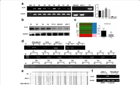
The expression status of ALX4 in breast cancer cell lines and normal breast tissues were detected by RT-PCR, qRT-PCR and WB. The results showed that ALX4 was down regulated in breast cancer cell lines compared with the nor-mal breast tissues both on mRNA and protein level (Fig. 1a, b). To further confirm this observation, the expression pat-tern of ALX4 was subsequently analyzed using The Cancer Genome Atlas (TCGA) database (111 normal breast tissue and 1097 breast cancer tissue). Consistent with our results, ALX4 expression was down regulated in TCGA breast can-cer tissues compared with normal breast tissues (P< 0.01) (Fig. 1c). Collectively these results suggested that ALX4 was down regulated in breast cancer, thus it may play an im-portant role in breast cancer development.
Methylation of ALX4 is associated with its down regulation in breast cancer
the randomly selected normal breast tissue, while sig-nificant CpG island methylation was detected in the two breast cancer cell lines MDA-MB-231 and MCF-7 (Fig. 1e). These above results demonstrated that the promoter region of ALX4 exhibited hyper-methylation status in breast cancer. To investigate the relationship between low expression and methylation status of ALX4, we first analyzed the TCGA data using cbiopor-tal website (www.cbioporcbiopor-tal.com). After downloading the original data, we analyzed the relationship between methylation and expression, and found the expression of ALX4 gene was negatively correlated with the methylation degree (P< 0.00) (Additional file: Fig. S1). In order to further verify that down regulation of ALX4 in breast cancer is related to methylation, breast cancer cell lines were treated with 5-aza-dc, a pharmacological inhibitor of DNA methylation as previously described [9, 28] and the results showed that ALX4 expression was restored (Fig. 1f ). These results indicated that
promoter methylation is responsible for the down regu-lation of ALX4 in breast cancer.
ALX4 over expression inhibits breast cancer cell proliferation, migration and invasion in vitro
To investigate the function of ALX4 in breast cancer, we first transfected MDA-MB-231 and MCF-7 cell lines using control vector and ALX4 expression vector and verified the expression of ALX4 by WB and RT-PCR (Fig. 2a). The effects of ALX4 over expression on cell proliferation and viability were firstly detected. A four days growth curve showed that ALX4 over expression inhibited the prolifera-tion of MDA-MB-231 and MCF-7 cells (Fig. 2b). The sup-pressive function of ALX4 was further verified by colony formation assay and EDU assay. ALX4 over expression inhibited the colony formation and the EDU positive rate of MDA- MB-231 and MCF-7 cell lines (Fig. 2c, d). Fur-thermore, we found that ALX4 over expression induced G1 / S blockade of breast cancer cell lines (Fig. 2e). We
a
b
d
e
f
c
Fig. 1The down regulation of ALX4 in breast cancer is caused by promoter methylation.aAnalysis of ALX4 mRNA expression levels in normal breast tissues and breast cancer cells using RT-PCR and qPCR. ALX4 was down regulated in breast cancer cells compared with normal breast tissues.bThe protein expression of ALX4 was detected in normal breast tissues and breast cancer cells.cDown regulation of ALX4 in breast cancer patients was further confirmed using TCGA database. ALX4 mRNA expression in the TCGA breast cancer RNAseq (IlluminaHiSeq;n= 1208, normal tissue = 111, breast cancer tissue = 1097) data sets. Error bars indicate s.d. **P< 0.01.dALX4 promoter methylation was analyzed by MSP method. ALX4 gene promoter was methylated in breast cancer cell lines, unmethylated in all normal breast tissues and frequently methylated in breast tumor tissues. M: PCR product amplifed by methylated -specific primers; U: PCR product amplifed by unmethylated specific primers; N: normal tissue; T: tumor tissues; PC: positive control, including fully unmethylated or fully methylated control; NC: negative control.eMethylation status of ALX4 promoter region of normal breast tissue and breast cancer cell lines was analyzed by BSP method. CpG islands were represented as open dots (unmethylated) or filled dots (methylated).
further investigated the effect of ALX4 on breast cancer cell metastasis. Transwell assays with or without matrix gel showed that ALX4 suppressed the migration and inva-sion ability of MDA-MB-231 and MCF-7 (Fig. 2f). These data suggested that ALX4 inhibited the proliferation and metastasis of breast cancer cell lines.
Knockdown of ALX4 expression recover cell proliferation, migration and invasion of breast cancer cells
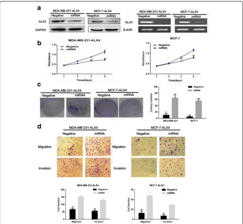
To further confirm the role of ALX4 in breast cancer cell growth and metastasis. We knocked down ALX4 expression in the MDA-MB-231-ALX4 and MCF-7-ALX4 stably cell lines with ALX4-miRNA. ALX4 expression was reduced in cells
a
b
c
d
e
f
Fig. 2ALX4 suppresses breast cancer cell proliferation and metastasis in vitro.aTransfectants of the control vector and ALX4 were identified WB and RT-PCR in MDA-MB-231 and MCF-7 cell lines.bMTS assays were performed to examine the effect of ALX4 over expression on the relative number of viable cells based on the absorbance.cOver expression of ALX4 reduced the colony formation ability of MDA-MB-231 and MCF-7 cell lines. Error bars indicate s.d. (n= 3).*P< 0.05, ** P < 0.01.dEDU assay was performed to detect the effect of ALX4 over expression on proliferation.
transfected with ALX4-miRNA as shown by WB and RT-PCR (Fig. 3a). Knockdown of ALX4 markedly enhanced cell viability and colony formation ability compared with the control group (Fig. 3b, c). Furthermore, knock-down of ALX4 reversed the inhibitory effect on cell metastasis of MDA-MB-231-ALX4 and MCF7-ALX4 stably cells (Fig. 3d). These knockdown results, to-gether with the above obtained data from ALX4 over expression study suggested that ALX4 might function as a potential tumor suppressor in breast cancer.
Overexpression of ALX4 inhibits tumor formation and metastasis in nude mice
To further investigate the tumor suppression function in vivo, nude mice xenograft tumor model was applied to determine the oncogenic role of ALX4 in tumorigen-icity of breast cancer cells. MDA-MB-231-ALX4 and vector control stably over expression cells were sub-cutaneously injected into nude mice to evaluate the effect on tumor formation. The result showed that mice receiving MDA-MB-231-ALX4 stably cells exhibited
a
b
c
d
significantly reduced tumor formation ability compared with vector control cells (Fig. 4A-C). Furthermore, we investigated the effect of ALX4 over expression on breast cancer cells metastasis in vivo via tail veins injection method. H&E staining results showed that ALX4 could suppress the liver metastasis (Fig. 4D) and this observation was further confirmed by qPCR using human-specificβ2
-MG (beta-2-microglobulin) [23] with mouse-specific β2
-MG as internal control (Fig. 4E). Collectively, these data indicated that ALX4 had a significant effect on impeding tumor formation and metastasis, supporting ALX4 as a tumor suppressor in breast cancer in vivo.
ALX4 exerts its anti-cancer function via Wnt/β-catenin pathway by degradation ofβ-catenin in breast cancer We next went to explore the possible mechanism by which ALX4 suppressed breast cancer progression. Accumulating evidence suggested that abnormally activated Wnt/β-catenin pathway contributed to the
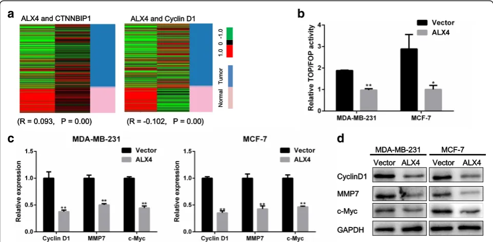
malignant phenotype of breast cancer [29–31]. Using the TCGA data base, we found that ALX4 expression was positively correlated with the negative regulator of Wnt/β -catenin pathway, such as WIF1 and CTNNBIP1, but nega-tively correlated with Wnt/β-catenin activated genes, LEF1 and Cyclin D1 (Fig. 5a) (Additional file 1: Figure S2 A, B). In light of this observation, we hypothesized that ALX4 may exerted its inhibitory function by disrupting the Wnt/β-catenin signal. To test this presumption, we first performed Top/Fop flash reporter assay, the results showed that ALX4 could significantly inhibit the tran-scription activity of β-catenin/T-cell factor (TCF) com-pared with the vector control group in MDA-MB-231 and MCF-7 cell lines (Fig. 5b). We subsequently analyzed the expression of the downstream target genes of Wnt/β -ca-tenin pathway, and found that ALX4 could decrease the mRNA and protein level of CyclinD1, c-Myc and MMP7 (Fig. 5c, d). Next we thought to figure out the possible mechanism by which ALX4 could interrupt the Wnt/β
-a
b
c
d
e
Fig. 4ALX4 inhibits tumor formation and metastasis in nude mice.aControl vector and ALX4 stably expressing MDA-MB-231 cells (8 × 10⁶) were
subcutaneously injected into the right flank of nude mice. Pictures of BALB/c-nude mice and solid tumor tissues were taken after 33 days.bThe tumor growth curve of ALX4 over expressing cells was compared with vector control cells.cTumor weights in the vector control and ALX4 groups were determined. Error bars indicate s.d. (n = 3).dLiver metastases were observed by H&E staining in the control group but not in the ALX4 group by tail veins injection method. Arrows indicate the metastatic loci.eLiver metastasis was further quantified using RT-qPCR. Human-specificβ2-MG levels were
used to quantify metastatic human cancer cells with the mouse-specificβ2-MG level as an internal control. The nude mice that were injected with
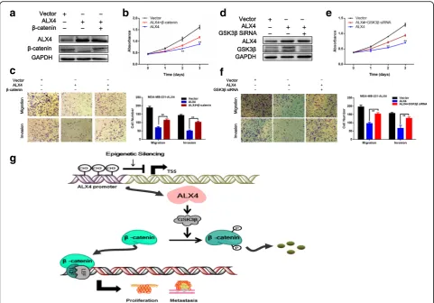
catenin signaling. As a transcription factor, we first exam-ine the effect of ALX4 over expression on the transcrip-tion of β-catenin (the key signal transmitter involved in Wnt/β-catenin pathway). However, luciferase reporter assay showed that ALX4 had no significant inhibition ef-fects on the transcription of β-catenin in the two breast cancer cell lines (Fig. 6a). Furthermore no significant change was observed for the mRNA expression of β -catenin after ALX4 overexpression (data not shown). Nevertheless we found that the protein level ofβ-catenin was decreased after ALX4 expression (Fig. 6b). Accumu-lating evidence indicated that the protein level of β -catenin was tightly controlled by the degradation complex composed by AXIN, ICAT, APC and GSK-3β by which GSK-3βphosphorylatesβ-catenin leading to its proteolytic degradation [32–35]. Thus we speculated that ALX4 may promote the phosphorylation degradation of β-catenin. To test this hypothesis, we detected the relative protein expression and the results showed that ALX4 induced the level of p-β-catenin and GSK-3β(Fig. 6b). To further in-vestigate whether GSK3β play important role in ALX4 mediated β-catenin phosphorylation and degradation, we
knocked down GSK3β expression in
MDA-MB-231-ALX4 stably cell line using siRNA and the level of p-β -catenin was reduced while the protein level of β-catenin was recovered (Fig. 6c). We further consolidated our ob-servation by detecting the expression of ALX4,β-catenin, p-β-catenin and GSK-3β in xenograft tumor tissues by
WB (Fig. 6d). Furthermore, by analyzing the TCGA breast cancer data, we found that the expression of ALX4 was positively correlated with the expression of the key mem-bers of the “β-catenin degradation complex” (Additional file 1: Figure S2C). To define whether β-catenin was re-quired for the anti-tumorigenesis function of ALX4, we re-expressedβ-catenin in MDA-MB-231-ALX4 stably cell line (Fig. 7a).β-catenin re-expression reversed the inhibi-tory effect of ALX4 on cell proliferation and metastasis (Fig. 7b, c). These data indicated that β-catenin was im-portant for the tumor suppression function of ALX4 in breast cancer. We further examine the effect of GSK3β knock down on the phenotypes of MDA-MB-231-ALX4 stably cell line. Knock down of GSK3βwas confirmed by WB (Fig. 7d), and the results showed that knock down of GSK3β could reverse the inhibitory effects of ALX4 on cell proliferation and metastasis at least in part (Fig. 7e, f). Taken together our results supported that ALX4 inhibited the progression of breast cancer through interfering Wnt/
β-catenin signaling via promoting phosphorylation degrad-ation ofβ-catenin in a GSK3βdependent manner (Fig. 7g).
The low expression of ALX4 is associated with poor survival outcomes of breast cancer patients
To investigate the clinical significance of ALX4 expres-sion in breast cancer patients, we first conducted IHC on a TMA containing 142 breast cancer patients with overall survival clinical statistics. After IHC, we used the
a
b
c
d
Fig. 5ALX4 suppresses the Wnt/β-catenin pathway.aALX4 was positively correlated with CTNNBIP1 expression (R= 0.093,P= 0.00) and negatively correlated with Cyclin D1 expression (R=−0.102, P = 0.00).bALX4 decreased the transcriptional activity of the Wnt/β-catenin pathway as determined by a TOP flash/FOP flash reporter luciferase activity assay in MDA-MB-231 and MCF-7 cell lines. TOP and FOP flash plasmids were co-transfected with control vector or ALX4 and PRL-TK plasmids in MDA-MB-231 and MCF-7 cells.c, dALX4 inhibited the expression of Wnt/
scoring system to consolidate the results for intensity and positive staining percentage. Based on the results, positive staining of tumor cell was quantified and classi-fied into two groups: high and low according to the me-dian score of 142 breast cancer patients (Fig. 8a). Survival analysis using Kaplan-Meier method and log rank test showed that patients with high ALX4 expres-sion (n= 42) presented a longer survival time than that of low ALX4 expression (n= 100) (P< 0.01) (Fig. 8b). To correct for bias caused by univariate analysis, ALX4 ex-pression as well as other parameters were examined in a multivariate Cox-regression analysis (after adjusting for age, histological grade, clinical stage, tumor size and lymph node status). In addition to tumor size (HR = 1.268,P= 0.00), ALX4 expression was found to be an in-dependent prognostic factor (HR = 0.345, P= 0.01) for the overall survival of breast cancer patients (Fig. 8c).
To further confirm these results, we performed a meta-analysis of the association between ALX4 gene ex-pression and prognostic outcomes among 1080 breast cancer patients using the TCGA data base. The results showed that high expression of ALX4 predicted longer survival time (n= 1080, p= 0.00, 2 group; n = 1080, p= 0.03, 3 group) (Additional file 1: Figure S4). Next, we sought to determine the impact of ALX4 expression on
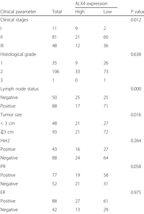
patient survival considering tumor size, clinical stage, lymph node and histological grade. After stratifying pa-tients based on ALX4 expression, we analyzed papa-tients’ survival data and found a significant association between ALX4 expression and patients at clinical stages II + III (P = 0.00) (Fig. 8d, e). We further investigated the rela-tionship between ALX4 expression and clinical parame-ters, and found that ALX4 expression was evidently correlated to clinical stages (n= 140, P = 0.01), tumor size (n= 142, P = 0.01) and lymph node status (n= 138, P = 0.00) of breast cancer patients (Table 1). Taken to-gether our data showed that ALX4 is an independent fa-vorable prognostic factor for breast cancer patients and its expression is associated with clinical parameters.
Discussion
Homeobox genes are a family of genes that share highly conserved structure while maintaining a high degree of di-versity [36, 37]. Its conserved sequences encode proteins containing homologous domains that are capable of bind-ing DNA which endow them with the ability to be in-volved extensively in the process of embryos, tissues and organs development and human diseases [36, 37]. ALX4, a paired-like homedomain transcription factor, is mainly expressed in the mesenchymal compartment of variety of
a
b
c
d
developing tissues such as skull and limbs [11–17]. Recent studies showed its opposing roles in HCC and ovarian cancers via distinct mechanisms [20, 38] indicating the functions and regulation mechanisms of ALX4 in the pro-gression of different tumor remain largely uninvestigated.
In the present study we firstly showed that ALX4 was down regulated in breast cancer. Using MSP and BSP methods we found that the promoter region of ALX4 was frequently methylated in breast cancer and demethylation treatment could recover the expression of ALX4. These results indicated that hyper-methylation contribute to the down regulation of ALX4 in breast cancer. Studies have shown that DNA methylation patterns in tumourigenesis is comprised of genome-wide hypo-methylation and CpG islands hyper-methylation and its main significance may be the molecular basis for tumor suppressor gene inacti-vation, proto-oncogene activation and genomic instability
[7, 8, 39]. Recently, numbers of epigenetically silenced genes were proved to be tumor suppressor in different types of cancer [9, 25, 40, 41]. These evidences indicted that ALX4 may be involved in the tumorigenesis of breast cancer.
Thus gain and loss of function studies were carried out to examine the function of ALX4 in breast cancer. Ectopic expression of ALX4 induced apoptosis (data not shown) and G1/S blocking thus inhibited the proliferation of breast cancer cells in vitro. Distance invasiveness is an-other malignant phenotype which contributes to the high mortality of breast cancer [5, 6] and we found that ALX4 could attenuate the metastasis ability of breast cancer cell lines in vitro and in vivo. These data suggested that ALX4 function as a tumor suppressor in breast cancer. Previous study found that ALX4 promoted ovarian cancer invasion by forming a complex with HOXB13 [20] but our data showed that ALX4 inhibited breast cancer metastasis.
a
b
d
e
f
c
g
Fig. 7ALX4 inhibited the progression of breast cancer via promoting phosphorylation degradation ofβ-catenin in a GSK3βdependent manner.
aThe expression of ALX4 andβ-catenin was analyzed by WB 48 h after transfection with control vector orβ-catenin over expression vector in MDA-MB-231-ALX4 cells.bMTS assays were used to examine the effect ofβ-catenin re-expression on cell proliferation in MDA-MB-231-ALX4 cells.
These results suggested that the function of ALX4 varied depending on different cancer types indicating its import-ant roles in tumorigenesis.
We further determined the possible mechanisms of the tumor inhibition effect of ALX4 in breast cancer. Abnormal activating of Wnt/β-catenin signaling has been recognized as an important mechanism of breast cancer initiation and progression [29–31]. We found that ectopic expression of ALX4 could inhibit the canonical Wnt signaling activity in breast cancer by TOP/FOP flash reporter assay and the Wnt target functional genes MMP7, c-Myc and Cyclin D1 were down regulated both at protein and mRNA level. These results demonstrated that ALX4 suppressed breast cancer progression by inhibiting the Wnt/β-catenin
pathway. We have previously showed that ALX4 sup-pressed lung cancer proliferation by activating the caspase cascade [18] and a recent study showed that ALX4 sup-pressed the EMT of liver cancer by inhibiting the sonic hedgehog (Shh) pathway [38]. These results indicated that the molecular mechanisms of ALX4 as tumor suppressor may be varied among different cancer types.
We further investigated the possible mechanism by which ALX4 inhibiting the Wnt/β-catenin pathway. In the canonical Wnt/β-catenin signaling, β-catenin func-tions as a key mediator to activate the expression of Wnt target genes thus promoting cell proliferation and metastasis [42–45] and a previous study found that β -catenin is required for the tumorigenic behavior of
a
b
c
d
e
Fig. 8High expression of ALX4 predicts a longer survival time in breast cancer patients.a142 breast cancer patients were divided into two groups according to the expression of ALX4 by identified by IHC. Scale bar, 100μm.bKaplan-Meier survival analysis of ALX4 expression in breast cancer patients. Long OS was observed in the high ALX4 expression group compared with the low ALX4 group.cMultivariate Coxregression analysis of the relationship between ALX4 expression and OS of breast cancer patients. ALX4 was determined to be an independent favorable prognostic factor.
breast cancer cells [46]. Our data showed that ectopic ex-pression of ALX4 reduced the protein level of β-catenin but had no significant effects on the transcription of β -catenin. Previous studies have suggested that the protein level of β-catenin is tightly controlled by the destruction complex composed by APC AXIN, ICAT and GSK-3β[32, 44, 47, 48] through which β-catenin is finally phosphory-lated by GSK-3βat the Ser33 and Ser37 leading to its pro-teolytic degradation [49–51]. We found that ALX4 could decrease the protein level of β-catenin by promoting its phosphorylation via upregulating the GSK3β. Accumulating evidence indicated that GSK3β mediated β-catenin phos-phorylation is the key step to generate theβ-TrCP-binding site for the subsequent degradation [35, 52, 53]. A recent study in HCC also showed that GSK-3β suppresses HCC cell dissociation in vitro by inhibiting Wnt/β-catenin sig-naling pathway [19]. Furthermore we found that the ex-pression of ALX4 was positively correlated with the expression of the key members of the “β-catenin degrad-ation complex”. Collectively, our data suggested for the first time that ALX4 exerted its inhibitory function by suppress-ing the Wnt/β-catenin pathway through promoting the
phosphorylation degradation of β-catenin in a GSK3β dependent manner.
Accumulating studies have indicated that the different expression pattern of gene showed tight correlation with the clinical progression of cancer patients [22, 25]. Despite the dramatic progress achieved in breast cancer related diagnostic and treatment techniques, the prognosis for breast cancer patients has not increased significantly, thus identification of novel biomarker is in urgent need [3, 4]. Therefore, to evaluate the clinical significance of ALX4 in breast cancer patient, we performed a tissue microarray on 142 breast cancer patients with clinical and survival data. We found that ALX4 expression is an independent favorable prognostic factor and is in tight relationship with clinical stages, tumor size and lymph node status in breast cancer patients. These data suggested that the expression of ALX4 may provide information for predicting the sur-vival of breast cancer patients.
Conclusions
In summary, our study indicates for the first time that ALX4 is an epigenetically inactivated tumor suppressor in breast cancer acting through inhibition the Wnt/β-catenin pathway. This adds to our current knowledge of the tumorigenesis process of breast cancer. Furthermore the expression of ALX4 is an independent favorable prognostic factor in breast cancer patients and is in tightly relationship with tumor progression. This may provide a new biomarker to predict the survival of breast cancer patients.
Additional file
Additional file 1: Figure S1.The correlation between ALX4 expression and methylation was analyzed using TCGA data set
(www.cbioportal.com). A: Cohort1:TCGA, Cell 2015,n= 552;Cohort2. B: TCGA, Provisional 2012,n= 656Figure S2.Analysis the relationship between ALX4 and Wnt/β-catenin suppression and activation genes in breast cancer using TCGA Database. (A) ALX4 was positively correlated with WIF1 expression (R= 0.503,P= 0.00). (B) ALX4 was negatively correlated with LEF1 expression (R=−0.062,P= 0.03). (C) AXL4 was positively correlated with the expression of the key members of“β -catenin degradation complex”Figure S3.The illustration of full length humanβ-catenin promoter (−2760 bp to +27 bp) that was cloned into the PGL3-Basic luciferase reporter vectorFigure S4.Relationship between ALX4 expression and survival of breast cancer patients retrieved from public datasets(https://genome-cancer.soe.ucsc.edu/proj/site/xena). (A) Survival analysis using TCGA dataset, the results showed that high expression of ALX4 predicted longer survival time (n= 1080,p= 0.00, 2 group). (B) Survival analysis using TCGA dataset the results showed that high expression of ALX4 predicted longer survival time (n = 1080,
p= 0.03, 3 group)Table S1.Primers used in this study. (DOCX 895 kb)
Abbreviations
AlX4:Aristaless-like homeobox-4; BSP: Bisulfite genomic sequence; H&E: Hematoxylin-eosin staining; IHC: Immunohistochemistry; MSP: Methylation specific PCR; OS: overall survival; TCGA: The Cancer Genome Atlas; TMA: Tissue microarray analysis
Table 1Correlation between ALX4 expression and clinical parameters from TMA data
ALX4 expression
Clinical parameter Total High Low Pvalue
Clinical stages 0.012
I 11 9 2
II 81 21 60
III 48 12 36
Histological grade 0.638
1 35 9 26
2 106 33 73
3 1 0 1
Lymph node status 0.000
Negative 50 25 25
Positive 88 17 71
Tumor size 0.016
< 3 cm 48 21 27
≧3 cm 93 21 72
Her2 0.264
Positive 43 16 27
Negative 88 24 64
PR 0.058
Positive 77 19 58
Negative 52 21 31
ER 0.975
Positive 88 27 61
Acknowledgements
The authors thank the Institute of Cancer, Southwest Hospital for providing human breast normal and tumor tissues.
Funding
This work was supported by the National Natural Science Foundation of China [No. 81502551, 81172714 and 8157311].
Availability of data and materials
The datasets generated and/or analyzed during the current study are available in the: www.cbioportal.com and https://genome-cancer.soe.ucsc.edu/proj/site/ hgHeatmap/.
Authors’contributions
JY, FH and JL conceived the study; JY, FH and WL designed the experiments. JY, HC and XH collected the patients’samples and conducted the experiments; XJ, LY and YH analyzed the online database; JY and FH interpreted the data and wrote the manuscript; HZ and JL supervised the study. JC and JL provided with valuable advices, and proofread the manuscript. All authors read and approved the final manuscript.
Ethics approval and consent to participate
All subjects signed an informed consent form. This study was approved by the ethics committee of the Southwest Hospital, affiliated with Third Military Medical University. Moreover, written consent was received from patients. Animal research was approved by the Institutional Animal Care and Use Committee of Third Military Medical University.
Consent for publication
Not applicable.
Competing interests
The authors declare that they have no competing interests.
Publisher’s Note
Springer Nature remains neutral with regard to jurisdictional claims in published maps and institutional affiliations.
Received: 23 September 2017 Accepted: 20 November 2017
References
1. Chen W, Zheng R, Baade PD, Zhang S, Zeng H, Bray F, Jemal A, XQ Y, He J. Cancer statistics in China, 2015. CA Cancer J Clin. 2016;66(2):115–32. 2. Siegel RL, Miller KD, Jemal A. Cancer statistics, 2017. CA Cancer J Clin. 2017;
67(1):7–30.
3. MB A, VS R, ME J, AO A. Breast cancer biomarkers: risk assessment, diagnosis, prognosis, prediction of treatment efficacy and toxicity, and recurrence. Curr Pharm Des. 2014;20(30):4879–98.
4. Bertozzi S, Londero AP, Cedolini C, Uzzau A, Seriau L, Bernardi S, Bacchetti S, Pasqual EM, Risaliti A. Prevalence, risk factors, and prognosis of peritoneal metastasis from breast cancer. SpringerPlus. 2015;4:688.
5. Charafe-Jauffret E, Ginestier C, Iovino F, Tarpin C, Diebel M, Esterni B, Houvenaeghel G, Extra JM, Bertucci F, Jacquemier J, et al. Aldehyde
dehydrogenase 1-positive cancer stem cells mediate metastasis and poor clinical outcome in inflammatory breast cancer. Clin Cancer Res. 2010;16(1):45–55. 6. Mego M, Mani SA, Cristofanilli M. Molecular mechanisms of metastasis in
breast cancer–clinical applications. Nat Rev Clin Oncol. 2010;7(12):693–701. 7. Esteller M. Cancer epigenomics: DNA methylomes and histone-modification
maps. Nat Rev Genet. 2007;8(4):286–98.
8. Risch A, Plass C. Lung cancer epigenetics and genetics. Int J Cancer. 2008; 123(1):1–7.
9. Han F, Liu W, Jiang X, Shi X, Yin L, Ao L, Cui Z, Li Y, Huang C, Cao J, et al. SOX30, a novel epigenetic silenced tumor suppressor, promotes tumor cell apoptosis by transcriptional activating p53 in lung cancer. Oncogene. 2015; 34(33):4391–402.
10. Zhu H, Wu K, Yan W, Hu L, Yuan J, Dong Y, Li Y, Jing K, Yang Y, Guo M. Epigenetic silencing of DACH1 induces loss of transforming growth factor-beta1 antiproliferative response in human hepatocellular carcinoma. Hepatology. 2013;58(6):2012–22.
11. Wuyts W, Cleiren E, Homfray T, Rasore-Quartino A, Vanhoenacker F, Van Hul W. The ALX4 homeobox gene is mutated in patients with ossification defects of the skull (foramina parietalia permagna, OMIM 168500). J Med Genet. 2000;37(12):916–20.
12. Qu S, Tucker SC, Ehrlich JS, Levorse JM, Flaherty LA, Wisdom R, Vogt TF. Mutations in mouse Aristaless-like4 cause Strong's luxoid polydactyly. Development. 1998;125(14):2711–21.
13. Chang H, Mohabir N, Done S, Hamel PA. Loss of ALX4 expression in epithelial cells and adjacent stromal cells in breast cancer. J Clin Pathol. 2009;62(10):908–14.
14. Joshi PA, Chang H, Hamel PA. Loss of Alx4, a stromally-restricted homeodomain protein, impairs mammary epithelial morphogenesis. Dev Biol. 2006;297(1):284–94.
15. Kayserili H, Uz E, Niessen C, Vargel I, Alanay Y, Tuncbilek G, Yigit G, Uyguner O, Candan S, Okur H, et al. ALX4 dysfunction disrupts craniofacial and epidermal development. Hum Mol Genet. 2009;18(22):4357–66.
16. Kuijper S, Feitsma H, Sheth R, Korving J, Reijnen M, Meijlink F. Function and regulation of Alx4 in limb development: complex genetic interactions with Gli3 and Shh. Dev Biol. 2005;285(2):533–44.
17. Valente M, Valente KD, Sugayama SS, Kim CA. Malformation of cortical and vascular development in one family with parietal foramina determined by an ALX4 homeobox gene mutation. AJNR Am J Neuroradiol. 2004;25(10):1836–9. 18. Liu WB, Han F, XH D, Jiang X, Li YH, Liu Y, Chen HQ, Ao L, Cui ZH, Cao J, et
al. Epigenetic silencing of Aristaless-like homeobox-4, a potential tumor suppressor gene associated with lung cancer. Int J Cancer. 2014;134(6): 1311–22.
19. Zhang JH, Jiao LY, Li TJ, Zhu YY, Zhou JW, Tian J. GSK-3beta suppresses HCC cell dissociation in vitro by upregulating epithelial junction proteins and inhibiting Wnt/beta-catenin signaling pathway. J Cancer. 2017;8(9):1598–608. 20. Yuan H, Kajiyama H, Ito S, Chen D, Shibata K, Hamaguchi M, Kikkawa F, Senga
T. HOXB13 and ALX4 induce SLUG expression for the promotion of EMT and cell invasion in ovarian cancer cells. Oncotarget. 2015;6(15):13359–70. 21. Yang JT, Han F, Liu WB, Zhang MQ, Huang YS, Hao XL, Jiang X, Yin L, Chen
HQ, Cao J, et al. LHX6, an independent prognostic factor, inhibits lung adenocarcinoma progression through transcriptional silencing of beta-catenin. J Cancer. 2017;8(13):2561–74.
22. Han F, Liu W, Xiao H, Dong Y, Sun L, Mao C, Yin L, Jiang X, Ao L, Cui Z, et al. High expression of SOX30 is associated with favorable survival in human lung adenocarcinoma. Sci Rep. 2015;5:13630.
23. Lupberger J, Kreuzer KA, Baskaynak G, Peters UR, le Coutre P, Schmidt CA. Quantitative analysis of beta-actin, beta-2-microglobulin and
porphobilinogen deaminase mRNA and their comparison as control transcripts for RT-PCR. Mol Cell Probes. 2002;16(1):25–30.
24. Li Q, Dashwood WM, Zhong X, Al-Fageeh M, Dashwood RH. Cloning of the rat beta-catenin gene (Ctnnb1) promoter and its functional analysis compared with the Catnb and CTNNB1 promoters. Genomics. 2004;83(2):231–42. 25. Yuan S, Yu Z, Liu Q, Zhang M, Xiang Y, Wu N, Wu L, Hu Z, Xu B, Cai T, et al.
GPC5, a novel epigenetically silenced tumor suppressor, inhibits tumor growth by suppressing Wnt/beta-catenin signaling in lung adenocarcinoma. Oncogene. 2016;35(47):6120–31.
26. Shivakumar M, Lee Y, Bang L, Garg T, Sohn KA, Kim D. Identification of epigenetic interactions between miRNA and DNA methylation associated with gene expression as potential prognostic markers in bladder cancer. BMC Med Genet. 2017;10(Suppl 1):30.
27. Yang Y, Fuentes F, Shu L, Wang C, Pung D, Li W, Zhang C, Guo Y, Kong AN. Epigenetic CpG methylation of the promoter and reactivation of the expression of GSTP1 by Astaxanthin in human prostate LNCaP cells. AAPS J. 2017;19(2):421–30. 28. Holliday R, Ho T. DNA methylation and epigenetic inheritance. Methods.
2002;27(2):179–83.
29. Laezza C, D'Alessandro A, Paladino S, Maria Malfitano A, Chiara Proto M, Gazzerro P, Pisanti S, Santoro A, Ciaglia E, Bifulco M, et al. Anandamide inhibits the Wnt/beta-catenin signalling pathway in human breast cancer MDA MB 231 cells. Eur J Cancer. 2012;48(16):3112–22.
30. Li Y, Jin K, van Pelt GW, van Dam H, Yu X, Mesker WE, Ten Dijke P, Zhou F, Zhang L. C-Myb enhances breast cancer invasion and metastasis through the Wnt/beta-catenin/Axin2 pathway. Cancer Res. 2016;76(11):3364–75. 31. Zhang ZM, JF W, Luo QC, Liu QF, QW W, Ye GD, She HQ, Li BA. Pygo2
activates MDR1 expression and mediates chemoresistance in breast cancer via the Wnt/beta-catenin pathway. Oncogene. 2016;35(36):4787–97. 32. Kimelman D, Xu W. Beta-catenin destruction complex: insights and
33. Roberts DM, Pronobis MI, Poulton JS, Waldmann JD, Stephenson EM, Hanna S, Peifer M. Deconstructing the sscatenin destruction complex: mechanistic roles for the tumor suppressor APC in regulating Wnt signaling. Mol Biol Cell. 2011;22(11):1845–63.
34. Stamos JL, Weis WI. The beta-catenin destruction complex. Cold Spring Harb Perspect Biol. 2013;5(1):a007898.
35. Liu C, Li Y, Semenov M, Han C, Baeg GH, Tan Y, Zhang Z, Lin X, He X. Control of beta-catenin phosphorylation/degradation by a dual-kinase mechanism. Cell. 2002;108(6):837–47.
36. Rivera-Pomar R, Niessing D, Schmidt-Ott U, Gehring WJ, Jackle H. RNA binding and translational suppression by bicoid. Nature. 1996;379(6567): 746–9.
37. Zhang Z, Gutierrez D, Li X, Bidlack F, Cao H, Wang J, Andrade K, Margolis HC, Amendt BA. The LIM homeodomain transcription factor LHX6: a transcriptional repressor that interacts with pituitary homeobox 2 (PITX2) to regulate odontogenesis. J Biol Chem. 2013;288(4):2485–500.
38. Shi Y, Sun X, He X. Overexpression of Aristaless-like Homeobox-4 inhibits proliferation, invasion, and EMT in hepatocellular carcinoma cells. Oncol Res. 2017;25(1):11–8.
39. Rodriguez-Paredes M, Esteller M. Cancer epigenetics reaches mainstream oncology. Nat Med. 2011;17(3):330–9.
40. Ronsch K, Jagle S, Rose K, Seidl M, Baumgartner F, Freihen V, Yousaf A, Metzger E, Lassmann S, Schule R, et al. SNAIL1 combines competitive displacement of ASCL2 and epigenetic mechanisms to rapidly silence the EPHB3 tumor suppressor in colorectal cancer. Mol Oncol. 2015;9(2):335–54. 41. Yang J, Xu Z, Li J, Zhang R, Zhang G, Ji H, Song B, Chen Z. XPC epigenetic
silence coupled with p53 alteration has a significant impact on bladder cancer outcome. J Urol. 2010;184(1):336–43.
42. MacDonald BT, Tamai K, He X. Wnt/beta-catenin signaling: components, mechanisms, and diseases. Dev Cell. 2009;17(1):9–26.
43. Takahashi-Yanaga F, Kahn M. Targeting Wnt signaling: can we safely eradicate cancer stem cells? Clin Cancer Res. 2010;16(12):3153–62. 44. Voronkov A, Krauss S. Wnt/beta-catenin signaling and small molecule
inhibitors. Curr Pharm Des. 2013;19(4):634–64.
45. Wang Y, Pan P, Wang Z, Zhang Y, Xie P, Geng D, Jiang Y, Yu R, Zhou X. Beta-catenin-mediated YAP signaling promotes human glioma growth. J Exp Clin Cancer Res. 2017;36(1):136.
46. Xu J, Prosperi JR, Choudhury N, Olopade OI, Goss KH. Beta-catenin is required for the tumorigenic behavior of triple-negative breast cancer cells. PLoS One. 2015;10(2):e0117097.
47. Amit S, Hatzubai A, Birman Y, Andersen JS, Shushan E, Mann M, Ben-Neriah Y, Alkalay I. Axin-mediated CKI phosphorylation of beta-catenin at Ser 45: a molecular switch for the Wnt pathway. Genes Dev. 2002;16(9):1066–76. 48. Morin PJ, Sparks AB, Korinek V, Barker N, Clevers H, Vogelstein B, Kinzler KW.
Activation of beta-catenin-Tcf signaling in colon cancer by mutations in beta-catenin or APC. Science. 1997;275(5307):1787–90.
49. He X, Semenov M, Tamai K, Zeng X. LDL receptor-related proteins 5 and 6 in Wnt/beta-catenin signaling: arrows point the way. Development. 2004; 131(8):1663–77.
50. Yost C, Torres M, Miller JR, Huang E, Kimelman D, Moon RT. The axis-inducing activity, stability, and subcellular distribution of beta-catenin is regulated in Xenopus embryos by glycogen synthase kinase 3. Genes Dev. 1996;10(12):1443–54.
51. Hagen T, Di Daniel E, Culbert AA, Reith AD. Expression and characterization of GSK-3 mutants and their effect on beta-catenin phosphorylation in intact cells. J Biol Chem. 2002;277(26):23330–5.
52. Frame S, Cohen P, Biondi RM. A common phosphate binding site explains the unique substrate specificity of GSK3 and its inactivation by
phosphorylation. Mol Cell. 2001;7(6):1321–7.
53. Wu G, Xu G, Schulman BA, Jeffrey PD, Harper JW, Pavletich NP. Structure of a beta-TrCP1-Skp1-beta-catenin complex: destruction motif binding and lysine specificity of the SCF(beta-TrCP1) ubiquitin ligase. Mol Cell. 2003;11(6): 1445–56.
• We accept pre-submission inquiries
• Our selector tool helps you to find the most relevant journal
• We provide round the clock customer support
• Convenient online submission
• Thorough peer review
• Inclusion in PubMed and all major indexing services
• Maximum visibility for your research
Submit your manuscript at www.biomedcentral.com/submit