R E S E A R C H
Open Access
Amyloid-
β
peptide-induced extracellular S100A9
depletion is associated with decrease of
antimicrobial peptide activity in human THP-1
monocytes
Eun Ok Lee
1, Ji Hye Yang
1, Keun-A Chang
3, Yoo-Hun Suh
2and Young Hae Chong
1*Abstract
Background:S100A9 protein (myeloid-related protein MRP14, also referred to as calgranulin B) is a reliable marker of inflammation, an important proinflammatory factor of innate immunity and acts as an additional antimicrobial peptide in the innate immune system. Evidence indicates that S100A9 contributes to Alzheimer’s disease (AD) pathology, although the precise mechanisms are not clear.
Methods:We were interested to study the mechanisms of S100A9 release upon Aβ1-42 stimulation, the potential roles of extracellular S100A9 depletion in Aβ-induced cytotoxicity, and the interaction with innate immune
response in THP-1 monocytic cells that have been challenged with mostly Aβ1-42 monomers instead of oligomers. We used protein preparation, Ca2+influx fluorescence imaging, MTT assay, siRNA knockdown, colony forming units (CFUs) assay and western blotting techniques to perform our study.
Results:Aβ1-42 monomers elicited a marked decrease of S100A9 release into the cell culture supernatant in a dose-dependent manner in human THP-1 monocytes. This reduction of S100A9 release was accompanied by an increase of intracellular Ca2+level. Aβ1-42-mediated decrease of S100A9 release was not associated with Aβ 1-42-induced cytotoxicity as measured by MTT reduction assay. This observation was confirmed with the recombinant S100A9, which had little effect on Aβ1-42-induced cytotoxicity. Moreover, depletion of S100A9 with siRNA did not significantly evoke the cell toxicity. On the other hand, Aβ1-42-induced extracellular S100A9 depletion resulted in decreased antimicrobial activity of the culture supernatant after Aβ1-42 stimulation. Immunodepletion of S100A9 with anti-S100A9 also decreased the antimicrobial peptide activity of the vehicle treated culture supernatant. Consistently, the recombinant S100A9 clearly elicited the antimicrobial peptide activityin vitro,confirming the observed antimicrobial activity of S100A9 in the culture supernatant.
Conclusion:Collectively, our findings suggest that the mostly monomeric form of Aβ1-42 negatively regulates the innate immune system by down-regulating the secretion of S100A9, which is likely a main mediator of
antimicrobial activity in the conditioned media of human THP-1 monocytes.
Keywords:Alzheimer’s disease, Aβ1-42, cytotoxicity, S100A9, Antimicrobial activity, Innate immune response
* Correspondence:[email protected]
1Department of Microbiology, School of Medicine, Ewha Medical Research
Institute, Ewha Womans University, 911-1, Mok-6-dong, Yangcheonku, Seoul 158-710, Republic of Korea
Full list of author information is available at the end of the article
Background
Alzheimer’s disease (AD) is the most common and still incurable form of dementia, which primarily affects the population over the age of 60 years. Amyloid beta (Aβ) deposition, neurofibrillary tangle formation and neuro-inflammation are the major pathogenetic mechanisms that, in concert, lead to neocortical and hippocampal at-rophy, memory dysfunction and decline of cognition in AD [1,2]. There are currently no curative or effective clinical treatments for AD [3].
The innate immune response and inflammatory sig-naling play determinant roles in brain homeostasis, neuroprotection and repair. However, altered or exces-sive signaling in these injury defense systems contributes to neuroinflammation and the irreversible degeneration of brain cells [4]. Extensive innate immune gene activa-tion reflecting chronic innate immune activaactiva-tion could accompany brain aging, increasing vulnerability to cog-nitive decline and neurodegeneration, consistent with the emerging idea of a critical involvement of inflam-mation in the earliest stages of AD [5]. Thus, clinical pharmaceutical trials aimed at modulating the immune system in AD have largely focused on dampening down central proinflammatory innate immunity and the ma-nipulation of systemic immunity, and its communication with the central nervous system (CNS) [6].
Calgranulins reflecting calcium-binding properties and high expression in granulocytes are comprised of three proteins: S100A8 (calgranulin A, also termed as MRP8), S100A9 (calgranulin B, also termed as MRP14) and S100A12 (Calgranulin C). They are predominantly expressed by neutrophils, monocytes and activated mac-rophages in inflamed tissue [7]. These S100 calcium-binding proteins are important molecular mediators in a range of diseases, including microbial infections. In particular, S100A9 protein is a reliable marker of in-flammation and an important proinflammatory factor of innate immunity. Elevated plasma levels of S100A9 are associated with inflammatory disorders such as chronic bronchitis, cystic fibrosis and rheumatoid arthritis [8].
The extracellular roles of S100A9 in leukocyte migra-tion and chemotaxis, leukocyte activamigra-tion, oxidant scavenging, and their relevance in inflammatory pro-cesses are in particular implicated [7,9,10]. Recent re-ports have also suggested that S100A9 acts as an additional antimicrobial peptide in the innate immune system, which provides immediate protection for the host against microbial challenge by recognizing the presence of microorganisms and preventing their tissue invasion, thus limiting microbial proliferation and inflammation [11,12].
Altered expression/function of these S100 protein members [13] has been associated with neurological dis-eases such as cerebral ischemia [14] and traumatic brain
injury [15]. Earlier studies demonstrated that S100 pro-teins assemble within neuritic plaques and reactive glia, which may serve to prolong neuroinflammation associ-ated with the pathogenesis of AD [16,17]. Our recent study showed that S100A9 expression was increased in the brains of Tg2576 mice, as well as in AD brains, which proposed its potential role in the neuroin-flammation related to the pathogenesis of AD [18,19]. Another recent study reported that S100A9 interacts with Aβand induces fibrillization, further supporting its association with AD [20]. However, a mechanistic link between S100A9 and AD pathology, and the detailed molecular mechanism have not been clearly shown.
We focused our research on the mechanisms of S100A9 release upon stimulation with mostly Aβ1-42 monomers, the potential roles of extracellular S100A9 depletion in Aβ-induced cytotoxicity, and the interaction with innate immune response in THP-1 monocytic cells that have been challenged with Aβ1-42 monomers in-stead of oligomers. The results of the present study show that the mostly monomeric form of Aβ1-42 negatively regulates the innate immune system by down-regulating the release of S100A9, which is likely a main mediator for the antimicrobial action in the culture media of hu-man THP-1 monocytes.
Materials and methods Materials
Synthetic siRNA for S100A9 and the non-specific con-trol pool were purchased from Santa Cruz Biotechnol-ogy (Santa Cruz, CA, USA). Lipofectamine 2000 was purchased from Invitrogen (Carlsbad, CA, USA). Anti-S100A9 was acquired from R&D Systems (Minneapolis, MN, USA). Horseradish peroxidase-conjugated anti-mouse IgG and anti-rabbit IgG were obtained from Jackson ImmunoResearch (West Grove, PA, USA). Acti-nomycin, inhibitor of de novo mRNA expression, and cycloheximide, inhibitor of protein synthesis, were obtained from Calbiochem (La Jolla, CA, USA). The 3 (4,5-dimethylthiazol-2-yl)-2,5-diphenyltetrazolium brom-ide (MTT) was obtained from United States Biochemical (Cleveland, OH, USA). The Ca2+ionophore, ionomycin, and an endoplasmic reticulum Ca2+ pump inhibitor, thapsigargin, were acquired from Sigma-Aldrich (St Louis, MO, USA). Anti-β-actin antibody and other chemicals, including 1,2-bis(o-aminophenoxy)ethane-N, N,N',N'-tetraacetic acid (BAPTA) and ethylene glycol tetraacetic acid (EGTA), were also acquired from Sigma-Aldrich.
Preparation of Aβpeptides
dissolved at 5 mM in dimethyl sulfoxide and diluted at 250 μM in double-distilled water before experiments. This preparation contains the mostly monomeric form of Aβ1-42 and very small amounts of dimers with larger oligomers up to 6-mers [21].
Preparation of recombinant S100A9 protein
Human recombinant (r) S100A9 was obtained from Dr Tessier at Laval University Hospital Center (Sainte-Foy, Québec, Canada), expressed in Escherichia coli and purified by previously defined protocols [22]. The purity of protein was verified by sodium dodecyl sulfate poly-acrylamide gel electrophoresis (SDS-PAGE). Specificity of S100A9-mediated effect was controlled by THP-1 cell treatment with heat-inactivated rS100A9 (rS100A9hi)
pre-pared by incubation at 85°C for 2 hours.
Cell culture
The human monocytic cell line THP-1 was obtained from ATCC (Rockville, MD, USA) and maintained in RPMI-1640 containing 10% heat-inactivated fetal calf serum as previously described [21]. THP-1, a mono-nuclear cell line of human origin, has been widely used as a model of human monocytes/macrophages or micro-glia not only because of its functional and morphological similarities, including its capacity to activate signal transduction pathways, but also because of functional differences in the metabolism of rodent and human microglial cells as previously described [23].
Experimental treatment
After being washed, THP-1 cells were incubated with serum-free RPMI-1640 supplemented with 0.5% glucose for 1 hour at 37°C before stimulation. The cells were then stimulated by the addition of the mostly mono-meric form of Aβ1-42 peptide for 24 hours in the pres-ence or abspres-ence of rS100A9 or rS100A9hi. In some
experiments, cells were incubated with ionomycin or thapsigargin to determine the effect of increase of intra-cellular Ca2+level. To deplete extracellular or intracellu-lar Ca2+, cells were pretreated for 1 hour with ethylene glycol tetraacetic acid (EGTA) or BAPTA, and further incubated for 24 hours in the presence or absence of Aβ1-42 monomers. All concentrations were selected on the basis of the maximal effects of the drugs on their specified targets. Vehicles were treated identically, but did not contain Aβ1-42 or pharmacological agents as de-scribed above. Vehicle alone exerted no detectable ef-fects on cell viability. After stimulation with Aβ1-42 and/or the specific agents for 24 hours, total cell lysate and the supernatant were prepared and stored at−20°C until use for quantification of S100A9 release by western blot analysis. The supernatant was also analyzed in par-allel for antimicrobial activities.
MTT assay
The viability of cells was analyzed by the MTT assay to assess mitochondrial dehydrogenase activity as previ-ously described [24]. Only viable cells are able to reduce MTT into a formazan product by mitochondrial de-hydrogenase. After 24 hours of treatment of THP-1 cells with Aβ1-42 and/or rS100A9, MTT was added to the medium (1 mg/ml) and incubated for 4 hours at 37°C. The medium was removed and the cells were diluted in 120μl of 1 N HCl:isopropyl alcohol (1:24) and incubated for 30 minutes at room temperature with shaking. The relative formazan concentration of each supernatant was measured by determination of the absorbance at 570 nm in a microplate reader.
Calcium imaging and fluorescence measurements
To visualize intracellular steady-state Ca2+levels, THP-1 cells were stained by adding Fluo 3A in its ace-toxymethyl ester form (Fluo-3 AM) to 5 μg/ml culture media throughout Aβ1-42 or vehicle treatment as previ-ously described [18]. Ca2+ influx fluorescence images were captured after treatment as indicated. Images were recorded using an Axiovert 200 inverted microscope and analyzed with the KS 300 analysis program (Zeiss, Oberkochen, Germany). An increase in intracellular Ca2+ level in the different cultures was expressed as fold of the response of the vehicle treated controls for each individual experiment.
siRNA studies
Synthetic siRNA for S100A9 and the non-specific con-trol pool were purchased from Santa Cruz Biotechnol-ogy, and transfection of the RNA oligonucleotide was performed using Lipofectamine 2000. THP-1 cells were treated with Lipofectamine 2000 (mock transfection), siRNA or non-specific RNA pool at the concentrations indicated. After 24 hours of transfection, the cell viability was measured by the MTT method.
E coliculture and treatment
E coli strain LE392 was used throughout this study. Colonies from agar were transferred by sterile loop to growth media and incubated aerobically in Luria Broth (Conda, Madrid, Spain) for 2 hours at 37°C, to generate mid-logarithmic growth cultures for use as inoculates in experiments. Bacteria inoculum cell densities were normalized to 5 × 10⁵cells/ml immediately before use. After stimulation with Aβ1-42, the supernatant col-lected was mixed with E coli in the ratio of 1:1. rS100A9 and rS100A9hi were directly diluted into
Experiments included control serial dilutions of medium or buffer vehicle alone.
Colony forming unit (CFU) assay
Serial dilutions of incubants were prepared and streaked onto the surface of Luria broth agar (Miller’s LB agar). The agar plates were then incubated overnight at 37°C and CFUs counted as previously described [25].
Preparation of human peripheral blood mononuclear cells (PBMC)
Human PBMC were isolated from peripheral blood of healthy subjects as previously described [24] and used as a positive control for S100A9 in western blot analysis. Preparations contained approximately 10% monocytes, 90% lymphocytes and <1.5% granulocytes.
Electrophoresis and western blotting
Immunoblotting was conducted as previously described [24,26]. Equal quantities of sample proteins were separated on the basis of molecular weight by 10% SDS-PAGE and transferred to polyvinylidene difluoride membranes (Millipore, Bedford, MA, USA), which were subsequently blocked for 0.5 hours with 3% milk in Tris-buffered saline with Tween 20. The membranes were then probed with primary antibody diluted with 1% milk and incubated over-night at 4°C. Signals were acquired with an enhanced chemiluminescence system after incubation with horserad-ish peroxidase-conjugated secondary antibodies (Jackson ImmunoResearch). Densitometric values were normalized versusβ-actin.
Statistical analyses
Differences between groups were evaluated for statistical significance using one-way analysis of variance (ANOVA) with a Student’s t-test. Null hypotheses of no difference were rejected ifPvalues were less than 0.05.
Results
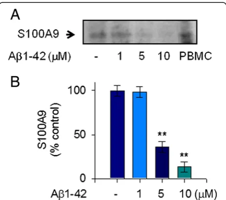
Aβ1-42 reduced extracellular release of S100A9 in human THP-1 monocytes
To clarify the pathological mechanism related to S100A9 in AD, we measured the extracellular release of S100A9 in response to stimulation with Aβ1-42 in human THP-1 monocytes. We used Aβ1-42 monomers instead of oligo-mers. The treatment of THP-1 cells with Aβ1-42 mono-mers significantly reduced the release of S100A9 at 24 hours in the conditioned media of THP-1 cells. This Aβ1-42-mediated decrease of S100A9 secretion occurred in a dose-dependent manner and maximal reduction of S100A9 secretion was found to occur at a concentration of 10 μM Aβ1-42 (Figure 1A, B). Notably, S100A9 se-cretion was consistently reduced when de novomRNA expression and protein synthesis were inhibited by
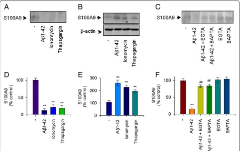
actinomycin D and cycloheximide, respectively. Thus, our data confirmed that Aβ1-42 elicited a marked de-crease of the extracellular S100A9 release in a dose-dependent manner in human THP-1 monocytes, and that reduction of S100A9 release is dependent on both transcriptional and translational activities (Figure 2A,B).
Intracellular Ca2+level is involved in Aβ1-42-induced depletion of extracellular S100A9
The increase of [Ca2+]imay initiate the inflammatory
re-sponse in activated microglia [27]. We observed that 10 μM Aβoligomers extensively increased the level of [Ca2+]i
in murine microglial BV2 cells as evaluated using the Fluo-3 AM method [18]. Thus, we investigated the role of intracellular Ca2+ levels in Aβ1-42-mediated reduction of S100A9 release and found that intracellular Ca2+ level is involved in human monocytic cells. We also observed that 10μM Aβ1-42 monomers significantly increased intracel-lular Ca2+ levels in THP-1 cells as measured by Fluo-3 AM (Figure 3A,B). Furthermore, treatment of THP-1 cells with the Ca2+ionophore, ionomycin, which induces [Ca2+]i
Figure 1S100A9 release in response to Aβ1-42 monomers in human monocytic THP-1 cells.To measure the extracellular release of S100A9 in response to mostly monomeric Aβ1-42 stimulation, THP-1 cells were incubated with either vehicle only (−) or increasing amounts of Aβ1-42 for 24 hours in serum-free RPMI-1640 medium supplemented with glucose (0.5%). (A) The cell-free conditioned media were examined for S100A9 via protein immunoblot. Positive control for S100A9 was shown in human PBMC whole cell lysate. Aβ1-42 decreased S100A9 release in conditioned media in a dose-dependent manner. Results are representative of three independent experiments. (B) Densitometric quantification of analyses of (A), showing the levels of S100A9 release. All data are presented as the means ± SEM (n = 3).
elevating intracellular Ca2+ concentration, induced the Aβ1-42-evoked response decreasing the release of S100A9 (Figure 4A,D). Moreover, thapsigargin, an endoplasmic reticulum Ca2+pump inhibitor, which induces an increase of intracellular Ca2+ level, also mimicked the Aβ1-42 -evoked effects. Concomitantly, the intracellular levels of S100A9 were increased in THP-1 cells treated with either ionomycin or thapsigargin as observed in Aβ1-42 treated cells (Figure 4B,E). However, the Aβ1-42-evoked response was significantly attenuated by either depletion of extracel-lular Ca2+with EGTA or chelation of intracellular Ca2+by BAPTA (Figure 4C,F). Together, these findings suggest that extracellular depletion of S100A9 in response to Aβ1-42 monomers is dependent on an increase of intracellular Ca2+and S100A9 levels in human THP-1 monocytes.
Aβ1-42-induced depletion of extracellular S100A9 was not associated with Aβ1-42-dependent cytotoxicity
To further describe the pathological mechanism related to S100A9 in AD, the role of extracellular S100A9 deple-tion related to the Aβ1-42-induced cytotoxicity was in-vestigated. As shown in Figure 5, Aβ1-42 treatment significantly increased cytotoxicity as measured by MTT reduction assay. Addition of rS100A9 protein into the cell culture supernatant did not significantly attenuate the Aβ1-42-induced cytotoxicity (Figure 5A). Treatment with rS100A9 alone in the absence of Aβ1-42 at concen-trations up to 10μg/ml had little effect on the cell viabil-ity and, as expected, a similar effect was observed with rS100A9hi(Figure 5B). In addition, depletion of S100A9
with siRNA did not significantly evoke cell toxicity (Figure 5C). These results demonstrate that extracellular depletion of S100A9 was not directly associated with the Figure 2S100A9 release process was dependent on
transcription and translation processes in human monocytic THP-1 cells.THP-1 cells were incubated with Aβ1-42 (10μM), actinomycin (ActD, 100 nM) or cycloheximide (CHX, 1μM) for 24 hours. (A) Western blot analyses were conducted to determine the effects of actinomycin or cycloheximide on S100A9 release in the conditioned media, as described in Figure 1. S100A9 secretion was reduced whende novomRNA expression and protein synthesis were inhibited. Representative gels from three experiments are shown. (B) Quantitative analysis of (A), showing the levels of S100A9 release. All data are presented as the means ± SEM (n = 3).**P<0.01, versus vehicle treated samples. ActD, actinomycin; CHX, cycloheximide.
Figure 3Aβ1-42 monomers increased [Ca2+]
iin human
monocytic THP-1 cells.(A) [Ca2+]iimages obtained by Fluo-3 AM at 24 hours after treatment with either vehicle only (−) or 10μM Aβ1-42 monomers (Aβ) in THP-1 cells. Scale bars represent 50μm. (B) The histogram showing the ratio of [Ca2+]ilevels to vehicle treated group. All data are presented as the means ± SEM (n = 3).
cytotoxicity in response to mostly Aβ1-42 monomers in human monocytic THP-1 cells.
Aβ1-42-induced extracellular S100A9 depletion resulted in decreased antimicrobial activity
Recent reports suggested that S100A9 acts as an additional antimicrobial peptide in the innate immune system, which provides immediate protection for the host against micro-bial challenge by recognizing the presence of microorgan-isms and preventing their tissue invasion, thus limiting microbial proliferation and inflammation [11,12]. We fur-ther investigated the antimicrobial activity of S100A9, which was released into the cell culture supernatants of THP-1 monocytes. The antimicrobial activities against
E coliwere assessed with the cell culture supernatants from vehicle or Aβ-42 treated THP-1 cells. Vehicle treated super-natant, which contained a significant amount of S100A9, demonstrated antimicrobial activity againstE coli. However, microbial growth was not decreased by the supernatant from Aβ1-42 treated THP-1 cells in which the S100A9 level was significantly reduced (Figure 6A). Moreover, rS100A9 protein clearly elicited the antimicrobial peptide activity
in vitro (Figure 6B), whereas rS100A9hihad little activity.
Consistently, immunodepletion of S100A9 with anti-S100A9 antibodies blocked antimicrobial activity of the ve-hicle treated supernatant (Figure 6C), confirming that the antimicrobial activity in the vehicle treated supernatant is S100A9-specific.
Figure 4The decreased extracellular S1009A in response to Aβ1-42 monomers is dependent on increase of intracellular Ca2+level in
Discussion
The present study has four main findings concerning a mechanistic link between S100A9 and AD pathology. First, the mostly monomeric form of Aβ1-42 markedly de-creased S100A9 release into the cell culture supernatant of human THP-1 monocytes in parallel with increased intra-cellular S100A9. Second, this reduction of S100A9 release was accompanied by increased intracellular Ca2+ level.
Third, depletion of extracellular S100A9 in response to Aβ1-42 monomers was not associated with Aβ1-42-induced cytotoxicity. Finally, Aβ1-42-Aβ1-42-induced extracellular S100A9 depletion decreased antimicrobial activity of the culture supernatant from human monocytes, which was pathogenically challenged with Aβ1-42. Our findings sug-gest that mostly Aβ1-42 monomers negatively regulates the innate immune system by down-regulating the Figure 5Extracellular S100A9 depletion by Aβ1-42 monomers was not associated with Aβ1-42-induced cytotoxicity.(A) To investigate the role of extracellular S100A9 depletion related to the Aβ1-42-induced cytotoxicity, THP-1 cells were incubated for 24 hours with Aβ1-42 (10μM) in the presence of 10μg/ml recombinant S100A9 (rS100A9) or heat inactivated rS100A9 (rS100A9hi). (B) THP-1 cells were also incubated with increasing amounts of rS100A9 alone as indicated for 24 hours. (C) THP-1 cells were transfected with S100A9 siRNA (100 ng/ml) or control siRNA (100 ng/ml) for 24 hours. The cell viability was measured by MTT reduction activity. rS100A9 protein did not attenuate the Aβ1-42-induced cytotoxicity. rS100A9 alone in the absence of Aβ1-42 also had little effect on the cell viability. Extracellular depletion of S100A9 with siRNA did not induce the cytotoxicity. Values are expressed as the means ± SEM of triplicate experiments. MTT, 3(4,5-dimethylthiazol-2-yl)-2,5-diphenyltetrazolium bromide.
secretion of S100A9, which is likely a main mediator of the antimicrobial activity in the culture supernatants of human THP-1 monocytes.
S100A8, S100A9 and S100A12, as endogenous pro-teins associated with inflammation, are proposed to act as damage-associated molecular pattern (DAMP) initia-tors of innate immunity [28]. They are found at high concentrations in inflamed tissue, where neutrophils and monocytes are the most abundant cell types, and are re-leased following neutrophil necrosis [29]. S100A8/S100A9 secretion may occur during interaction of phagocytes with endothelial cells and/or stimulation by lipopolysaccharide; IL-1βand TNF can promote S100A8/S100A9 release from monocytes [30,31]. Secretion may involve an energy-dependent process requiring protein kinase C activation in combination with a second calcium-dependent signal and interactions with microtubules [31,32]. Consistent with previous results that activated murine macrophages and human monocytes secreted significant amounts of S100A8 [33,34], this study has shown that human THP-1 monocytes secreted significant amounts of S100A9, which might be involved in autocrine/paracrine activities under-lining the inflammatory process; although underlying mo-lecular mechanisms of S100A9 secretion in human THP-1 monocytes remains to be determined.
S100A9 was increased within neuritic plaques and reactive glia, and was proposed to participate in the neuroinflammation associated with the pathogenesis of AD [17]. A recent study also reported that S100A9 inter-acts with Aβ1-40 and induces its fibrillization, further supporting its association with AD [20]. Consistent with previous observations, our recent study has shown that S100A9 expression was increased in the brains of Tg2576 mice and AD patients [18]. The toxic oligomeric forms of Aβ increased intracellular S100A9 levels in parallel with increases of [Ca2+]i and up-regulated
S100A9 was found to be involved in the production of proinflammatory cytokines in BV2 cells [18]. Together, these findings propose the potential role of excessive S100A9 expression elicited by Aβ oligomers in the neuroinflammation related to the learning and memory impairment in AD patients, and suggest S100A9 as a possible target for the pathogenesis of AD [18,19]. On the other hand, it is noteworthy that the present study has shown for the first time, to our knowledge, that the mostly monomeric form of Aβ1-42 led to a marked de-crease of S100A9 secretion, accompanied by a mild increase of intracellular Ca2+ level in human THP-1 monocytes. Furthermore, since S100A9 has Ca2+binding capacity, this extracellular depletion of S100A9 in re-sponse to Aβ1-42 monomers appears to be a conse-quence of increased intracellular S100A9 in parallel with the increased [Ca2+]i. A recent study has demonstrated a
link between extracellular Ca2+entry and a formation of
Ca2+-dependent heterocomplexes of S100A9, which is a probable prerequisite for its intracellular biological activities such as nicotinamide adenine dinucleotide phosphate-oxidase (NADPH oxidase) activation in myeloid cells [35]. This association of increased Ca2+ level with increased intracellular heterotetramers of S100A9 strongly supports our study.
The oligomeric forms of Aβ exhibit stronger cytotox-icity than the monomeric form or the less toxic insoluble fibrillary form through their ability to bind lipid bilayers and cause uncontrolled influx of extracellular Ca2+, with devastating consequences for cellular Ca2+ homeostasis [36-38]. The present study, in which mostly Aβ1-42 monomers instead of oligomers were used, has demon-strated that Aβ1-42 monomers as measured by MTT assay exhibited cell toxicity in human THP-1 cells. Im-portantly, depletion of extracellular S100A9 release by Aβ1-42 monomers or siRNA was found to have little ef-fect on the cell viability of human monocytic cells. Moreover, the recombinant S100A9 did not significantly evoke cell toxicity and had little effect on Aβ1-42-induced cytotoxicity in human THP-1 monocytes.
While some aspects of excessive S100A9 could drive disease progression through the inflammation-induced up-regulation of proinflammatory cytokines, as shown in our previous study [18], there is also evidence that S100A9 may exert neuroprotective action. According to published reports, the proinflammatory functions of S100A9 tended to underplay important regulatory, anti-oxidant and protective properties [9,10]. S100A9 in-teraction with Aβ1-40 resulted in reduced S100A9 cytotoxicity by the binding of S100A9 toxic species to Aβ1-40 amyloid structures [20]. Consequently, it was implied that secreted S100A9 during inflammation pro-moted the formation of amyloid plaques and that plaque formation may be the result of a protective response within the brain of AD patients, in part mediated by S100A9 [20]. Taken together, these findings suggest that S100A9 could mediate proinflammatory and anti-inflammatory effects, depending on the monomeric or oligomeric forms of Aβspecies [39], the precise protocol used, including the excess or depleted concentrations, dur-ation of exposure, overall immune environment, different cell types and species studied, and disease states; although the reason why S100A9 apparently mediated different ef-fects on cell toxicity is not yet understood. Further studies are needed to clarify the apparent controversy, and to de-termine both intracellular and extracellular S100A9 using the toxic oligomeric form of Aβ1-42.
Consequently, acting as an antimicrobial peptide in the innate immune system, S100A9 could provide immedi-ate protection for the host against microbial challenge by recognizing the presence of microorganisms and preventing their tissue invasion, thus limiting microbial proliferation and inflammation. It is noteworthy that S100A9 is released more from damaged cells and may play a major antimicrobial role [42]. Importantly, our results have shown that Aβ1-42-induced extracellular S100A9 depletion resulted in decreased antimicrobial activity of the culture supernatant of human THP-1 monocytes. This observation was confirmed by im-munodepletion of S100A9 with anti-S100A9, which decreased the antimicrobial activity of the culture supernatant of the vehicle treated cells. Furthermore, the recombinant S100A9 elicited the antimicrobial pep-tide activityin vitro.
This is the first report to demonstrate that the mostly monomeric soluble form of Aβ1-42 negatively regulates the innate immune system by down-regulating the se-cretion of S100A9, which subsequently reduces the S100A9-dependent antimicrobial peptide activity in the culture supernatants of human THP-1 monocytes. This finding stands in stark contrast to recent reports dem-onstrating that Aβ1-42 possess antimicrobial activity to kill bacteria under the appropriate conditions, which favor the formation of oligomers of Aβpeptide [25,43]. Further research will be required to demonstrate whether the oligomeric form of Aβ1-42 would act to-gether or in parallel with S100A9 to exert its antimicro-bial property, and how different forms of Aβ species such as the toxic oligomeric form of Aβ1-42 versus the less toxic monomeric form of Aβ1-42 dysregulate or play a host defense rolein vivo.
A large body of data supports a central role for neuroinflammation in AD neuropathology and Aβ as the source of AD-associated inflammation [4,6]. Given that inflammatory response in the immunologically privileged CNS is mediated by the innate immune sys-tem, our data raise the possibility that rather than Aβ acting as a sole independent initiator of neuro-inflammation, increased Aβ may trigger dysregulation of the innate immune system through depletion of extracellular S100A9 release from monocytes and de-crease of its antimicrobial activity to protect against in-vading microbes. Increased microbial infection may further trigger a self-perpetuating innate immune sponse leading to an inappropriate inflammatory re-sponse in the CNS and subsequent production of Aβ, although the underlying cause of the aberrant neuro-inflammation in AD patients still remains unclear. A number of studies reporting infection of the CNS of AD patients with various microbial pathogens [44-48] strongly support our study.
Conclusion
Collectively, our data indicate that Aβ1-42 monomers decrease the secretion of S100A9 in situations where Aβ1-42 enhances cytotoxicity. Furthermore, our findings suggest that the mostly monomeric form of Aβ1-42 negatively regulates the innate immune system by down-regulating the extracellular release of S100A9, which possesses antimicrobial peptide activity in human mono-cytes. The results of this study, at least in part, support the notion that increased amounts of Aβ1-42 are not only toxic to human monocyte but also disrupt its nor-mal physiological role for a host defense in the innate immune system, thereby contributing to an increased microbial infection in AD patients. Consequently, the re-sults of this study have important implications for on-going and future AD treatment strategies. However, the relevance of these findings in vivoremains to be clearly elucidated.
Abbreviations
ActD:Actinomycin; AD: Alzheimer’s disease; ANOVA: Analysis of variance; Aβ: Amyloid beta; BAPTA: 1,2-bis(o-aminophenoxy)ethane-N,N,N',N'-tetraacetic acid; CFU: Colony forming unit; CHX: Cycloheximide; CNS: Central nervous system; DAMP: Damage-associated molecular pattern; EGTA: Ethylene glycol tetraacetic acid; MTT: 3(4,5-dimethylthiazol-2-yl)-2, 5-diphenyltetrazolium bromide; NADPH oxidase: Nicotinamide adenine dinucleotide phosphate-oxidase; PBMC: Peripheral blood mononuclear cells; rS100A9: Recombinant S100A9; rS100A9hi: Heat-inactivated rS100A9; SDS-PAGE: Sodium dodecyl sulfate polyacrylamide gel electrophoresis; siRNA: Small interfering RNA.
Competing interests
The authors declare that they have no competing interests.
Authors’contributions
The work presented here was carried out in collaboration between all authors. EL and JY carried out most of the laboratory experiments, analyzed the data and interpreted the results. KC helped in preparation of the manuscript. YS and YC conceived the idea for the study, and helped in designing methods and experiments. YC critically supervised the complete study. All the authors read and approved the final revised manuscript.
Acknowledgements
We are grateful to Dr Tessier at Laval University Hospital Center for providing human rS100A9 protein. This research was supported by Mid-Career Researcher Program through National Research Foundation grant funded by the Ministry of Education, Science and Technology (MEST) (2009–0086201), and by Basic Science Research Program through the National Research Foundation of Korea funded by the MEST (2010–0022658).
Author details
1Department of Microbiology, School of Medicine, Ewha Medical Research
Institute, Ewha Womans University, 911-1, Mok-6-dong, Yangcheonku, Seoul 158-710, Republic of Korea.2Department of Pharmacology, College of
Medicine, Seoul National University, Seoul, Republic of Korea.3Department of
Pharmacology, College of Medicine, Gachon University, Incheon, Republic of Korea.
Received: 28 January 2013 Accepted: 24 April 2013 Published: 30 May 2013
References
2. Chételat G, Villemagne VL, Pike KE, Ellis KA, Bourgeat P, Jones G, O’Keefe GJ, Salvado O, Szoeke C, Martins RN, Ames D, Masters CL, Rowe CC, Australian Imaging Biomarkers and Lifestyle Study of ageing (AIBL) Research Group: Independent contribution of temporal beta-amyloid deposition to memory decline in the pre-dementia phase of Alzheimer’s disease. Brain2011,134:798–807.
3. Lukiw WJ:Amyloid beta (Aβ) peptide modulators and other current treatment strategies for Alzheimer's disease (AD).Expert Opin Emerg Drugs2012,17:43–60.
4. Eikelenboom P, Veerhuis R, van Exel E, Hoozemans JJ, Rozemuller AJ, van Gool WA:The early involvement of the innate immunity in the pathogenesis of late-onset Alzheimer’s disease: neuropathological, epidemiological and genetic evidence.Curr Alzheimer Res2011,8:142–150. 5. Cribbs DH, Berchtold NC, Perreau V, Coleman PD, Rogers J, Tenner AJ,
Cotman CW:Extensive innate immune gene activation accompanies brain aging, increasing vulnerability to cognitive decline and neurodegeneration: a microarray study.J Neuroinflammation2012,9:179. 6. Butchart J, Holmes C:Systemic and central immunity in Alzheimer’s
disease: therapeutic implications.CNS Neurosci Ther2012,18:64–76. 7. Perera C, McNeil HP, Geczy CL:S100 Calgranulins in inflammatory arthritis.
Immunol Cell Biol2010,88:41–49.
8. van Lent PL, Grevers L, Blom AB, Sloetjes A, Mort JS, Vogl T, Nacken W, van den Berg WB, Roth J:Myeloid-related proteins S100A8/S100A9 regulate joint inflammation and cartilage destruction during antigen-induced arthritis.Ann Rheum Dis2008,67:1750–1758.
9. Sroussi HY, Berline J, Palefsky JM:Oxidation of methionine 63 and 83 regulates the effect of S100A9 on the migration of neutrophils in vitro. J Leukoc Biol2007,81:818–824.
10. Sroussi HY, Lu Y, Villines D, Sun Y:The down regulation of neutrophil oxidative metabolism by S100A8 and S100A9: implication of the protease-activated receptor-2.Mol Immunol2012,50:42–48.
11. Erez O, Romero R, Tarca AL, Chaiworapongsa T, Kim YM, Than NG, Vaisbuch E, Draghici S, Tromp G:Differential expression pattern of genes encoding for antimicrobial peptides in the fetal membranes of patients with spontaneous preterm labor and intact membranes and those with preterm prelabor rupture of the membranes.J Matern Fetal Neonatal Med2009,22:1103–1115. 12. Hiroshima Y, Bando M, Kataoka M, Inagaki Y, Herzberg MC, Ross KF, Hosoi K,
Nagata T, Kido J:Regulation of antimicrobial peptide expression in human gingival keratinocytes by interleukin-1α.Arch Oral Biol2011, 56:761–767.
13. Zimmer DB, Chaplin J, Baldwin A, Rast M:S100-mediated signal transduction in the nervous system and neurological diseases.Cell Mol Biol2005,51:201–214. 14. Postler E, Lehr A, Schluesener H, Meyermann R:Expression of the S-100
proteins MRP-8 and−14 in ischemic brain lesions.Glia1997,19:27–34. 15. Engel S, Schluesener H, Mittelbronn M, Seid K, Adjodah D, Wehner HD,
Meyermann R:Dynamics of microglial activation after human traumatic brain injury are revealed by delayed expression of macrophage-related proteins MRP8 and MRP14.Acta Neuropathol2000,100:313–322. 16. Heneka MT, Wiesinger H, Dumitrescu OL, Riederer P, Feinstein DL, Klockgether
T:Neuronal and glial coexpression of argininosuccinatesynthetase and inducible nitric oxide synthase in Alzheimer disease.J Neuropathol Exp Neurol2001,60:906–916.
17. Shepherd CE, Goyette J, Utter V, Rahimi F, Yang Z, Geczy CL, Halliday GM: Inflammatory S100A9 and S100A12 proteins in Alzheimer’s disease. Neurobiol Aging2006,27:1554–1563.
18. Ha TY, Chang KA, Kim J, Kim HS, Kim S, Chong YH, Suh YH:S100a9 knockdown decreases the memory impairment and the neuropathology in Tg2576 mice. AD animal model.PLoS One2010,5:e8840.
19. Chang KA, Kim HJ, Suh YH:The role of S100a9 in the pathogenesis of Alzheimer’s disease: the therapeutic effects of S100a9 knockdown or knockout.Neurodegener Dis2012,10:27–29.
20. Zhang C, Liu Y, Gilthorpe J, van der Maarel JR:MRP14 (S100A9) protein interacts with Alzheimer beta-amyloid peptide and induces its fibrillization.PLoS One2012,7:e32953.
21. Lee EO, Kang JL, Chong YH:The amyloid-beta peptide suppresses transforming growth factor-beta1-induced matrix metalloproteinase-2 production via Smad7 expression in human monocytic THP-1 cells. J Biol Chem2005,280:7845–7853.
22. Ryckman C, Vandal K, Rouleau P, Talbot M, Tessier PA:Proinflammatory activities of S100: proteins S100A8, S100A9, and S100A8/A9 induce neutrophil chemotaxis and adhesion.J Immunol2003,170:3233–3242.
23. Chong YH, Sung JH, Shin SA, Chung JH, Suh YH:Effects of the beta-amyloid and carboxyl-terminal fragment of Alzheimer’s amyloid precursor protein on the production of the tumor necrosis factor-alpha and matrix metalloproteinase-9 by human monocytic THP-1.J Biol Chem
2001,276:23511–23517.
24. Lee EO, Kim SE, Park HK, Kang JL, Chong YH:Extracellular HIV-1 Tat upregulates TNF-αdependent MCP-1/CCL2 production via activation of ERK1/2 pathway in rat hippocampal slice cultures: inhibition by resveratrol, a polyphenolicphytostilbene.Exp Neurol2011,229:399–408. 25. Soscia SJ, Kirby JE, Washicosky KJ, Tucker SM, Ingelsson M, Hyman B, Burton
MA, Goldstein LE, Duong S, Tanzi RE, Moir RD:The Alzheimer’s disease-associated amyloid beta-protein is an antimicrobial peptide.PLoS One
2010,5:e9505.
26. Chong YH, Shin YJ, Lee EO, Kayed R, Glabe CG, Tenner AJ:ERK1/2 activation mediates Abeta oligomer-induced neurotoxicity via caspase-3 activation and tau cleavage in rat organotypic hippocampal slice cultures.J Biol Chem2006,281:20315–20325.
27. Malm TM, Koistinaho M, Parepalo M, Vatanen T, Ooka A, Karlsson S, Koistinaho J:Bone-marrow-derived cells contribute to the recruitment of microglial cells in response to beta-amyloid deposition in APP/PS1 double transgenic Alzheimer mice.Neurobiol Dis2005,18:134–142. 28. Foell D, Wittkowski H, Vogl T, Roth J:S100 proteins expressed in
phagocytes: a novel group of damage-associated molecular pattern molecules.J Leukoc Biol2007,81:28–37.
29. Voganatsi A, Panyutich A, Miyasaki KT, Murthy RK:Mechanism of extracellular release of human neutrophil calprotectin complex.J Leukoc Biol2001,70:130–134.
30. Kido J, Hayashi N, Kataoka M, Nagata T:Calprotectin expression in human monocytes:induction by porphyromonasgingivalis lipopolysaccharide, tumor necrosis factor-alpha, and interleukin-1beta.J Periodontol2005, 76:437–442.
31. Rammes A, Roth J, Goebeler M, Klempt M, Hartmann M, Sorg C: Myeloid-related protein (MRP) 8 and MRP14, calcium-binding proteins of the S100 family, are secreted by activated monocytes via a novel, tubulin-dependent pathway.J Biol Chem1997,272:9496–9502.
32. Vogl T, Ludwig S, Goebeler M, Strey A, Thorey IS, Reichelt R, Foell D, Gerke V, Manitz MP, Nacken W, Werner S, Sorg C, Roth J:MRP8 and MRP14 control microtubule reorganization during transendothelial migration of phagocytes.Blood2004,104:4260–4268.
33. Xu K, Geczy CL:IFN-gamma and TNF regulate macrophage expression of the chemotactic S100 protein S100A8.J Immunol2000,164:4916–4923. 34. Xu K, Yen T, Geczy CL:IL-10 up-regulates macrophage expression of the
S100 protein S100A8.J Immunol2001,166:6358–6366.
35. Bréchard S, Plançon S, Tschirhart EJ:New insights into the regulation of neutrophil NADPH oxidase activity in the phagosome: a focus on the role of lipid and Ca(2+) signaling.Antioxid Redox Signal2013,18:661–676. 36. Demuro A, Parker I, Stutzmann GE:Calcium signaling and amyloid toxicity
in Alzheimer disease.J Biol Chem2010,285:12463–12468.
37. Demuro A, Smith M, Parker I:Single-channel Ca(2+) imaging implicates Aβ1-42 amyloid pores in Alzheimer’s disease pathology.J Cell Biol2011, 195:515–524.
38. Camandola S, Mattson MP:Aberrant subcellular neuronal calcium regulation in aging and Alzheimer’s disease.Biochim Biophys Acta2011, 1813:965–973.
39. Ladiwala AR, Litt J, Kane RS, Aucoin DS, Smith SO, Ranjan S, Davis J, Van Nostrand WE, Tessier PM:Conformational differences between two amyloidβoligomers of similar size and dissimilar toxicity.J Biol Chem
2012,287:24765–24773.
40. Medzhitov R:Recognition of microorganisms and activation of the immune response.Nature2007,449:819–826.
41. Striz I, Trebichavsky I:Calprotectin–a pleiotropic molecule in acute and chronic inflammation.Physiol Res2004,53:245–253.
42. Swangchan-Uthai T, Lavender CR, Cheng Z, Fouladi-Nashta AA, Wathes DC: Time course of defense mechanisms in bovine endometrium in response to lipopolysaccharide.Biol Reprod2012,87:135.
43. Kagan BL, Jang H, Capone R, TeranArce F, Ramachandran S, Lal R, Nussinov R:Antimicrobial properties of amyloid peptides.Mol Pharm2012, 9:708–717.
45. Itzhaki RF, Wozniak MA, Appelt DM, Balin BJ:Infiltration of the brain by pathogens causes Alzheimer’s disease.Neurobiol Aging2004,25:619–627. 46. Miklossy J:Emerging roles of pathogens in Alzheimer disease.Expert Rev
Mol Med2011,13:e30.
47. Youngsteadt E:Virology. Alzheimer’s risk factor also aids HIV.Science
2008,320:1577.
48. Miklossy J:Alzheimer’s disease - a neurospirochetosis. Analysis of the evidence following Koch’s and Hill’s criteria.J Neuroinflammation2011,8:90.
doi:10.1186/1742-2094-10-68
Cite this article as:Leeet al.:Amyloid-βpeptide-induced extracellular
S100A9 depletion is associated with decrease of antimicrobial peptide
activity in human THP-1 monocytes.Journal of Neuroinflammation2013
10:68.
Submit your next manuscript to BioMed Central and take full advantage of:
• Convenient online submission
• Thorough peer review
• No space constraints or color figure charges
• Immediate publication on acceptance
• Inclusion in PubMed, CAS, Scopus and Google Scholar
• Research which is freely available for redistribution
![Figure 3 Amonocytic THP-1 cells.β1-42 monomers increased [Ca2+]i in human (A) [Ca2+]i images obtained by Fluo-3 AMat 24 hours after treatment with either vehicle only (−) or 10 μMAβ1-42 monomers (Aβ) in THP-1 cells](https://thumb-us.123doks.com/thumbv2/123dok_us/9119920.1904925/5.595.308.540.89.443/figure-amonocytic-monomers-increased-obtained-treatment-vehicle-monomers.webp)