R E V I E W
Open Access
The biological function of the cellular prion
protein: an update
Marie-Angela Wulf, Assunta Senatore and Adriano Aguzzi
*Abstract
The misfolding of the cellular prion protein (PrP
C)
causes fatal neurodegenerative diseases. Yet PrP
Cis
highly conserved in mammals, suggesting that it
exerts beneficial functions preventing its evolutionary
elimination. Ablation of PrP
Cin mice results in
well-defined structural and functional alterations
in the peripheral nervous system. Many additional
phenotypes were ascribed to the lack of PrP
C, but
some of these were found to arise from genetic
artifacts of the underlying mouse models. Here, we
revisit the proposed physiological roles of PrP
Cin the
central and peripheral nervous systems and highlight
the need for their critical reassessment using new,
rigorously controlled animal models.
The cellular prion protein (PrP
C) is a cell surface protein
expressed in a variety of different organs and tissues
with high expression levels in the central and peripheral
nervous systems [1]. It is mainly known for its infamous
role in prion diseases, where its misfolding and
aggrega-tion cause inevitably fatal neurodegenerative condiaggrega-tions
[2]. Prion diseases are transmissible and misfolded prion
protein (PrP
Sc) is—according to the
“protein-only
hypothesis’”—the only disease-causing agent [3]. Under
this view, it is puzzling that a protein underlying such
severe diseases is highly conserved throughout mammals
[4]. This suggests the existence of distinct benefits and,
potentially, important physiological functions.
A definitive, fully satisfactory understanding of the
physiological function of PrP
Chas been lacking for a
long time. Very recently, we identified a native function
of PrP
Cin the peripheral nervous system and the
underlying mechanism of that function [5]. However,
PrP
Cis also highly expressed in the central nervous
* Correspondence:[email protected]
Institute of Neuropathology, University of Zurich, Rämistrasse 100, CH-8091 Zürich, Switzerland
system (CNS) and its biological activity there is still far
from being clear. This review will focus on the
pro-posed roles of cellular prion protein in the central and
peripheral nervous systems.
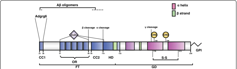
The prion protein undergoes post-translational
proteolytic processing
The cellular prion protein is encoded by the
Prnp
gene.
In mice, the entire protein-coding open-reading frame is
encoded within the third exon of
Prnp
[6–8]. After
translation and cotranslational extrusion into the lumen
of the endoplasmic reticulum, PrP
Cadopts its
physio-logical structure with a C-terminal globular domain and
an N-terminal flexible tail [9] (Fig. 1). The N-terminal
tail consists of two charged clusters (CC1 and CC2), the
octarepeat region (OR) and a hydrophobic domain (HD).
Additionally, two N-glycosylation sites are located in the
globular domain upstream of the sialylated GPI-anchor
at the C-terminus [10, 11].
After being transported to the cell membrane, PrP
Cresides extracellularly in lipid rafts, where it is attached
to the outer leaflet by a glycosyl phosphoinosityl (GPI)
anchor [12]. It undergoes rapid constitutive endocytosis
and subsequently either recycling or degradation [13, 14].
PrP
Cendocytosis can occur by both clathrin-dependent
[15] and caveolin pathways [16].
As part of its post-translational metabolism, PrP
Ccan
undergo proteolytic cleavage events termed, in analogy
to amyloid precursor protein processing,
α-,
β-, and
possibly also
γ-cleavage (Fig. 1). These cleavage events
release the so-called N1 + C1, N2 + C2, and C3 fragments,
respectively [17–20]. These events may be important for
both physiology and pathology. Alpha cleavage prevents
the C1 fragment from being converted into PrP
Sc[21], the
rate of the beta cleavage is increased in the disease [19],
and PrP
Cdeletion mutants lacking the alpha-cleavage
site show spontaneous neurodegeneration exhibiting
pathological features distinct from those of prion
dis-eases (reviewed in [22]).
The enzyme responsible for PrP
Ccleavage may not be
unique, and several members of the ADAM
(a_disinte-grin and metalloproteinase) family have been implicated
[23–26]. PrP
Ccan bind divalent cations such as copper
and zinc [27] by the octarepeat-containing flexible tail
and it has been reported to interact with a plethora of
different proteins. These interactions have been taken to
reflect its putative role in several cellular processes, but
they may also simply be a consequence of the
unstruc-tured, flexible conformation of the N-terminus of PrP
C.
Genetic pitfalls of PrP
Cgene ablation
Soon after PrP
Scwas proposed to be the causative agent
of prion diseases,
Prnp
knockout mice lacking PrP
Cwere
generated in order to answer the question whether the
loss of physiological PrP
Cfunction would lead to
neu-rodegeneration in prion diseases. The first
Prnp
null
mouse strain, designated
Prnp
-/-, or Zurich I (ZrchI,
Prnp
ZH1/ZH1), was produced in a mixed C57BL/6 J ×
129/Sv(ev) background [28] and a second line of PrP
C-deficient mice, known as Npu or Edinburgh (Edbg),
was produced with a pure 129/Ola genetic background
[29]. In a first round of characterization, these mice
were not found to show any clear abnormality except
for their resistance to prion infection [30]. They
devel-oped and bred normally, and although they displayed
subtle alterations in behavior [28], their otherwise
ap-parent normality seemed to rule out a physiological
function of PrP
Cthat is essential for life. If there is one,
it is highly redundant or it can be compensated for.
PrP
C-deficient mice from which the entire
Prnp
gene
was removed [31–34] develop progressive cerebellar
ataxia, which was originally attributed to the loss of
PrP
Cbut was later discovered to be due to the deletion
of a splice acceptor site in exon 3 of
Prnp
[35]. This led
to aberrant overexpression of the PrP
Cparalogue gene
(
Prnd
) encoding Doppel (Dpl) [36, 37], causing selective
neurodegeneration of cerebellar Purkinje cells. Notably,
the reintroduction of
Prnp
in mice overexpressing
Prnd
in the brain rescued the phenotype, suggesting a
func-tional link between the two proteins [38].
Later, using the Cre-loxP system, conditional PrP
Cknockout NFH-Cre/tg37 mice were generated to examine
the effects of acute PrP
Cdepletion on neuronal viability
and function in the brain of 9-week-old adults. This
ap-proach was thought to avoid compensatory mechanisms
active at the embryonal stage that would have masked
PrP
Closs of function phenotypes [39]. Again, depleting
neuronal PrP
Cin adult mice did not result in
neurodegen-eration or histopathological changes, but it led to subtle
electrophysiological abnormalities in the hippocampus
(Table 1). A closer look at different neuronal and other
cell functions in PrP
C-ablated mice revealed a number
of differences from wild-type mice that were attributed
to the physiological function of PrP
C. While some of
these studies were consistent among different PrP
C-ficient lines, others yielded contradictory results
de-pending on methodologies and the mouse models that
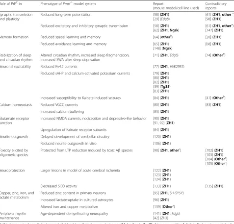
were used (Table 2).
A genetic confounder has been shown to underlie
some of these inconsistencies [40, 41]. For many years,
knockout alleles were usually created in embryonic stem
cells from the Sv129 strain of mice, and the resulting
mice were backcrossed to C57BL/6 mice [42]. This
prac-tice typically leads to variable, poorly controlled Mendelian
segregation of polymorphic alleles whose distribution
depends on their genetic linkage to the knockout allele.
All
Prnp
knockout mouse lines have been generated in
this way with the exception of the
“Edinburgh”
mouse,
which was maintained in a pure 129 background [42].
230
51 90
OR
94 110 133
CC1
FT GD
CC2 HD
GPI
180
CHO
196
CHO Adgrg6
23 28
Cu2+
178 213
S-S
Fig. 1.Structural organization of PrPC. Schematic representation of mature mouse PrPC, showing protein domains, sites of post-translational
Table 1
Lines of PrP
C-ablated mice covered in this review
Name Year produced Reference Genetic background “Doppel artifact“
Zurich I (ZrchI, ZH1) 1992 [28] Mixed C57BL/6J x 129/Sv(ev) No
Edinburgh (Edbg) 1994 [29] 129/Ola No
Nagasaki (Ngsk) 1996 [31] Mixed C57BL/6J x 129/Sv(ev) Yes
Rcm0 1997 [33] Mixed C57BL/6J x 129/Sv(ev) Yes
Zurich II (ZrchII, ZH2) 2001 [34] Mixed C57BL/6J x 129/Sv(ev) Yes
NFH-Cre/tg37 (adult onset) 2002 [39] Mixed C57BL/6J x 129/Sv(ev) No
Zurich III (ZrchIII, ZH3) 2016 [42] C57BL/6J No
Boldindicates mixed genetic background of at least two distinct mouse strains, possibly leading to the“flanking-gene problem”.Italicindicates mice maintained on single, pure genetic background
Table 2
Proposed physiological roles of cellular prion protein
Role of PrPCin Phenotype ofPrnp-/-model system Report
(mouse model/cell line used)
Contradictory reports
Synaptic transmission and plasticity
Reduced long-term potentiation [58](ZH1)
[29] (Edgb)
[61] (ZH1,othera) [98] (ZH1)
Reduced excitatory and inhibitory synaptic transmission [58] (ZH1) [62] (ZH1,Ngsk)
[61] (ZH1,othera) [147] (ZH1)
Memory formation Reduced spatial learning and memory [64] (otherb) [28] (ZH1)
Reduced avoidance learning and memory [65] (ZH1) [148] (Ngsk)
[68] (ZH1)
Stabilization of sleep and circadian rhythm
Altered circadian rhythm, increased sleep fragmentation, increased SWA after sleep deprivation
[71] (ZH1,Edgb) [74] (Otherb)
Neuronal excitability Reduced Kv4.2 currents [77] (ZH1, HEK293T)
Reduced sAHP and calcium-activated potassium currents [79] (ZH1) [80] (ZH1) [82] (ZH1) [39] (Tg35) [83] (ZH1)
Increased susceptibility to Kainate-induced seizures [84] (ZH1) [41] (Otherb)
Calcium homeostasis Reduced VGCC currents [80] (ZH1) [83] (ZH1)
Increased calcium buffering [83] (ZH1)
Glutamate receptor function
Increased NMDA currents, nociception and depressive-like behavior [90] (ZH1) [91,92] (ZH1)
Upregulation of Kainate receptor subunits [84] (ZH1)
Neurite outgrowth Delayed development of cerebellar circuitry [120] (ZH1)
Reduced neurite outgrowth in vitro [106] (ZH1)
Toxicity elicited by oligomeric species
Protected from LTP reduction induced by toxic Aβspecies [98] (ZH1,otherc) [102] (ZH1) [103] (ZH1) [104] (Otherb) [105] (Othera)
Neuroprotection Larger lesions in model of acute cerebral ischemia [122] (ZH1) [123] (ZH1) [124] (ZH1)
Decreased SOD activity [133] (ZH1) [135] (ZH1)
Copper, zinc, iron, and lactate metabolism
Reduced zinc content in primary neurons [95] (ZH1, SH-SY5Y)
Increased lactate-uptake in cultured astrocytes [96] (ZH1)
Altered iron and copper metabolism [139] (Othera)
Peripheral myelin maintenance
Age-dependent demyelinating neuropathy [141] (ZH1,Edgb) [42] (ZH3)
Boldindicates mixed genetic background of at least two distinct mouse strains, possibly leading to the“flanking-gene problem”.Italicindicates mice maintained on single, pure genetic background. Mouse lines specified as“other”are:a
ZH1 backcrossed to FVB;b
Edgb (back-)crossed to C57BL/10;c
Even after more than 12 generations of backcrossing, a
small part of the chromosome around the
Prnp
locus
still stems from the 129 strain, raising the question
whether any observed phenotypes were actually due to
polymorphisms in genes flanking
Prnp
. Indeed, we found
that SIRPα, a polymorphic
Prnp
-flanking gene, is
actu-ally responsible for an alleged
Prnp
-/-phenotype: the
in-hibition of macrophage phagocytosis of apoptotic cells
that was observed in PrP
C-deficient mice with mixed
genetic background but not in co-isogenic
Prnp
-/-mice
[40]. Recently, a new PrP
C-deficient mouse strain,
Prnp
ZH3/ZH3, was produced in our lab using
TALEN-mediated genome editing in fertilized mouse oocytes
and maintained in a pure C57BL/6 J genetic background
[42]. These strictly co-isogenic C57BL/6 J-
Prnp
ZH3/ZH3mice differ from wild-type mice only by eight deleted
nucleotides in the
Prnp
reading frame. In an effort to
improve the quality of studies on the function of the
cel-lular prion protein, we are distributing
Prnp
ZH3/ZH3mice
without requesting any kind of Material Transfer
Agree-ment, hence enabling better-controlled future studies. In
view of the broad availability of
Prnp
ZH3/ZH3mice, we
contend that the use of mixed-background PrP
C-defi-cient mice is obsolete and liable to artifacts.
Do further mammal species teach us more about the
function of PrP
C? The gene encoding PrP
Chas been
ablated experimentally in cattle [43] and goats [44], and
a naturally occurring
Prnp
knockout goat has been
reported [45]. While no pathological phenotypes were
reported in any of these animals, it may be rewarding
to perform specific investigations of these animals, e.g.,
concerning the integrity of the peripheral nervous
sys-tem in advanced age.
A large set of human genomic data was analyzed to
quantify the penetrance of variants of the human PrP
Cgene (
PRNP
) in prion disease [46]. Surprisingly,
hetero-zygous loss-of-function variants were identified in three
individuals. These individuals in their 50s and 70s are
probably healthy, and no evidence of any neurological
defect or peripheral neuropathy was documented. This
result suggests that heterozygous loss of
PRNP
in
humans may not be haploinsufficient. It remains to be
assessed, however, whether homozygous deletion and
therefore complete loss of PrP
Cmay create a disease
in humans.
Evidence for a role of synaptic PrP
Cin memory
and sleep
PrP
Cis strongly expressed in both neurons and glial cells
of the CNS [1]. In neurons, PrP
Cis preferentially localized
in the pre- and postsynaptic compartments of nerve
ter-minals. Immunocytochemical studies by light and electron
microscopy in primate and rodent brains [47, 48], as well
as examination of an EGFP-tagged PrP
Cin transgenic
mice, showed that PrP
Cis enriched along axons and in
pre-synaptic terminals [49, 50] and that it undergoes
anterograde and retrograde axonal transport [51, 52].
PrP
Cis also present in postsynaptic structures [53, 54].
It has recently been shown that sialic acid within the
GPI-anchor is important for targeting PrP
Cto synapses
[55]. This expression pattern implies that PrP
Cmight
be involved in preserving normal synaptic structure and
function by regulating synaptic transmission and
plasti-city (Fig. 3). Supporting this notion, synaptic
dysfunc-tion and synaptic loss are a prominent and early event
in prion diseases [56, 57].
Early reports showed that PrP
C-deficient mice (both
ZH1 and Edbg) display reduced long-term potentiation
(LTP) in hippocampal Schaffer collaterals and weakened
inhibitory GABAergic synaptic transmission [58, 59].
These defects have been claimed to be rescued by a
human
PRNP
transgene [60]. However, none of these
results were reproduced in PrP
C-deficient mice of
three different genetic backgrounds (Table 2). These
discrepancies have remained essentially unexplained
for the past 20 years [61]. Later studies reported a
PrP
Cdosage-dependent facilitation of synaptic
trans-mission, with PrP
C-over-expressing mice exhibiting
supra-physiological synaptic transmission [62]. This
effect seemed to result from a more efficient
recruit-ment of pre-synaptic fibers that, in turn, may depend
on PrP
Cexpression levels.
LTP is one of the neurophysiological correlates of
syn-aptic plasticity, the ability of synapses to change their
strength in response to previous activity. Because
synap-tic plassynap-ticity (LTP) in the hippocampus underlies
learn-ing and memory formation [63], any LTP deficits may
result in cognitive defects. Initial reports did not show
any reduction in memory performance of
Prnp
ZH1/ZH1mice in the Morris Water Maze test [28]. However, a
later study found deficiencies in spatial learning and
memory in PrP
C-deficient mouse lines on various
back-grounds [64]. These deficits were explained by reduced
LTP in the dentate gyrus in PrP
C-deficient mice in vivo
,
and were rescued by neuronal expression of PrP
C.
Prnp
ZH1/ZH1mice were reported to have impaired
mem-ory performance only when aged (9 months). Several
molecular mechanisms for this defect have been
pro-posed, but none were verified at the mechanistic level
[65–67]. These results stand in contrast to another study
carried out in
Prnp
ZH1/ZH1mice that did not reveal any
memory impairment up to an age of 2 years [68]. Thus,
a role for PrP
cin memory is still contentious.
like sporadic and familial fatal insomnia [69, 70]. Both
Prnp
ZH1/ZH1and co-isogenic
Prn
p
Edbg/Edbgmice were
reported to display altered circadian rhythms, increased
sleep fragmentation, and increased slow wave activity
(SWA) following sleep deprivation [71]. The latter
phenotype was rescued by reintroduction of PrP
Cex-pression [72]. It was suggested that the difference in
SWA between wild-type and
Prnp
null mice reflected a
function of PrP
Cin neurotransmission or a protective
role on synapses [73]. Another study confirmed sleep
disturbance in
Prnp
knockout mice. However, using a
prolonged sleep deprivation protocol, the defect was
found to be associated with reduced slow-wave activity
and to altered hormonal reactivity to prolonged stress
[74]. Interestingly, deterioration of slow-wave activity
was found to contribute to sleep deficits in Alzheimer’s
disease (AD) and was reversed by enhancing GABA
Aergic
inhibition [75].
The molecular bases of sleep regulation are not
com-pletely understood. Tantalizingly, recent work indicates
that calcium-dependent hyperpolarization is critical to
sleep duration, and that sleep deterioration is associated
with impairment of calcium-dependent potassium
chan-nels, voltage-gated calcium channels (VGCC), and
N-methyl-D-aspartate (NMDA) glutamate receptors [76].
Since both hyperpolarization linked to calcium
dysho-meostasis and NMDA receptor-related hyperexcitability
were documented in
Prnp
ablated mice (discussed in the
next section), loss of PrP
C-dependent control of these
ion channels may underlie the sleep disruption in PrP
C-deficient mice and perhaps also in prion diseases.
Possible functions for PrP
Care suggested by
interaction partners
Albeit controversial, the participation of PrP
Cin
neuro-biological processes, and particularly in sleep regulation
and memory, raises the question whether the cellular
prion protein modulates synaptic mechanisms and
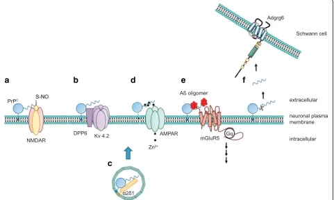
neur-onal excitability at a molecular level. Insights into possible
mechanisms may be provided by the documented
inter-action of PrP
Cwith several ion channels and metabotropic
glutamate receptors (Fig. 2). However, caution is needed
a
b
d
e
f
c
Fig. 2.PrPCexerts its functions via distinct mechanisms. The cellular prion protein may utilize several mechanisms to modulate cellular functions.
As schematically depicted ina, PrPCmay directly alter the function of its target protein by mediating posttranslational modifications, for example,
by promoting the S-nitrosylation of the NMDA receptor. Alternatively, PrPCmodulates auxiliary proteins of ion channels, thereby regulating the
biophysical properties of the channel (b) or its trafficking (c). Another function of PrPCarises from its ability to bind divalent cations such as zinc
(Zn2+) or copper (Cu2+). It was claimed that PrPCmay buffer these cations within the synaptic cleft and may facilitate their uptake (d) via AMPA
receptors. Some better-defined actions of PrPCinclude its binding to misfolded oligomeric protein species and signaling in complex with
other membrane receptors (e). Additionally, PrPCcan signal intransby its N-terminal cleavage products, which may bind to other receptors,
while screening the crowded PrP
Cinteractome: only
inter-actions with molecular partners displaying a functional
correlate to PrP
Cbinding should be considered of
poten-tial biological relevance.
PrP
Cinteracts with Dipeptidyl peptidase-like 6 (DPP6),
an auxiliary subunit of the voltage-gated potassium
channel 4.2 (Kv4.2), which leads to increased and
pro-longed currents through this channel, thereby reducing
cellular excitability [77]. Also, PrP
Cand a mutant PrP
Cversion that is linked to a genetic prion disease have
been shown to co-immunoprecipitate with the auxiliary
subunit
α2δ-1 of VGCCs in transgenic mice [78].
Mutant PrP
Caffected
α2δ-1 trafficking and function at
the synapses. The molecular role of PrP
Cin VGCC
function under physiological conditions remains
un-clear (Fig. 3). However, deficits in VGCC currents and
calcium homeostasis were reported in
Prnp
ZH1/ZH1neurons [79, 80] and were proposed to underlie the
reduced slow after hyperpolarization (sAHP) seen in
PrP
C-deficient mice. The sAHP is a property of many
neurons that is evoked by repetitive action potentials
and controls subsequent action potential firing. The
intermediate-conductance, calcium-activated potassium
channel (IkCa) has been claimed to control this
neuro-physiological parameter [81].
Not only could the sAHP defect be reproduced by
inde-pendent research groups in
Prnp
ZH1/ZH1mice [80, 82, 83],
it has also been shown in an adult-onset model of PrP
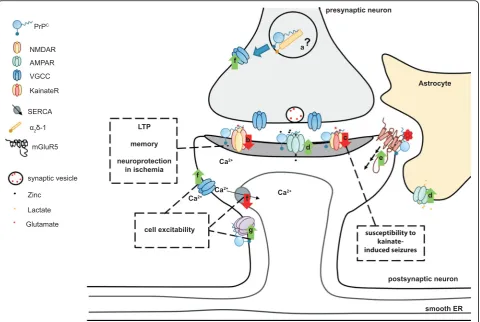
CFig. 3.Schematic overview of possible physiological functions of PrPCand their effect in the central nervous system. PrPCregulates ion channels
and neurotransmitter receptors at the pre- and postsynaptic levels.aPrPCmight modulate VGCC trafficking at the presynapse via interaction
with theα2δ-1 VGCC subunit.bPostsynaptically, PrPCdampens NMDA receptor-mediated currents by modulating various receptor subunits of
this channel. It was speculated that control of NMDA receptor function might be related to certain reported phenotypes of PrPC-ablated mice.
cPrPCmay also control the glutamatergic system by modulating the subunit composition of kainate receptors. This possibly relates to increased
susceptibility of PrPC-ablated mice to kainate-induced seizures.dPrPCassociates with, and promotes cell surface localization of, AMPA receptor
subunits. This facilitates zinc uptake at the synaptic cleft via AMPA receptors. On astrocytes, a PrPC–AMPA complex may be involved in the uptake
of lactate.ePrPCbinds to toxic oligomeric protein species. PrPCbinds to Aβoligomers and, in complex with metabotropic glutamate receptor 5
(mGluR5), was proposed to trigger intracellular signaling related to Alzheimer's disease pathology.fPrPCcontrols calcium influx via interaction
with different ion channels. Additionally, PrPCwas claimed to regulate calcium storage via the sarcoplasmic/endoplasmic reticulum calcium
ATPase (SERCA).gPrPCpositively modulates potassium currents as exemplified by association with DPP6, an auxiliary subunit of the Kv 4.2
potassium channel. Control of calcium and potassium channels might be related to the alleged function of PrPCin neuronal excitability.ER
depletion, indicating that this is not a developmental
phenotype [39]. At the cellular level, reduced sAHP is
ex-pected to result in increased neuronal firing. Increased
neuronal excitability in the PrP
C-deficient hippocampus
has also been reported in the context of higher
vulnerabil-ity of PrP
C-deficient mice to kainate-induced seizures
(Fig. 3). This was related to the interaction of PrP
Cwith
the kainate receptor subunit GluR6/7 [84]. Additionally,
in the absence of PrP
C, GluR6/7-containing KA receptors
were upregulated and the neurotoxic signaling was
en-hanced [84–86]. This suggests a neuroprotective role of
PrP
Cagainst excitotoxic insults.
However, whether this represents a bona fide PrP
Cphenotype remains controversial.
Prnp
-/-mice on a
differ-ent genetic background displayed a reduced susceptibility
to kainate-induced seizures in the absence of PrP
C. As this
phenotype could not be rescued by reintroduction of an
exogenous
Prnp
gene, it is suspected that polymorphisms
in some unidentified
Prnp
-flanking genes may underlie
the discrepant phenotypes [41]. More recently, however,
although the effect of the
“
Prnp
-flanking gene”
in the
KA-mediated responses was confirmed in PrP
C-deficient mice
of a mixed background (B6129 and B6.129), enhanced
sensitivity to epileptogenic drugs was found in the
co-isogenic
Prnp
Edbg/Edbgmouse
[87].
The
molecular
mechanism underlying this phenotype is unclear but the
aa32–93 region of PrP
C(spanning the octapeptide repeats)
and its glycosylphosphoinositol anchor may be involved.
Consistent with the higher neuronal excitability of PrP
C-deficient mice, anatomical changes within the
hippocam-pus indicated a reorganization of neuronal circuitry
similar to the
“epileptic neuronal network”
seen in certain
human epilepsies [88].
PrP
Cmight also act as a modulator of glutamate
recep-tors of the NMDA subtype. These are heterotetramers
composed of two GluN1 subunits and two GluN2
sub-units of different subtypes [89]. It was found that PrP
Cinhibited NMDA receptors and prevented potentially
exci-totoxic calcium influx through these channels by
associ-ation with NMDA receptors containing the GluN2D
subunit [90], as shown by co-immunoprecipitation and
immunofluorescence imaging. Absence of PrP
Cled to
up-regulation of GluN2D-containing NMDA receptors and
enhanced signaling due to prolonged kinetics of
NMDA-mediated currents [90]. This was subsequently linked to
increased depressive-like behavior and increased
noci-ception in PrP
C-deficient mice. Both phenotypes were
rescued by pharmacological inhibition of NMDA
recep-tors [91, 92]. Additionally, copper-dependent interaction
of PrP
Cwith the GluN1 subunit was documented,
which was involved in nitrosylation of NMDA subunits
GluN2A and GluN1. This represents a second mechanism
by which the presence of PrP
Creduces NMDA currents
and signaling [93, 94].
The putative control of ionotropic glutamate receptors
by PrP
Cmay be even more complex, since PrP
Calso
interacts with
α-amino-3-hydroxy-5-methyl-4-isoxazo-lepropionic acid (AMPA) receptor subunits GluA1 and
GluA2. This interaction may be relevant to the PrP
C-mediated cellular uptake of zinc through AMPA
recep-tors [95] and to the regulation of lactate transport in
astrocytes [96]. Whether the binding of PrP
Cwith
AMPA receptor subunits also plays a role in AMPA
re-ceptor function remains to be elucidated.
PrP
Cinteractions with metabotropic glutamate
receptors and A
β
Recently, PrP
Chas been shown to interact not only
with ionotropic but also with metabotropic glutamate
receptors of group I, mGluR1 and mGluR5 (Fig. 2).
Me-tabotropic glutamate receptors are members of the G
protein-coupled receptor (GPCR) superfamily of seven
transmembrane-domain proteins that are activated by
glutamate and transduce intracellular signals via G
pro-teins. PrP
Cbinds to and signals through mGluR5 in
disease-related conditions [97].
Several studies found that Aβ
oligomers, the
neuro-toxic protein species involved in AD, can bind to PrP
C[98, 99] (Figs. 1 and 2) and activate the Fyn kinase
through mGluR5 [97]. Aβ–PrP
C–mGluR5 complexes
are responsible for facilitation of long-term depression
(LTD) in vivo [100] and dendritic spine loss in cultured
neurons [97]. The Aβ–PrP
C–mGlu5R complex might act
upstream of the phosphorylation of the NMDA receptor
subunit GluN2B. This event within the pathogenic
cas-cade triggered by Aβ
oligomers requires PrP
C-dependent
Fyn activation [54] and underlies the Aβ
oligomer-induced disruption of LTP in AD.
Focusing on the role of PrP
C, two main considerations
can be drawn from these studies. First, PrP
Cappears to
function as a cell surface receptor for synaptotoxic
oligo-mers of the Aβ
peptide and, as reported by Resenberger
and colleagues [101], of other
β-sheet-rich neurotoxic
proteins. However, whereas the physical interaction of
PrP
Cwith Aβ
oligomers was confirmed [99, 102], it is
unclear whether PrP
Cis necessary for the synaptotoxic
effect of Aβ
oligomers [102–105]. Secondly, although
the interplay between mGluR5 and PrP
Cmay be relevant
to AD pathology, it remains unclear whether the binding
to PrP
Caffects physiological functions of group I
me-tabotropic glutamate receptors. Intriguingly, a role for
the PrP
C–mGluR1 complex in neurite outgrowth has
been reported [106].
Role of PrP
Cin development
analysis of PrP
Cknockout embryos showed several
differ-entially expressed genes (DEGs), while the number of
DEGs in brains of adult PrP
C-deficient mice is almost
neg-ligible [42, 109]. Interestingly, the number of DEGs is
higher in brains where PrP
Chad been knocked out
postna-tally, suggesting that the function of PrP
Cmay be
compen-sated for by other proteins during development [110]. To
date, only a few in vivo studies on the role of PrP
Cin CNS
development are available. They suggest that PrP
C-ablated
mice exhibit reduced proliferation rates of neuronal
pro-genitor cells in the embryonic, newborn, and adult CNS
[111, 112]. Additionally, increased proliferation of
oligo-dendrocyte precursor cells with a concomitant maturation
delay of oligodendrocytes and astrocytes has been reported
[111, 113]. These observations were supported by the
results of several in vitro experiments [111, 113, 114].
However, as the genetic background can influence brain
development and adult neurogenesis [115], these studies
should be confirmed in coisogenic PrP
C-ablated mice.
Additionally, in vitro studies suggest that PrP
Cis
im-plicated in the regulation of neuritogenesis [116, 117] as
well as axonal growth [48, 118, 119]. Also, there is some
evidence that PrP
Cis involved in the development of the
cerebellar circuitry, leading to delayed motor
develop-ment of PrP
C-deficient mice [120].
Possible neuroprotective roles of PrP
CPrP
Cmay have a neuroprotective role in a mouse model
of cerebral ischemia, as PrP
C-deficient mice show larger
lesions in acute cerebral ischemia. Furthermore,
overex-pression of PrP
Ccan reduce the lesion size compared to
wild-type mice [121–124]. Attenuation of NMDA
signal-ing by PrP
Chas been proposed to be the basis of a
neu-roprotective role of PrP
Cagainst NMDA-mediated
toxicity in ischemia [125]. Additionally, it was found that
cleavage of PrP
Cinto its N- and C-terminal fragments is
enhanced under ischemic conditions and these cleavage
products can themselves be neuroprotective [124]. In
particular, the N-terminal cleavage fragment (N1) might be
neuroprotective against staurosporine-induced Caspase-3
activation in a model of pressure-induced ischemia in the
rat retina [126]. These results are supported by several
in vitro studies, where expression of PrP
Cwas protective
against staurosporine or anisomycin-induced apoptosis
[127, 128]. Conversely, loss of PrP
Cwas beneficial against
glutamate-induced excitotoxicity in vitro, an effect
sup-posedly mediated by increased uptake of glutamate in
PrP
C-ablated astrocytes [129].
The protective function of the N1 fragment is also
very intriguing in the context of the Aβ
oligomer-related
synaptotoxicity. This intrinsically disordered N-terminal
portion of PrP
Cis involved in binding to
β-sheet-rich
peptides like Aβ
oligomers [99, 101] and mediates the
detrimental effects of Aβ
oligomers on synaptic function
as mentioned before. However, in its soluble form as
secreted upon PrP
Ccleavage, N1 acted in a decoy
receptor-like mode: it prevented Aβ
peptide fibrillization
and reduced the neurotoxicity of amyloid-β
oligomers
in vitro and in vivo [130]. Additionally, the rate of PrP
Calpha-cleavage is increased in brain tissue from patients
suffering from AD and it was proposed that
alpha-cleavage represents an endogenous protective mechanism
against amyloid-β
toxicity in humans [131].
However, PrP
C-deficient mice do not exhibit altered
amyloid-β
toxicity [102–105] and there was no protective
effect of PrP
Cin mouse models of other
neurodegenera-tive diseases, including Parkinson's and Huntington's
dis-ease, as well as a mouse model of tauopathy [124, 132].
Based on in vitro studies, by virtue of its ability to bind
copper, PrP
Chas been proposed to participate in
resist-ance to oxidative stress by preventing reactive oxygen
species (ROS) generation via free copper-mediated redox
reactions. Also, PrP
Cwas at some point thought to
regu-late the function of superoxide dismutase (SOD) [133].
It was even proposed that PrP
Ccould act as a SOD by
it-self [27, 134]. However, a function of PrP
Cin copper
me-tabolism is still controversial and the influence of PrP
Con
either SOD level or the intrinsic dismutase activity of PrP
Cwas shown by us and others to be artifactual [135, 136].
There might be, however, alternative ways in which
PrP
Cprotects against ROS toxicity. For instance, PrP
C-dependent expression of antioxidant enzymes was
sug-gested as an explanation for resistance to oxidative stress
mediated by PrP
C[137, 138] as well as a conjectured
PrP
Cfunction in iron metabolism and control of
redox-iron balance in cell lines [139, 140].
Role of PrP
Cin the peripheral nervous system
PrP
C-deficient mice of five different PrP
C-knockout
strains, including the
Prnp
ZH3/ZH3mice (coisogenic to
BL/6 mice), develop a late-onset peripheral neuropathy,
indicating that peripheral myelin maintenance is a bona
fide physiological function of PrP
C[42, 141, 142]. PrP
Cneuronal
expression
and
amino-proximal
cleavage
(Fig. 2) are necessary for the promyelinating signal [141].
It was then discovered that the very N-terminal
polyca-tionic cluster of PrP
Cbinds to the G-protein-coupled
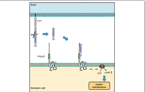
re-ceptor Adgrg6 (Gpr126) on Schwann cells (Fig. 1),
eliciting a promyelinating cAMP response in vitro and
in vivo in mice and zebrafish (Fig. 4) [5]. This pointed to
the N-terminal fragment of PrP
Cas a promyelinating
factor that might serve as a possible treatment in other
peripheral chronic demyelinating polyneuropathies.
also showed severe demyelination in both the spinal
cord and cerebellar white matter in vivo [144, 145].
Thus, although in the CNS an eventual PrP
Cfunction
in myelin homeostasis is dispensable, a contribution of
aberrant PrP
Cfunction in demyelinating diseases in the
brain is a conceivable scenario. Moreover, whether the
N-terminal cleavage product of PrP
Cis also signaling
via other G-protein-coupled receptors in distinct
bio-logical processes is likely, but remains to be elucidated.
PrP
Cfunction: the next chapters
All available data point to PrP
Cexerting its function in
concert with additional membrane proteins. On one hand,
PrP
Ccan regulate the cellular transport and localization of
its binding partners. On the other hand, PrP
Ccan directly
modulate the functionality of the binding partner—as seen
for certain ion channels and ionotropic glutamate
recep-tors. Also, PrP
Ccan signal in
trans
via its N-terminal
cleav-age products. Finally, PrP
Cappears to scavenge amyloid
aggregates of Aβ, and it will be interesting to see whether
further pathological aggregates can also be recognized by
PrP
C. Given these findings, the question arises whether the
cellular prion protein needs its misfolding-prone structure
with a disordered flexible tail to fulfill its physiological
function. The fact that the functional domains of PrP
Care
conserved from avians to mammals speaks in favor of this
hypothesis [4, 146]. Even though the interpretation of
many studies is hampered by the genetic impurity of the
mouse models used, there is enough evidence that PrP
Cplays a role in several physiological functions in the central
and peripheral nervous systems. Nevertheless, it appears
implausible that PrP
Cis involved in such a large number of
cellular functions, particularly in view of the small number
of validated pathological phenotypes in PrP
C-deficient
mice. The emergence of new, rigorously controlled animal
models will be of help for revisiting and critically assessing
some of these phenotypes.
Acknowledgements
AA is the recipient of support from an Advanced Grant of the European Research Council (Prion2020), a European Union Framework 7 Grant (NEURINOX), the Swiss National Research Foundation, Sinergia grant #147660, the Clinical Research Priority Programs“Small RNAs”and “Human Hemato-Lymphatic Diseases”, SystemsX.ch and the EU Joint Programme on Neurodegenerative Disease Research (JPND) CureALS and REfrAME. M-AW was supported by an MD/PhD fellowship from the Swiss National Foundation. AS is a recipient of a Forschungskredit of the University of Zurich, grant number [FK–16–048].
PrPC
Adgrg6
cAMP ATP
myelin maintenance Axon
Schwann cell
AC
Fig. 4.Axonal PrPCpromotes myelin maintenance intransvia Adgrg6 on Schwann cells. Mice devoid of PrPCdevelop a chronic demyelinating
neuropathy, which suggested a pro-myelinating function of PrPC. In the peripheral nervous system, the N1 fragment of axonal PrPCinteracts with
Authors’contributions
M-AW, AS, and AA wrote the manuscript. All authors have read the manuscript and agree with its content.
Competing interests
The authors declare that they have no competing interests.
Publisher
’
s Note
Springer Nature remains neutral with regard to jurisdictional claims in published maps and institutional affiliations.
References
1. Bendheim PE, Brown HR, Rudelli RD, Scala LJ, Goller NL, Wen GY, et al. Nearly ubiquitous tissue distribution of the scrapie agent precursor protein. Neurology. 1992;42:149.
2. Prusiner SB. Novel proteinaceous infectious particles cause scrapie. Science. 1982;216:136–44. http://dx.doi.org/10.1126/science.6801762.
3. Prusiner SB. Prions. Proc Natl Acad Sci U S A. 1998;95:13363–83. 4. Wopfner F, Weidenhöfer G, Schneider R, von Brunn A, Gilch S, Schwarz TF,
et al. Analysis of 27 mammalian and 9 avian PrPs reveals high conservation of flexible regions of the prion protein. J Mol Biol. 1999;289:1163–78. 5. Küffer A, Lakkaraju AKK, Mogha A, Petersen SC, Airich K, Doucerain C, et al.
The prion protein is an agonistic ligand of the G protein-coupled receptor Adgrg6. Nature. 2016;536:464–8.
6. Chesebro B, Race R, Wehrly K, Nishio J, Bloom M, Lechner D, et al. Identification of scrapie prion protein-specific mRNA in scrapie-infected and uninfected brain. Nature. 1985;315:331–3.
7. Oesch B, Westaway D, Wälchli M, McKinley MP, Kent SBH, Aebersold R, et al. A cellular gene encodes scrapie PrP 27-30 protein. Cell. 1985;40:735–46. 8. Westaway D, Cooper C, Turner S, Da Costa M, Carlson GA, Prusiner SB.
Structure and polymorphism of the mouse prion protein gene. Proc Natl Acad Sci U S A. 1994;91:6418–22.
9. Riek R, Hornemann S, Wider G, Glockshuber R, Wüthrich K. NMR characterization of the full-length recombinant murine prion protein, mPrP (23–231). FEBS Lett. 1997;413:282–8.
10. Stahl N, Borchelt DR, Hsiao K, Prusiner SB. Scrapie prion protein contains a phosphatidylinositol glycolipid. Cell. 1987;51:229–40.
11. Stahl N, Baldwin M, Hecker R, Pan K-M, Burlingame A, Prusiner S. Glycosylinositol phospholipid anchors of the scrapie and cellular prion proteins contain sialic acid. Biochemistry (Mosc). 1992;31:5043–53. 12. Naslavsky N, Stein R, Yanai A, Friedlander G, Taraboulos A. Characterization
of detergent-insoluble complexes containing the cellular prion protein and its scrapie isoform. J Biol Chem. 1997;272:6324–31.
13. Morris RJ, Parkyn CJ, Jen A. Traffic of prion protein between different compartments on the neuronal surface, and the propagation of prion disease. FEBS Lett. 2006;580:5565–71.
14. Parizek P. Similar turnover and shedding of the cellular prion protein in primary lymphoid and neuronal cells. J Biol Chem. 2001;276:44627–32. 15. Taylor DR. Assigning functions to distinct regions of the N-terminus of the
prion protein that are involved in its copper-stimulated, clathrin-dependent endocytosis. J Cell Sci. 2005;118:5141–53.
16. Peters PJ, Mironov A, Peretz D, van Donselaar E, Leclerc E, Erpel S, et al. Trafficking of prion proteins through a caveolae-mediated endosomal pathway. J Cell Biol. 2003;162:703–17.
17. Harris DA, Huber MT, Van Dijken P, Shyng SL, Chait BT, Wang R. Processing of a cellular prion protein: identification of N-and C-terminal cleavage sites. Biochemistry (Mosc). 1993;32:1009–16.
18. Walmsley AR, Watt NT, Taylor DR, Perera WSS, Hooper NM.α-cleavage of the prion protein occurs in a late compartment of the secretory pathway and is independent of lipid rafts. Mol Cell Neurosci. 2009;40:242–8. 19. Chen SG, Teplow DB, Parchi P, Teller JK, Gambetti P, Autilio-Gambetti L.
Truncated forms of the human prion protein in normal brain and in prion diseases. J Biol Chem. 1995;270:19173–80.
20. Lewis V, Johanssen VA, Crouch PJ, Klug GM, Hooper NM, Collins SJ. Prion protein“gamma-cleavage”: characterizing a novel endoproteolytic processing event. Cell Mol Life Sci. 2016;73:667–83.
21. Westergard L, Turnbaugh JA, Harris DA. A naturally occurring C-terminal fragment of the prion protein (PrP) delays disease and acts as a dominant-negative inhibitor of PrPSc formation. J Biol Chem. 2011;286:44234–42.
22. Yusa S, Oliveira-Martins JB, Sugita-Konishi Y, Kikuchi Y. Cellular prion protein: from physiology to pathology. Viruses. 2012;4:3109–31. 23. Taylor DR, Parkin ET, Cocklin SL, Ault JR, Ashcroft AE, Turner AJ, et al. Role
of ADAMs in the ectodomain shedding and conformational conversion of the prion protein. J Biol Chem. 2009;284:22590–600. http://dx.doi.org/10. 1074/jbc.M109.032599.
24. Vincent B, Paitel E, Saftig P, Frobert Y, Hartmann D, De Strooper B, et al. The disintegrins ADAM10 and TACE contribute to the constitutive and phorbol ester-regulated normal cleavage of the cellular prion protein. J Biol Chem. 2001;276:37743–6.
25. Altmeppen HC, Prox J, Puig B, Kluth MA, Bernreuther C, Thurm D, et al. Lack of a-disintegrin-and-metalloproteinase ADAM10 leads to intracellular accumulation and loss of shedding of the cellular prion protein in vivo. Mol Neurodegener. 2011;6:36.
26. Altmeppen HC, Prox J, Krasemann S, Puig B, Kruszewski K, Dohler F, et al. The sheddase ADAM10 is a potent modulator of prion disease. Elife. 2015;4, e04260.
27. Brown DR, Clive C, Haswell SJ. Antioxidant activity related to copper binding of native prion protein. J Neurochem. 2001;76:69–76.
28. Büeler HR, Fischer M, Lang Y, Bluethmann H, Lipp HP, DeArmond SJ, et al. Normal development and behaviour of mice lacking the neuronal cell-surface PrP protein. Nature. 1992;356:577–82. http://dx.doi.org/10. 1038/356577a0.
29. Manson JC, Clarke AR, Hooper ML, Aitchison L, McConnell I, Hope J. 129/Ola mice carrying a null mutation in PrP that abolishes mRNA production are developmentally normal. Mol Neurobiol. 1994;8:121–7.
30. Büeler H, Aguzzi A, Sailer A, Greiner R-A, Autenried P, Aguet M, et al. Mice devoid of PrP are resistant to scrapie. Cell. 1993;73:1339–47.
31. Sakaguchi S, Katamine S, Nishida N, Moriuchi R, Shigematsu K, Sugimoto T, et al. Loss of cerebellar Purkinje cells in aged mice homozygous for a disrupted PrP gene. Nature. 1996;380:528–31.
32. Katamine S, Nishida N, Sugimoto T, Noda T, Sakaguchi S, Shigematsu K, et al. Impaired motor coordination in mice lacking prion protein. Cell Mol Neurobiol. 1998;18:731–2.
33. Moore RC. Gene targeting studies at the mouse prion protein locus [PhD Thesis]. Edinburgh, Scotland: University of Edinburgh; 1997. 34. Rossi D, Cozzio A, Flechsig E, Klein MA, Rülicke T, Aguzzi A, et al. Onset of
ataxia and Purkinje cell loss in PrP null mice inversely correlated with Dpl level in brain. EMBO J. 2001;20:694.
35. Weissmann C, Aguzzi A. PrP’s double causes trouble. Science. 1999;286:914. 36. Moore RC, Lee IY, Silverman GL, Harrison PM, Strome R, Heinrich C, et al.
Ataxia in prion protein (PrP)-deficient mice is associated with upregulation of the novel PrP-like protein doppel. J Mol Biol. 1999;292:797–817. 37. Lu K, Wang W, Xie Z, Wong B-S, Li R, Petersen RB, et al. Expression and
Structural characterization of the recombinant human doppel protein. Biochemistry (Mosc). 2000;39:13575–83.
38. Moore RC, Mastrangelo P, Bouzamondo E, Heinrich C, Legname G, Prusiner SB, et al. Doppel-induced cerebellar degeneration in transgenic mice. Proc Natl Acad Sci U S A. 2001;98:15288–93.
39. Mallucci GR, Ratte S, Asante EA, Linehan J, Gowland I, Jefferys JGR, et al. Post-natal knockout of prion protein alters hippocampal CA1 properties, but does not result in neurodegeneration. EMBO J. 2002;21:202–10. 40. Nuvolone M, Kana V, Hutter G, Sakata D, Mortin-Toth SM, Russo G, et al.
SIRP polymorphisms, but not the prion protein, control phagocytosis of apoptotic cells. J Exp Med. 2013;210:2539–52.
41. Striebel JF, Race B, Pathmajeyan M, Rangel A, Chesebro B. Lack of influence of prion protein gene expression on kainate-induced seizures in mice: studies using congenic, coisogenic and transgenic strains. Neuroscience. 2013;238:11–8.
42. Nuvolone M, Hermann M, Sorce S, Russo G, Tiberi C, Schwarz P, et al. Strictly co-isogenic C57BL/6 J-Prnp−/−mice: A rigorous resource for prion science. J Exp Med. 2016;213:313–27.
43. Richt JA, Kasinathan P, Hamir AN, Castilla J, Sathiyaseelan T, Vargas F, et al. Production of cattle lacking prion protein. Nat Biotechnol. 2007;25:132–8. 44. Yu G. Functional disruption of the prion protein gene in cloned goats.
J Gen Virol. 2006;87:1019–27.
45. Benestad SL, Austbø L, Tranulis MA, Espenes A, Olsaker I. Healthy goats naturally devoid of prion protein. Vet Res. 2012;43:87.
47. Salès N, Rodolfo K, Hässig R, Faucheux B, Di Giamberardino L, Moya KL. Cellular prion protein localization in rodent and primate brain. Eur J Neurosci. 1998;10:2464–71.
48. Salès N, Hässig R, Rodolfo K, Di Giamberardino L, Traiffort E, Ruat M, et al. Developmental expression of the cellular prion protein in elongating axons. Eur J Neurosci. 2002;15:1163–77.
49. Herms J, Tings T, Gall S, Madlung A, Giese A, Siebert H, et al. Evidence of presynaptic location and function of the prion protein. J Neurosci. 1999;19:8866–75.
50. Mironov A, Latawiec D, Wille H, Bouzamondo-Bernstein E, Legname G, Williamson RA, et al. Cytosolic prion protein in neurons. J Neurosci. 2003;23:7183–93.
51. Borchelt DR, Koliatsos VE, Guarnieri M, Pardo CA, Sisodia SS, Price DL. Rapid anterograde axonal transport of the cellular prion glycoprotein in the peripheral and central nervous systems. J Biol Chem. 1994;269:14711–4. 52. Moya KL, Hässig R, Créminon C, Laffont I, Di Giamberardino L. Enhanced
detection and retrograde axonal transport of PrPc in peripheral nerve: cellular prion protein in peripheral nerve. J Neurochem. 2003;88:155–60. 53. Haeberle AM, Ribaut-Barassin C, Bombarde G, Mariani J, Hunsmann G, Grassi J,
et al. Synaptic prion protein immuno-reactivity in the rodent cerebellum. Microsc Res Tech. 2000;50:66–75.
54. Um JW, Nygaard HB, Heiss JK, Kostylev MA, Stagi M, Vortmeyer A, et al. Alzheimer amyloid-βoligomer bound to postsynaptic prion protein activates Fyn to impair neurons. Nat Neurosci. 2012;15:1227–35. 55. Bate C, Nolan W, McHale-Owen H, Williams A. Sialic acid within the
glycosylphosphatidylinositol anchor targets the cellular prion protein to synapses. J Biol Chem. 2016;291:17093–101.
56. Jeffrey M, Halliday WG, Bell J, Johnston AR, MacLeod NK, Ingham C, et al. Synapse loss associated with abnormal PrP precedes neuronal degeneration in the scrapie-infected murine hippocampus. Neuropathol Appl Neurobiol. 2008;26:41–54.
57. Šišková Z, Reynolds RA, O’Connor V, Perry VH. Brain region specific pre-synaptic and post-synaptic degeneration are early components of neuropathology in prion disease. PLoS One. 2013;8:e55004. Mallucci GR, editor.
58. Collinge J, Whittington MA, Sidle KCL, Smith CJ, Palmer MS, Clarke AR, et al. Prion protein is necessary for normal synaptic function. Nature. 1994;370:295–7.
59. Manson J, Hope J, Clarke AR, Johnston A, Black C, MacLeod N. PrP Gene dosage and long term potentiation. Neurodegeneration. 1995;4:113–4. 60. Whittington MA, Sidle KCL, Gowland I, Meads J, Hill AF, Palmer MS, et al.
Rescue of neurophysiological phenotype seen in PrP null mice by transgene encoding human prion protein. Nat Genet. 1995;9:197–201. 61. Lledo PM, Tremblay P, DeArmond SJ, Prusiner SB, Nicoll RA. Mice deficient
for prion protein exhibit normal neuronal excitability and synaptic transmission in the hippocampus. Proc Natl Acad Sci U S A. 1996;93:2403–7. 62. Carleton A, Tremblay P, Vincent JD, Lledo PM. Dose-dependent, prion
protein (PrP)-mediated facilitation of excitatory synaptic transmission in the mouse hippocampus. Pflüg Arch Eur J Physiol. 2001;442:223–9. 63. Bliss TVP, Collingridge GL. A synaptic model of memory: long-term
potentiation in the hippocampus. Nature. 1993;361:31–9.
64. Criado JR, Sánchez-Alavez M, Conti B, Giacchino JL, Wills DN, Henriksen SJ, et al. Mice devoid of prion protein have cognitive deficits that are rescued by reconstitution of PrP in neurons. Neurobiol Dis. 2005;19:255–65. 65. Coitinho AS, Roesler R, Martins VR, Brentani RR, Izquierdo I. Cellular prion
protein ablation impairs behavior as a function of age. NeuroReport. 2003;14:1375–9.
66. Coitinho AS, Freitas ARO, Lopes MH, Hajj GNM, Roesler R, Walz R, et al. The interaction between prion protein and laminin modulates memory consolidation. Eur J Neurosci. 2006;24:3255–64.
67. Coitinho AS, Lopes MH, Hajj GNM, Rossato JI, Freitas AR, Castro CC, et al. Short-term memory formation and long-term memory consolidation are enhanced by cellular prion association to stress-inducible protein 1. Neurobiol Dis. 2007;26:282–90.
68. Lipp H-P, Stagliar-Bozicevic M, Fischer M, Wolfer DP. A 2-year longitudinal study of swimming navigation in mice devoid of the prion protein: no evidence for neurological anomalies or spatial learning impairments. Behav Brain Res. 1998;95:47–54.
69. Lugaresi E, Medori R, Montagna P, Baruzzi A, Cortelli P, Lugaresi A, et al. Fatal familial insomnia and dysautonomia with selective degeneration of thalamic nuclei. N Engl J Med. 1986;315:997–1003.
70. Mastrianni JA, Nixon R, Layzer R, Telling GC, Han D, DeArmond SJ, et al. Prion protein conformation in a patient with sporadic fatal insomnia. N Engl J Med. 1999;340:1630–8.
71. Tobler I, Gaus SE, Deboer T, Achermann P, Fischer M, Rulicke T, et al. Altered circadian activity rhythms and sleep in mice devoid of prion protein. Nature. 1996;380:639–42.
72. Huber R, Deboer T, Tobler I. Prion protein: a role in sleep regulation? J Sleep Res. 1999;8:30–6.
73. Huber R, Deboer T, Tobler I. Sleep deprivation in prion protein deficient mice and control mice: genotype dependent regional rebound. Neuroreport. 2002;13:1–4.
74. Sánchez-Alavez M, Conti B, Moroncini G, Criado JR. Contributions of neuronal prion protein on sleep recovery and stress response following sleep deprivation. Brain Res. 2007;1158:71–80.
75. Busche MA, KekušM, Adelsberger H, Noda T, Förstl H, Nelken I, et al. Rescue of long-range circuit dysfunction in Alzheimer’s disease models. Nat Neurosci. 2015;18:1623–30.
76. Tatsuki F, Sunagawa GA, Shi S, Susaki EA, Yukinaga H, Perrin D, et al. Involvement of Ca2 + -dependent hyperpolarization in sleep duration in mammals. Neuron. 2016;90:70–85.
77. Mercer RCC, Ma L, Watts JC, Strome R, Wohlgemuth S, Yang J, et al. The Prion protein modulates A-type K+ currents mediated by Kv4.2 complexes through dipeptidyl aminopeptidase-like protein 6. J Biol Chem. 2013;288:37241–55.
78. Senatore A, Colleoni S, Verderio C, Restelli E, Morini R, Condliffe SB, et al. Mutant PrP suppresses glutamatergic neurotransmission in cerebellar granule neurons by impairing membrane delivery of VGCCα2δ-1 subunit. Neuron. 2012;74:300–13.
79. Herms JW, Korte S, Gall S, Schneider I, Dunker S, Kretzschmar HA. Altered intracellular calcium homeostasis in cerebellar granule cells of prion protein-deficient mice. J Neurochem. 2000;75:1487–92.
80. Fuhrmann M, Bittner T, Mitteregger G, Haider N, Moosmang S, Kretzschmar H, et al. Loss of the cellular prion protein affects the Ca2+ homeostasis in hippocampal CA1 neurons. J Neurochem. 2006;98:1876–85.
81. King B, Rizwan AP, Asmara H, Heath NC, Engbers JDT, Dykstra S, et al. IKCa channels are a critical determinant of the slow AHP in CA1 pyramidal neurons. Cell Rep. 2015;11:175–82.
82. Colling SB, Collinge J, Jefferys JGR. Hippocampal slices from prion protein null mice: disrupted Ca2+-activated K+currents. Neurosci Lett. 1996;209:49–52. 83. Powell AD, Toescu EC, Collinge J, Jefferys JGR. Alterations in Ca2 + -buffering
in prion-null mice: association with reduced afterhyperpolarizations in CA1 hippocampal neurons. J Neurosci. 2008;28:3877–86.
84. Carulla P, Bribián A, Rangel A, Gavín R, Ferrer I, Caelles C, et al. Neuroprotective role of PrPC against kainate-induced epileptic seizures and cell death depends on the modulation of JNK3 activation by GluR6/7–PSD-95 binding. Mol Biol Cell. 2011;22:3041–54.
85. Maglio LE, Perez MF, Martins VR, Brentani RR, Ramirez OA. Hippocampal synaptic plasticity in mice devoid of cellular prion protein. Mol Brain Res. 2004;131:58–64.
86. Rangel A, Burgaya F, Gavín R, Soriano E, Aguzzi A, del Río JA. Enhanced susceptibility ofPrnp-deficient mice to kainate-induced seizures, neuronal apoptosis, and death: Role of AMPA/kainate receptors. J Neurosci Res. 2007;85:2741–55.
87. Carulla P, Llorens F, Matamoros-Angles A, Aguilar-Calvo P, Espinosa JC, Gavín R, et al. Involvement of PrPC in kainate-induced excitotoxicity in several mouse strains. Sci Rep. 2015;5:11971.
88. Colling SB, Khana M, Collinge J, Jefferys JGR. Mossy fibre reorganization in the hippocampus of prion protein null mice. Brain Res. 1997;755:28–35. 89. Mayer ML. Structural biology of glutamate receptor ion channel complexes.
Curr Opin Struct Biol. 2016;41:119–27.
90. Khosravani H, Zhang Y, Tsutsui S, Hameed S, Altier C, Hamid J, et al. Prion protein attenuates excitotoxicity by inhibiting NMDA receptors. Sci Signal. 2008;181:551.
91. Gadotti VM, Bonfield SP, Zamponi GW. Depressive-like behaviour of mice lacking cellular prion protein. Behav Brain Res. 2012;227:319–23.
92. Gadotti VM, Zamponi GW. Cellular prion protein protects from inflammatory and neuropathic pain. Mol Pain. 2011;7:1.
94. Gasperini L, Meneghetti E, Pastore B, Benetti F, Legname G. Prion protein and copper cooperatively protect neurons by modulating NMDA receptor through S-nitrosylation. Antioxid Redox Signal. 2015;22:772–84. 95. Watt NT, Taylor DR, Kerrigan TL, Griffiths HH, Rushworth JV, Whitehouse IJ,
et al. Prion protein facilitates uptake of zinc into neuronal cells. Nat Commun. 2012;3:1134.
96. Kleene R, Loers G, Langer J, Frobert Y, Buck F, Schachner M. Prion protein regulates glutamate-dependent lactate transport of astrocytes. J Neurosci. 2007;27:12331–40.
97. Um JW, Kaufman AC, Kostylev M, Heiss JK, Stagi M, Takahashi H, et al. Metabotropic glutamate receptor 5 is a coreceptor for alzheimer Aβ oligomer bound to cellular prion protein. Neuron. 2013;79:887–902. 98. Laurén J, Gimbel DA, Nygaard HB, Gilbert JW, Strittmatter SM. Cellular prion
protein mediates impairment of synaptic plasticity by amyloid-βoligomers. Nature. 2009;457:1128–32.
99. Chen S, Yadav SP, Surewicz WK. Interaction between human prion protein and amyloid-β(Aβ) oligomers: role of N-terminal residues. J Biol Chem. 2010;285:26377–83.
100. Hu N-W, Nicoll AJ, Zhang D, Mably AJ, O’Malley T, Purro SA, et al. mGlu5 receptors and cellular prion protein mediate amyloid-β-facilitated synaptic long-term depression in vivo. Nat Commun. 2014;5:3374. http://www.nature. com/doifinder/10.1038/ncomms4374.
101. Resenberger UK, Harmeier A, Woerner AC, Goodman JL, Muller V, Krishnan R, et al. The cellular prion protein mediates neurotoxic signalling of [beta]-sheet-rich conformers independent of prion replication. EMBO J. 2011;30:2057–70.
102. Balducci C, Beeg M, Stravalaci M, Bastone A, Sclip A, Biasini E, et al. Synthetic amyloid- oligomers impair long-term memory independently of cellular prion protein. Proc Natl Acad Sci U S A. 2010;107:2295–300.
103. Calella AM, Farinelli M, Nuvolone M, Mirante O, Moos R, Falsig J, et al. Prion protein and Aβ-related synaptic toxicity impairment: Cellular prion protein and amyloid-β. EMBO Mol Med. 2010;2:306–14.
104. Kessels HW, Nguyen LN, Nabavi S, Malinow R. The prion protein as a receptor for amyloid-[bgr]. Nature. 2010;466:E3–4.
105. Cisse M, Sanchez PE, Kim DH, Ho K, Yu G-Q, Mucke L. Ablation of cellular prion protein does not ameliorate abnormal neural network activity or cognitive dysfunction in the J20 line of human amyloid precursor protein transgenic mice. J Neurosci. 2011;31:10427–31.
106. Beraldo FH, Arantes CP, Santos TG, Machado CF, Roffe M, Hajj GN, et al. Metabotropic glutamate receptors transduce signals for neurite outgrowth after binding of the prion protein to lamininγ1 chain. FASEB J. 2011;25:265–79. 107. Manson J, West JD, Thomson V, McBride P, Kaufman MH, Hope J. The prion
protein gene: a role in mouse embryogenesis? Development. 1992;115:117–22. 108. Tremblay P, Bouzamondo-Bernstein E, Heinrich C, Prusiner SB, DeArmond SJ.
Developmental expression of PrP in the post-implantation embryo. Brain Res. 2007;1139:60–7.
109. Benvegnu S, Roncaglia P, Agostini F, Casalone C, Corona C, Gustincich S, et al. Developmental influence of the cellular prion protein on the gene expression profile in mouse hippocampus. Physiol Genomics. 2011;43:711–25. 110. Chadi S, Young R, Le Guillou S, Tilly G, Bitton F, Martin-Magniette M-L, et al. Brain transcriptional stability upon prion protein-encoding gene invalidation in zygotic or adult mouse. BMC Genomics. 2010;11:1.
111. Bribián A, Fontana X, Llorens F, Gavín R, Reina M, García-Verdugo JM, et al. Role of the cellular prion protein in oligodendrocyte precursor cell proliferation and differentiation in the developing and adult mouse CNS. PLoS One. 2012;7, e33872.
112. Steele AD, Emsley JG, Ozdinler PH, Lindquist S, Macklis JD. Prion protein (PrPc) positively regulates neural precursor proliferation during developmental and adult mammalian neurogenesis. Proc Natl Acad Sci U S A. 2006;103:3416–21. http://dx.doi.org/10.1073/pnas.0511290103.
113. Arantes C, Nomizo R, Lopes MH, Hajj GNM, Lima FRS, Martins VR. Prion protein and its ligand stress inducible protein 1 regulate astrocyte development. Glia. 2009;57:1439–49.
114. Prodromidou K, Papastefanaki F, Sklaviadis T, Matsas R. Functional cross-talk between the cellular prion protein and the neural cell adhesion molecule is critical for neuronal differentiation of neural stem/precursor cells: PrP and NCAM in NPC neuronal differentiation. Stem Cells. 2014; 32:1674–87.
115. Kempermann G, Gage FH. Genetic influence on phenotypic differentiation in adult hippocampal neurogenesis. Stem Cells Mamm Brain. 2002;134:1–12.
116. Santuccione A, Sytnyk V, Leshchyns’ka I, Schachner M. Prion protein recruits its neuronal receptor NCAM to lipid rafts to activate p59fynand to enhance
neurite outgrowth. J Cell Biol. 2005;169:341–54.
117. Pantera B, Bini C, Cirri P, Paoli P, Camici G, Manao G, et al. PrPcactivation
induces neurite outgrowth and differentiation in PC12 cells: role for caveolin-1 in the signal transduction pathway. J Neurochem. 2009;110:194–207. 118. Hajj GNM, Lopes MH, Mercadante AF, Veiga SS, da Silveira RB, Santos TG,
et al. Cellular prion protein interaction with vitronectin supports axonal growth and is compensated by integrins. J Cell Sci. 2007;120:1915–26. 119. Kanaani J, Prusiner SB, Diacovo J, Baekkeskov S, Legname G. Recombinant
prion protein induces rapid polarization and development of synapses in embryonic rat hippocampal neurons in vitro: Prion protein enhances neuronal polarization. J Neurochem. 2005;95:1373–86.
120. Prestori F, Rossi P, Bearzatto B, Lainé J, Necchi D, Diwakar S, et al. Altered neuron excitability and synaptic plasticity in the cerebellar granular layer of juvenile prion protein knock-out mice with impaired motor control. J Neurosci. 2008;28:7091–103.
121. Shyu W-C. Overexpression of PrPC by adenovirus-mediated gene targeting reduces ischemic injury in a stroke rat model. J Neurosci. 2005;25:8967–77. 122. Weise J, Sandau R, Schwarting S, Crome O, Wrede A, Schulz-Schaeffer W,
et al. Deletion of cellular prion protein results in reduced Akt activation, enhanced postischemic caspase-3 activation, and exacerbation of ischemic brain injury. Stroke. 2006;37:1296–300.
123. Doeppner TR, Kaltwasser B, Schlechter J, Jaschke J, Kilic E, Bähr M, et al. Cellular prion protein promotes post-ischemic neuronal survival, angioneurogenesis and enhances neural progenitor cell homing via proteasome inhibition. Cell Death Dis. 2015;6, e2024.
124. Mitteregger G, Vosko M, Krebs B, Xiang W, Kohlmannsperger V, Nölting S, et al. The role of the octarepeat region in neuroprotective function of the cellular prion protein. Brain Pathol. 2007;17:174–83.
125. Black SAG, Stys PK, Zamponi GW, Tsutsui S. Cellular prion protein and NMDA receptor modulation: protecting against excitotoxicity. Front Cell Dev Biol. 2014;2:45. http://journal.frontiersin.org/article/10.3389/ fcell.2014.00045/abstract.
126. Guillot-Sestier MV, Sunyach C, Druon C, Scarzello S, Checler F. The alpha-secretase-derived N-terminal product of cellular prion, N1, displays neuroprotective function in vitro and in vivo. J Biol Chem. 2009;284:35973–86. http://dx.doi.org/10.1074/jbc.M109.051086.
127. Chiarini LB, Freitas ARO, Zanata SM, Brentani RR, Martins VR, Linden R. Cellular prion protein transduces neuroprotective signals. EMBO J. 2002;21:3317. 128. Lopes MH. Interaction of cellular prion and stress-inducible protein 1
promotes neuritogenesis and neuroprotection by distinct signaling pathways. J Neurosci. 2005;25:11330–9.
129. Pathmajeyan MS, Patel SA, Carroll JA, Seib T, Striebel JF, Bridges RJ, et al. Increased excitatory amino acid transport into murine prion protein knockout astrocytes cultured in vitro. Glia. 2011;59:1684–94.
130. Fluharty BR, Biasini E, Stravalaci M, Sclip A, Diomede L, Balducci C, et al. An N-terminal fragment of the prion protein binds to amyloid- oligomers and inhibits their neurotoxicity in vivo. J Biol Chem. 2013. http://www.jbc. org/cgi/doi/10.1074/jbc.M112.423954.
131. Béland M, Bédard M, Tremblay G, Lavigne P, Roucou X. Aβinduces its own prion protein N-terminal fragment (PrPN1)–mediated neutralization in amorphous aggregates. Neurobiol Aging. 2014;35:1537–48.
132. Steele AD, Zhou Z, Jackson WS, Zhu C, Auluck P, Moskowitz MA, et al. Context dependent neuroprotective properties of prion protein (PrP). Prion. 2009;3:240–9. 133. Brown DR, Schulz-Schaeffer WJ, Schmidt B, Kretzschmar HA. Prion
protein-deficient cells show altered response to oxidative stress due to decreased SOD-1 activity. Exp Neurol. 1997;146:104–12.
134. Brown DR, Boon-Seng W, Hafiz F, Clive C, Haswell SJ, Jones IM. Normal prion protein has an activity like that of superoxide dismutase. Biochem J. 1999;344:1–5.
135. Waggoner DJ, Drisaldi B, Bartnikas TB, Casareno RLB, Prohaska JR, Gitlin JD, et al. Brain copper content and cuproenzyme activity do not vary with prion protein expression level. J Biol Chem. 2000;275:7455–8.
136. Hutter G, Heppner FL, Aguzzi A. No superoxide dismutase activity of cellular prion protein in vivo. Biol Chem. 2005;384:1279.
137. Klamt F, Dal-Pizzol F, Conte da Frota Jr ML, Walz R, Andrades ME, da Silva EG, et al. Imbalance of antioxidant defense in mice lacking cellular prion protein. Free Radic Biol Med. 2001;30:1137–44.
and increased susceptibility to hydrogen peroxide toxicity. Am J Pathol. 1999;155:1723–30.
139. Gasperini L, Meneghetti E, Legname G, Benetti F. In absence of the cellular prion protein, alterations in copper metabolism and copper-dependent oxidase activity affect iron distribution. Front Neurosci. 2016;10:437. http://journal.frontiersin.org/Article/10.3389/fnins.2016.00437/abstract. 140. Singh A, Kong Q, Luo X, Petersen RB, Meyerson H, Singh N. Prion protein
(PrP) knock-out mice show altered iron metabolism: a functional role for PrP in iron uptake and transport. PLoS One. 2009;4, e6115.
141. Bremer J, Baumann F, Tiberi C, Wessig C, Fischer H, Schwarz P, et al. Axonal prion protein is required for peripheral myelin maintenance. Nat Neurosci. 2010;13:310–8.
142. Nishida N, Tremblay P, Sugimoto T, Shigematsu K, Shirabe S, Petromilli C, et al. A mouse prion protein transgene rescues mice deficient for the prion protein gene from purkinje cell degeneration and demyelination. Lab Invest. 1999;79:689–97.
143. Koirala S, Corfas G. Identification of novel glial genes by single-cell transcriptional profiling of bergmann glial cells from mouse cerebellum. PLoS One. 2010;5, e9198.
144. Radovanovic I. Truncated prion protein and Doppel are myelinotoxic in the absence of oligodendrocytic PrPC. J Neurosci. 2005;25:4879–88. 145. Baumann F, Tolnay M, Brabeck C, Pahnke J, Kloz U, Niemann HH, et al.
Lethal recessive myelin toxicity of prion protein lacking its central domain. EMBO J. 2007;26:538–47.
146. Rivera-Milla E, Oidtmann B, Panagiotidis CH, Baier M, Sklaviadis T, Hoffmann R, et al. Disparate evolution of prion protein domains and the distinct origin of Doppel- and prion-related loci revealed by fish-to-mammal comparisons. Faseb J. 2006;20:317–9.
147. Herms JW, Kretzschmar HA, Titz S, Keller BU. Patch-clamp analysis of synaptic transmission to cerebellar Purkinje cells of prion protein knockout mice. Eur J Neurosci. 1995;7:2508–12.