REVIEW
The effect of statin treatment
on circulating coenzyme Q10 concentrations:
an updated meta-analysis of randomized
controlled trials
Hua Qu
1,4†, Yan‑yan Meng
3†, Hua Chai
3,4, Fang Liang
3,4, Jia‑yi Zhang
4, Zhu‑ye Gao
2,4*and Da‑zhuo Shi
2,4*Abstract
Background: The effect of statin treatment on circulating coenzyme Q10 (CoQ10) has been studied in numer‑
ous randomized controlled trails (RCTs). However, whether statin treatment decreases circulating CoQ10 is still controversial.
Methods: PubMed, EMBASE, and the Cochrane Library were searched to identify RCTs to investigate the effect of statin treatment on circulating CoQ10. We calculated the pooled standard mean difference (SMD) using a fixed effect model or random effect model to assess the effect of statin treatment on circulating CoQ10. The methodological quality of the studies was determined according to the Cochrane Handbook. Publication bias was evaluated by a fun‑ nel plot, the Egger regression test, and the Begg–Mazumdar correlation test.
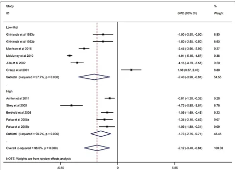
Results: Twelve RCTs with a total of 1776 participants were evaluated. Compared with placebo, statin treatment resulted in a reduction of circulating CoQ10 (SMD, − 2.12; 95% CI, − 3.40 to − 0.84; p = 0.001), which was not associ‑ ated with the duration of statin treatment (Exp, 1.00; 95% CI, 0.97 to 1.03; p = 0.994). Subgroup analysis demonstrated that both lipophilic statins (SMD, − 1.91; 95% CI, − 3.62 to 0.2; p = 0.017) and hydrophilic statins (SMD, − 2.36; 95% CI, − 4.30 to − 0.42; p = 0.028) decreased circulating CoQ10, and no obvious difference was observed between the two groups (SMD, − 0.20; 95% CI, − 0.208 to 0.618; p = 0.320). In addition, both low‑middle intensity statins (SMD, − 2.403; 95% CI, − 3.992 to − 0.813; p < 0.001) and high intensity statins (SMD, − 1.727; 95% CI, − 2.746 to − 0.709; p < 0.001) decreased circulating CoQ10. Meta‑regression showed that the effect of statin on decreasing circulating CoQ10 was not closely associated with the duration of statin treatment (Exp, 1.00; 95% CI, 0.97 to 1.03; p = 0.994).
Conclusions: Statin treatment decreased circulating CoQ10 but was not associated with the statin solution, intensity, or treatment time. The findings of this study provide a potential mechanism for statin‑associated muscle symptoms (SAMS) and suggest that CoQ10 supplementation may be a promising complementary approach for SAMS.
Keywords: Statin treatment, Circulating CoQ10, Meta‑analysis, Pharmaceutical metabolism
© The Author(s) 2018. This article is distributed under the terms of the Creative Commons Attribution 4.0 International License (http://creat iveco mmons .org/licen ses/by/4.0/), which permits unrestricted use, distribution, and reproduction in any medium, provided you give appropriate credit to the original author(s) and the source, provide a link to the Creative Commons license, and indicate if changes were made. The Creative Commons Public Domain Dedication waiver (http://creat iveco mmons .org/ publi cdoma in/zero/1.0/) applies to the data made available in this article, unless otherwise stated.
Open Access
*Correspondence: [email protected]; [email protected]
†Hua Qu and Yan‑yan Meng contributed equally to this article and are
co‑first authors
4 Xiyuan Hospital, China Academy of Chinese Medical Sciences, Beijing,
China
Introduction
Statins are widely used in the prevention and treatment
of coronary heart disease [1]. Numerous large-scale
studies have demonstrated that statins substantially reduce cardiovascular morbidity and mortality in both primary and secondary prevention, in both genders and in all age groups [2–4]. However, statin-associated muscle symptoms (SAMS), covering a broader range of muscle symptoms following statin treatment, are
an important reason for statin discontinuation [5]. A
previous study demonstrated that SAMS result in sig-nificantly high discontinuation rates of statin treat-ment (up to 75%) within 2 years of initiation [6], and in 65% of former statin users, the main reason for sta-tin non-adherence or disconsta-tinuation was the onset of
side effects, predominantly SAMS [7]. Non-adherence
or discontinuation of statin treatment contributes to adverse cardiovascular outcomes. A meta-analysis showed a 15% lower cardiovascular risk in patients who adhered to statin treatment compared with those with
low adherence [8]. Studying the possible mechanism
and therapeutic approach of SAMS could decrease the cardiovascular risk in patients who are intolerant
to statins due to SAMS [9]. The European
Atheroscle-rosis Society has proposed four strategies for treating SAMS, including re-challenge with alterative statin therapy, lower or intermittent statin therapy, non-sta-tin-based lipid-lowering therapy, and complementary therapies. Complementary therapies, such as coenzyme Q10 (CoQ10) supplementation, might be a
promis-ing method to manage SAMS [9]. The mechanism of
SAMS is currently unclear, but changes in circulating coenzyme Q10 concentration may be involved in the pathological process. Exploring the level of circulating CoQ10 following statin treatment may explain poten-tial mechanisms and suggest possible complementary approaches for SAMS.
CoQ10 is a naturally occurring, fat-soluble quinone located in the hydrophobic portions of cellular mem-branes [10], and it plays an important role in mitochon-drial energy metabolism and stabilization of muscle cell
membranes [11, 12]. A previous animal study
demon-strated that statin treatment could lead to the
reduc-tion of circulating CoQ10 [13]. However, the findings
concerning changes in circulating CoQ10 following statin therapy have been inconsistent in clinical studies. A previous meta-analysis [14] only included 6 clinical studies, and several RCTs, including statin treatment for a period of 24 weeks, have been published since, and these studies have provided new evidence. There-fore, the present meta-analysis of RCTs was designed to reassess the effect of statin treatment on circulating CoQ10.
Methods
This study was performed according to the guidelines of the 2009 Preferred Reporting Items for Systematic Reviews and Meta-Analysis statement (PRISM) (Addi-tional file 1: Table S1) [15].
Data source and search strategies
Two reviewers (Hua Qu and Hua Chai) searched Pub-Med, EMBASE, and the Cochrane Library with no lan-guage restrictions from inception to January 2018 to identify all existing literature. Mesh terms and free-text terms were used in each database with the following rele-vant keywords: “statin treatment” AND “coenzyme Q10” AND “randomized controlled trials.” A manual search was also performed to identify relevant references from the selected articles and published reviews. The studies were eligible if they met the following inclusion criteria: (1) randomized, controlled, parallel, or crossover trial, (2) the intervention group received statin and the compari-son group received placebo, or the intervention group received lipophilic statin and the comparison group received hydrophilic statin, and (3) the outcome regard-ing circulatregard-ing CoQ10 (plasma CoQ10 or serum CoQ10) was available.
Data extraction and assessment of study quality
Two reviewers (Hua Qu and Yan-yan Meng) extracted data independently. If a disagreement occurred, it was resolved by consulting with a third investigator (Da-zhuo Shi). We contacted the authors if the article was only published with an abstract, and the studies without origi-nal data were excluded. The following data were extracted from each individual eligible study: (1) the first author’s name and publication year, (2) intervention duration, (3) inclusion criteria, (4) participant number, (5) partici-pants’ age, (6) percentage of males, and (7) clinical out-comes. The methodological quality of eligible studies was determined according to the recommendation of the
Cochrane Handbook [16].
Statistical analysis
In this meta-analysis, continuous data were used to ana-lyze the standard mean difference (SMD) with a 95% confidence interval (CI) for the effect size. Heterogene-ity in the eligible studies was evaluated using the Chi-square test based on Cochran’s Q test and I2 statistic at
the p < 0.10 level of significance, and quantification of heterogeneity was calculated using the I2 metric, which
describes the percentage of total variation estimated to be due to heterogeneity rather than chance. When P for the heterogeneity was < 0.1 and I2≥ 50%, the inter-study
on the heterogeneity analysis. The fixed effect model was applied if I2 < 50%, and the random effect model was
chosen if I2≥ 50%. We performed subgroup analysis and
meta-regression to detect the potential sources of het-erogeneity in the condition of I2≥ 50%. Sensitivity
analy-sis was performed to assess the robustness of the pooled SMDs by eliminating one study at a time. The publication bias was evaluated by funnel plot, Egger regression, and
the Begg–Mazumdar correlation test. Statistical analysis was performed using Stata (version 12.0). There is no reg-istered protocol for the present meta-analysis.
Results
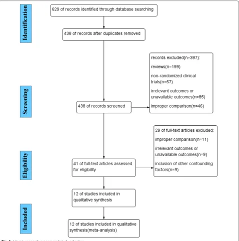
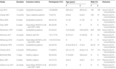
Description of included studies
were identified, and 191 articles were excluded as dupli-cated records. After the titles and abstracts of the arti-cles were screened, 397 artiarti-cles were excluded due to review format, improper study type, and/or improper comparisons. After the remaining 41 full-text articles were reviewed, 29 articles were excluded due to improper comparisons, irrelevant outcomes, and/or unavailable outcomes. Finally, 12 articles [17–28] with 1776 par-ticipants published in English from 1993 to 2018, with sample sizes ranging from 19 to 1103 participants and intervention durations ranging from 14 days to 26 weeks, were entered into our meta-analysis (Fig. 1, Table 1). Nine RCTs [17–21, 23–25], including 11 study arms (822 participants in the statin treatment group vs. 830 par-ticipants in the placebo group), evaluated the effect of statins (statins vs. placebo) on circulating CoQ10, and 4 RCTs [22, 25, 26, 28], including 7 study arms (94 partici-pants in a lipophilic statin group vs. 115 participartici-pants in a hydrophilic statin group), evaluated the effect of different
soluble statins (lipophilic statins vs. hydrophilic statins) on circulating CoQ10.
Quality assessment
“Low risk,” “high risk,” or “unclear risk” was categorized for all 12 included studies according to 7 risk biases presented in sequence generation, allocation sequence concealment, blinding of participants and personnel, blinding of outcome assessment, incomplete outcome data, selective outcome reporting, and other potential sources of bias (Additional file 2: Figure S1) [16]. No obvious attrition bias or reporting bias was observed. Additionally, the randomization and blinding in the included articles were considered adequate in the present study according to the Cochrane Handbook [16].
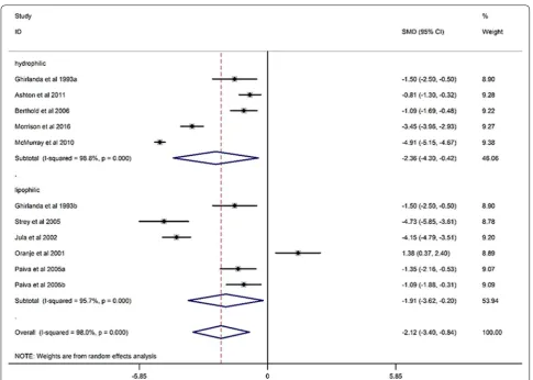
The effect of statin treatment on circulating CoQ10
When compared with placebo, statin treatment decreased
circulating CoQ10 (SMD, − 2.12; 95% CI, − 3.40 to
− 0.84; p=0.001) with a significant heterogeneity
Table 1 Basic characteristics of the participants
NYHA New York Heart Association, LVEF left ventricular ejection fraction, HIV human immunodeficiency virus, STEMI st segment elevation myocardial infarction, CAD coronary artery disease, CK creatine kinase, CoQ10 coenzyme Q 10, T treatment group, C control group, N not mentioned
Study Duration Inclusion criteria Participants (T/C) Age (years) Male (%) Outcome
T C T C
Jula 2015 12 weeks Dyslipidemia patients 120 (60/60) 48.4 ± 6.2 48.0 ± 6.2 100 100 Serum CoQ 10 concentrations Oranje 2001 3 months Type 2 diabetes patients 19 (9/10) 64 ± 8 63 ± 8 100 70 Plasma CoQ 10
concentrations Päivä 2005 8 weeks Dyslipidemia patients 48 (32/16) 31–69 31–69 N N Plasma CoQ 10
concentrations Strey 2005 6 weeks Heart failure (NYHA II or III)
with LVEF < 40% 48 (24/24) N N N N Plasma CoQ 10 concentrations Mortensen 1997 6 weeks Dyslipidemia patients 45 (23/22) 52.9 (32,69) 54.8 (42,67) 60.9 40.9 Serum CoQ 10
concentrations Morrison 2017 24 weeks Patients with HIV 147 (71/75) 45 (41,51) 47 (39,53) 81 76 Plasma CoQ 10
concentrations McMurray 2010 3 months Heart failure (NYHA II or III)
with LVEF < 40% 1103 (551/552) N N N N Serum CoQ 10 concentrations Ghirlanda 1993 3 months Dyslipidemia patients 30 (20/10) 47 ± 8; 49 ± 10 47 ± 8 60; 50 70 Plasma CoQ 10
concentrations Chitose 2014 6 months STEMI patients 75 (38/37) 64.1 ± 11.8 65.8 ± 12.4 73.7 75.7 Plasma CoQ 10
concentrations Berthold 2006 14 days Healthy subjects 48 (24/24) 31.9 ± 8.8 28.6 ± 6.6 N N Plasma CoQ 10
concentrations Barry 2001 4 weeks Healthy subjects 24 (12/12) 26 ± 5 26 ± 5 41.7 41.7 Plasma CoQ 10
concentrations Ashton et al. 2011 26 weeks Heart failure (NYHA II or III)
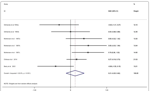
(I2= 98%, p < 0.001) (Fig. 2). The subgroup analysis
dem-onstrated that statins could decrease circulating CoQ10 with both lipophilic statins (SMD, − 1.91; 95% CI, − 3.62 to 0.2; p= 0.017) and hydrophilic statins (SMD, − 2.36; 95% CI, − 4.30 to − 0.42; p= 0.028) (Fig. 3), and no obvi-ous difference was observed between hydrophilic statins and lipophilic statins in the efficacy of decreasing circu-lating CoQ10 (SMD, − 0.20; 95% CI, − 0.208 to 0.618; p= 0.320) (Fig. 4). In addition, both low-middle inten-sity statins (SMD, − 2.403; 95% CI, − 3.992 to − 0.813; p < 0.001) and high intensity statins (SMD, − 1.727; 95% CI, − 2.746 to − 0.709; p < 0.001) could decrease circulat-ing CoQ10 (Fig. 5). The meta-regression showed that the effect of statins on decreasing circulating CoQ10 was not closely associated with the statin treatment time (Exp, 1.00; 95% CI, 0.97 to 1.03; p= 0.994) (Fig. 6).
Sensitivity analysis
To ensure the reliability of the present meta-analysis, we performed sensitivity analysis to evaluate the robustness of the pooled SMDs by eliminating each study one at a time sequentially, which indicated that the heterogeneity
among the studies did not significantly change regarding the effect of statins on circulating CoQ10. Thus, no one study showed a significant impact on the results of the present meta-analysis.
Publication bias
Three methods, including funnel plot, Egger regres-sion test and the Begg–Mazumdar correlation test, were used to evaluate publication bias regarding the effect of statin treatment on circulating CoQ10, which suggested potential publication bias (Begg–Mazumdar correlation test, Kendall’s score = 0, continuity corrected z= 0.08, continuity corrected p= 1; Egger regression test, Coef., 9.81; 95% CI, 2.52 to 0.17.11; p= 0.014) (Additional file 3: Figure S2). Using a “trim and fill” correction, 5 poten-tially missing studies were imputed leading to a cor-rected effect size (SMD, − 4.01, 95% CI, − 5.38 to − 2.63, p < 0.0001), which was consistent with the previous effect size.
NOTE: Weights are from random effects analysis Overall (I-squared = 98.0%, p = 0.000) Ghirlanda et al 1993a
Ghirlanda et al 1993b
McMurray et al 2010 Jula et al 2002
Paiva et al 2005b Paiva et al 2005a Berthold et al 2006 Ashton et al 2011 ID
Morrison et al 2016 Study
Strey et al 2005
Oranje et al 2001
-2.12 (-3.40, -0.84) -1.50 (-2.50, -0.50) -1.50 (-2.50, -0.50)
-4.91 (-5.15, -4.67) -4.15 (-4.79, -3.51)
-1.09 (-1.88, -0.31) -1.35 (-2.16, -0.53) -1.09 (-1.69, -0.48) -0.81 (-1.30, -0.32) SMD (95% CI)
-3.45 (-3.96, -2.93) -4.73 (-5.85, -3.61)
1.38 (0.37, 2.40)
100.00 8.90 8.90 9.38 9.20 9.09 9.07 9.22 9.28 Weight 9.27 % 8.78 8.89
-2.12 (-3.40, -0.84) -1.50 (-2.50, -0.50) -1.50 (-2.50, -0.50)
-4.91 (-5.15, -4.67) -4.15 (-4.79, -3.51)
-1.09 (-1.88, -0.31) -1.35 (-2.16, -0.53) -1.09 (-1.69, -0.48) -0.81 (-1.30, -0.32) SMD (95% CI)
-3.45 (-3.96, -2.93) -4.73 (-5.85, -3.61)
1.38 (0.37, 2.40)
100.00 8.90 8.90 9.38 9.20 9.09 9.07 9.22 9.28 Weight 9.27 % 8.78 8.89 0
-5.85 0 5.85
Discussion
In the previous meta-analysis performed by Banach et al. [14], only 6 studies were included. Additionally, subgroup analysis based on the intensity of statins and comparisons between lipophilic statins and hydrophilic statins were not performed due to the limited number of enrolled studies and their small sample sizes. In the present meta-analysis, the results from 12 RCTs validated a reduction of circulating CoQ10 following statin treatment. We also showed, for the first time, that both lipophilic statins and hydrophilic statins could decrease circulating CoQ10, and there was no significant difference between the two groups. In addition, a significant effect was observed for both low-middle intensity statins and high intensity statins in terms of decreasing the level of circulating CoQ10, and the effect of statins on circulating CoQ10 was not closely associated with the statin treatment time (from 14 days to 26 weeks).
regenerates active forms of the antioxidants ascorbic acid and tocopherol, which play important roles in maintain-ing mitochondrial energy metabolism and stabilizmaintain-ing muscle cell membranes [30–34]. CoQ10 deficiency pre-sents as increased oxidative stress, increased inflamma-tory responses, and an imbalanced serotonergic system,
which may contribute to SAMS [35]. The present
meta-analysis validated the effect of statin in decreasing cir-culating CoQ10, which will be helpful for studying the mechanism of SAMS and may provide a complementary approach for SAMS treatment.
Previous studies have suggested that hydrophilic statins may have a lower rate of SAMS compared with lipophilic statins and that clinicians should consider switching to a hydrophilic statin to manage SAMS [36, 37]. How-ever, in the present meta-analysis, no obvious difference was observed between lipophilic statins and hydrophilic statins in decreasing circulating CoQ10. Shi et al. also found that patients with SAMS who are intolerant to some hydrophilic statins may be successfully managed with simvastatin (lipophilic statin) monotherapy [38].
Therefore, whether hydrophilic statins have a lower rate of SAMS compared with lipophilic statins deserves fur-ther study. In addition, a lower dose of statin is always recommended to manage patients who tolerate statins
because of SAMS [9]. However, significant effects of
statins were observed in both low-moderate intensity statins and high intensity statins by decreasing circulat-ing CoQ10 in the present study. Therefore, perform-ing large-scale trials is necessary to compare the rate of SAMS between low-moderate intensity statins and high intensity statins. Several limitations in the present study should be noted. First, the eligible studies were hetero-geneous because of certain factors, such as population characteristics, study design, and duration of statin treat-ment. Thus, we performed subgroup analysis, sensitiv-ity analysis, and meta-regression to minimize the effect of heterogeneity on estimated effect size and assure the reliability of the outcomes. Second, there was potential publication bias for the effect of statin treatment on cir-culating CoQ10, so we used the trim and fill method to assure the robustness of the pooled results.
NOTE: Weights are from random effects analysis Overall (I-squared = 52.0%, p = 0.051) Mortensen et al 1997b
Chitose et al 2014 Study
Mortensen et al 1997c Ghirlanda et al 1993a
Barry et al 2001 ID
Mortensen et al 1997a Ghirlanda et al 1993b
0.21 (-0.20, 0.62) 0.80 (-0.03, 1.64)
0.27 (-0.18, 0.73) 0.79 (0.06, 1.52) -0.52 (-1.41, 0.37)
-0.68 (-1.50, 0.15) SMD (95% CI)
0.60 (-0.22, 1.42) 0.00 (-0.88, 0.88)
100.00 13.04
21.03 %
14.95 12.10
13.21 Weight
13.30 12.36
0.21 (-0.20, 0.62) 0.80 (-0.03, 1.64)
0.27 (-0.18, 0.73) 0.79 (0.06, 1.52) -0.52 (-1.41, 0.37)
-0.68 (-1.50, 0.15) SMD (95% CI)
0.60 (-0.22, 1.42) 0.00 (-0.88, 0.88)
100.00 13.04
21.03 %
14.95 12.10
13.21 Weight
13.30 12.36
0
-1.64 0 1.64
In conclusion, statin treatment decreases circulat-ing CoQ10, regardless of statin solution, intensity, or treatment time. The findings provide a potential mech-anism for SAMS and suggest that CoQ10 supplementa-tion might be a promising complementary approach for SAMS.
Additional files
Additional file 1: Table S1. PRISMA (Preferred Reporting Items for Sys‑ tematic Reviews and Meta‑Analyses) checklist [15].
Additional file 2: Figure S1. Risk of bias.
Additional file 3: Figure S2. Publication bias.
Authors’ contributions
D‑zS and Z‑yG designed the review and provided methodological perspec‑ tives. HQ and Y‑yM developed the search strategy, performed the literature search, and wrote the manuscript. HC, FL, and J‑yZ performed the study Fig. 5 Forest plot for circulating CoQ10, low‑mid intensity statin vs high intensity statin (subgroup analysis). CoQ10 coenzyme Q10, SMD standard mean difference, CI confidence interval, ID identity number
selection, data extraction, and data analyses. All authors read and approved the final manuscript.
Author details
1 China Academy of Chinese Medical Sciences, Beijing, China. 2 China Heart
Institute of Chinese Medicine, China Academy of Chinese Medical Sciences, Beijing, China. 3 Beijing University of Traditional Chinese Medicine, Beijing,
China. 4 Xiyuan Hospital, China Academy of Chinese Medical Sciences, Beijing,
China.
Competing interests
The authors declare that they have no competing interests.
Ethics approval and consent to participate
This article does not contain any studies with human participants or animals performed by any of the authors. Our study group worked under the over‑ sight of the institutional ethics committee and followed all ethical recommen‑ dations. Before submitting our article, all authors agreed to the publication of our work.
Funding
This work was supported by the National Natural Science Foundation of China (No. 81030063), the National Sci‑Tech Support Plan of China (No. 2013BAI02B01), the Major National Research and Development Project of China (No. 2018ZX09201009‑002‑006), the National Natural Science Founda‑ tion of China (No. 81303150), and the project of China Academy of Chinese Medical Sciences (No. ZZ11‑066).
Publisher’s Note
Springer Nature remains neutral with regard to jurisdictional claims in pub‑ lished maps and institutional affiliations.
Received: 21 July 2018 Accepted: 29 October 2018
References
1. Reiner Z, Catapano AL, De Backer G, Graham I, Taskinen M‑R, Wiklund O, et al. ESC/EAS Guidelines for the management of dyslipidaemias: the Task Force for the management of dyslipidaemias of the European Society of Cardiology (ESC) and the European Atherosclerosis Society (EAS). Eur Heart J. 2011;32:1769–818. https ://doi.org/10.1093/eurhe artj/ehr15 8. 2. Thavendiranathan P. Primary prevention of cardiovascular diseases with
statin therapy. Arch Intern Med. 2006;166:2307. https ://doi.org/10.1001/ archi nte.166.21.2307.
3. Mills EJ, Wu P, Chong G, Ghement I, Singh S, Akl EA, et al. Efficacy and safety of statin treatment for cardiovascular disease: a network meta‑anal‑ ysis of 170 255 patients from 76 randomized trials. QJM. 2011;104:109–24.
https ://doi.org/10.1093/qjmed /hcq16 5.
4. Gutierrez J, Ramirez G, Rundek T, Sacco RL. Statin therapy in the preven‑ tion of recurrent cardiovascular events. Arch Intern Med. 2012. https :// doi.org/10.1001/archi ntern med.2012.2145.
5. Law M, Rudnicka AR. Statin safety: a systematic review. Am J Cardiol. 2006;97:S52–60.
6. Chodick G, Shalev V, Gerber Y, Heymann AD, Silber H, Simah V, et al. Long‑term persistence with statin treatment in a not‑for‑profit health maintenance organization: a population‑based retrospective cohort study in Israel. Clin Ther. 2008;30:2167–79.
7. Cohen JD, Brinton EA, Ito MK, Jacobson TA. Understanding Statin Use in America and Gaps in Patient Education (USAGE): an internet‑ based survey of 10,138 current and former statin users. J Clin Lipidol. 2012;6:208–15.
8. Chowdhury R, Khan H, Heydon E, Shroufi A, Fahimi S, Moore C, et al. Adherence to cardiovascular therapy: a meta‑analysis of prevalence and clinical consequences. Eur Heart J. 2013;34:2940–8. https ://doi. org/10.1093/eurhe artj/eht29 5.
9. Stroes ES, Thompson PD, Corsini A, Vladutiu GD, Raal FJ, Ray KK, et al. Statin‑associated muscle symptoms: impact on statin
therapy—European Atherosclerosis Society Consensus Panel State‑ ment on Assessment, Aetiology and Management. Eur Heart J. 2015;36:1012–22.
10. Hernández‑Camacho JD, Bernier M, López‑Lluch G, Navas P. Coenzyme Q10 supplementation in aging and disease. Front Physiol. 2018. https ://doi.org/10.3389/fphys .2018.00044 /full.
11. Eriksson EK, Agmo Hernández V, Edwards K. Effect of ubiquinone‑10 on the stability of biomimetic membranes of relevance for the inner mitochondrial membrane. Biochim Biophys Acta Biomembr. 2018;1860:1205–15.
12. Xu Z, Huo J, Ding X, Yang M, Li L, Dai J, et al. Coenzyme Q10 improves lipid metabolism and ameliorates obesity by regulating CaMKII‑medi‑ ated PDE4 inhibition. Sci Rep. 2017;7:8253. http://www.natur e.com/ artic les/s4159 8‑017‑08899 ‑7.
13. Wang LW, Jabbour A, Hayward CS, Furlong TJ, Girgis L, Macdonald PS, et al. Potential role of coenzyme Q10 in facilitating recovery from statin‑induced rhabdomyolysis. Intern Med J. 2015;45:451–3. https :// doi.org/10.1111/imj.12712 .
14. Banach M, Serban C, Ursoniu S, Rysz J, Muntner P, Toth PP, et al. Statin therapy and plasma coenzyme Q10 concentrations—a systematic review and meta‑analysis of placebo‑controlled trials. Pharmacol Res. 2015;99:329–36.
15. Moher D, Liberati A, Tetzlaff J, Altman DG. Preferred reporting items for systematic reviews and meta‑analyses: the PRISMA statement. PLoS Med. 2009;6:e1000097. https ://doi.org/10.1371/journ al.pmed.10000 97. 16. Higgins JPT, Altman DG, Gøtzsche PC, Jüni P, Moher D, Oxman AD,
et al. The Cochrane Collaboration’s tool for assessing risk of bias in randomised trials. BMJ. 2011;343:d5928.
17. Jula A, Marniemi J, Huupponen R, Virtanen A, Rastas M, Rönnemaa T. Effects of diet and simvastatin on serum lipids, insulin, and antioxi‑ dants in hypercholesterolemic men: a randomized controlled trial. JAMA. 2002;287:598–605. http://www.ebsco host.com.
18. Oranje WA, Sels JP, Rondas‑Colbers GJ, Lemmens PJ, Wolffenbuttel BH. Effect of atorvastatin on LDL oxidation and antioxidants in normocho‑ lesterolemic type 2 diabetic patients. Clin Chim Acta. 2001;311:91–4. 19. Ashton E, Windebank E, Skiba M, Reid C, Schneider H, Rosenfeldt F,
et al. Why did high‑dose rosuvastatin not improve cardiac remodeling in chronic heart failure? Mechanistic insights from the UNIVERSE study. Int J Cardiol. 2011;146:404–7. https ://doi.org/10.1016/j.ijcar d.2009.12.028.
20. Strey CH, Young JM, Molyneux SL, George PM, Florkowski CM, Scott RS, et al. Endothelium‑ameliorating effects of statin therapy and coenzyme Q10 reductions in chronic heart failure. Atherosclerosis. 2005;179:201–6.
21. Päivä H, Thelen KM, Van Coster R, Smet J, De Paepe B, Mattila KM, et al. High‑dose statins and skeletal muscle metabolism in humans: a rand‑ omized, controlled trial. Clin Pharmacol Ther. 2005;78:60–8.
22. Mortensen SA, Leth A, Agner E, Ronde M. Dose‑related decrease of serum coenzyme Q10 during treatment with HMG‑CoA reductase inhibitors. Mol Aspects Med. 1997;18:137–44.
23. Morrison JT, Longenecker CT, Mittelsteadt A, Jiang Y, Debanne SM, McComsey GA. Effect of rosuvastatin on plasma coenzyme Q10 in HIV‑ infected individuals on antiretroviral therapy. HIV Clin Trials. 2016;17:140– 6. http://www.embas e.com/searc h/resul ts?subac tion=viewr ecord &from=expor t&id=L6108 35401 .
24. McMurray JJV, Dunselman P, Wedel H, Cleland JGF, Lindberg M, Hjalmar‑ son K, et al. Coenzyme Q10, rosuvastatin, and clinical outcomes in heart failure: a pre‑specified substudy of CORONA (Controlled Rosuvastatin Multinational Study in Heart Failure). J Am Coll Cardiol. 2010;56:1196–204.
https ://doi.org/10.1016/j.jacc.2010.02.075.
25. Ghirlanda G, Oradei A, Manto A, Lippa S, Uccioli L. Evidence of Plasma CoQ10—lowering effect by HMG‑CoA reductase inhibitors: a double blind, placebo‑controlled study. Clin Pharmocol. 1993;33:226–9. 26. Chitose T, Sugiyama S, Sakamoto K, Shimomura H, Yamashita T, Hokamaki
J, et al. Effect of a hydrophilic and a hydrophobic statin on cardiac salvage after ST‑elevated acute myocardial infarction—a pilot study. Atherosclerosis. 2014;237:251–8. https ://doi.org/10.1016/j.ather oscle rosis .2014.08.053.
•fast, convenient online submission •
thorough peer review by experienced researchers in your field • rapid publication on acceptance
• support for research data, including large and complex data types •
gold Open Access which fosters wider collaboration and increased citations maximum visibility for your research: over 100M website views per year •
At BMC, research is always in progress.
Learn more biomedcentral.com/submissions
Ready to submit your research? Choose BMC and benefit from:
28. Bleske BE, Willis RA, Anthony M, Casselberry N, Datwani M, Uhley VE, et al. The effect of pravastatin and atorvastatin on coenzyme Q10. Am Heart J. 2001;142:2.
29. Caparrós‑Martín JA, Lareu RR, Ramsay JP, Peplies J, Reen FJ, Headlam HA, et al. Statin therapy causes gut dysbiosis in mice through a PXR‑depend‑ ent mechanism. Microbiome. 2017;5:95. https ://doi.org/10.1186/s4016 8‑017‑0312‑4.
30. Paunović MG, Matić MM, Ognjanović BI, Saičić ZS. Antioxidative and haema‑ toprotective activity of coenzyme Q10 and vitamin E against cadmium‑ induced oxidative stress in Wistar rats. Toxicol Ind Health. 2017;33:746–56.
https ://doi.org/10.1177/07482 33717 72548 0.
31. Alkholy UM, Abdalmonem N, Zaki A, Elkoumi MA, Hashim MIA, Basset MAA, et al. The antioxidant status of coenzyme Q10 and vitamin E in children with type 1 diabetes. J Pediatr (Rio J). 2018. https ://doi.org/10.1016/j. jped.2017.12.005.
32. James AM, Smith RAJ, Murphy MP. Antioxidant and prooxidant properties of mitochondrial coenzyme Q. Arch Biochem Biophys. 2004;423:47–56. 33. Yubero D, Adin A, Montero R, Jou C, Jiménez‑Mallebrera C, García‑Cazorla
A, et al. A statistical algorithm showing coenzyme Q10 and citrate synthase
as biomarkers for mitochondrial respiratory chain enzyme activities. Sci Rep. 2016;6:15. http://www.natur e.com/artic les/s4159 8‑016‑0008‑1.
34. Ong S‑B, Kalkhoran SB, Hernández‑Reséndiz S, Samangouei P, Ong S‑G, Hausenloy DJ. Mitochondrial‑shaping proteins in cardiac health and dis‑ ease—the long and the short of it! Cardiovasc Drugs Ther. 2017;31:87–107.
https ://doi.org/10.1007/s1055 7‑016‑6710‑1.
35. Alcocer‑Gómez E, Sánchez‑Alcázar JA, Cordero MD. Coenzyme Q10 regu‑ lates serotonin levels and depressive symptoms in fibromyalgia patients. J Clin Psychopharmacol. 2014;34:277–8. https ://insig hts.ovid.com/cross ref?an=00004 714‑20140 4000‑00023 .
36. Mampuya WM, Frid D, Rocco M, Huang J, Brennan DM, Hazen SL, et al. Treatment strategies in patients with statin intolerance: the Cleveland Clinic experience. Am Heart J. 2013;166:597–603.
37. Rallidis LS, Fountoulaki K, Anastasiou‑Nana M. Managing the underesti‑ mated risk of statin‑associated myopathy. Int J Cardiol. 2012;159:169–76. 38. Qu H, Guo M, Kou N, Wu H‑T, Zhang Y, Gao Z‑Y, et al. Simvastatin mono‑