Teresa Sicińska-Werner
1, Marek Mędraś
2, 3Hormonal Status, Tobacco Addiction,
and Coronary Arteriosclerosis
in 40- to 60-Year-Old Non-Diabetic Men*
Stan hormonalny i nałóg palenia tytoniu
a występowanie miażdżycy naczyń wieńcowych
u mężczyzn w wieku 40–60 lat niechorujących na cukrzycę
1 Provincial Endocrinology Clinic, Provincial Medical Centre, Opole, Poland
2 Department and Clinic Endocrinology, Diabetology and Isotope Therapy, Wroclaw Medical University, Poland 3 Sports Medicine and Nutrition Department of Biostructure Chair University School of Physical Education
in Wroclaw, Poland
Abstract
Background. Tobacco addiction undoubtedly plays a major role in coronary atherosclerosis formation in men. An age-related decrease in androgenic hormones is probably also involved. Since the coexistence of diabetes dra-matically increases cardiovascular risk and mortality from these causes, it is important to study this issue in men without diabetes in assessing the impact of hormonal changes and tobacco consumption on atherogenesis.
Objectives. Assessment of impact of hormonal changes and tobacco consumption on artheriosclerosis in the coro-nary arteries in non-diabetic men aged 40–60.
Material and Methods. One hundred two men, including 62 smokers, aged 40–60 years without diabetes in whom angi-ography due to cardiac indications was performed were involved in the study. From this group, subgroups of patients, 42 with coronary atherosclerosis and 20 without changes in coronary angiography, were established. A control group of 40 healthy men with a negative medical history and normal physical examination and exercise test included 20 smokers and 20 nonsmokers. The concentrations of total and free testosterone, calculated free and bioavailable testosterone, SHBG, DHEAS, E2, IGF-1, GH, and the free androgen index (FAI) were studied in the men. Hormonal status and tobacco
addic-tion parameters were compared with atherogenesis development in the coronary arteries.
Results. The presence of atherosclerosis in the coronary arteries correlated with increased tobacco consumption, reduced concentration of free testosterone (assessed by an RIA test), and increased SHBG compared with the men without atherosclerosis. Smokers with atherosclerosis of the coronary arteries also showed lower levels of DHEAS than the healthy smokers and higher LH than the healthy nonsmokers. There were no differences in the mean concentrations of total testosterone, FAI, free and calculated bioavailable testosterone, estradiol, growth hormone, and IGF-1 in the studied groups. No relationship was observed between hormone levels tested with the parameters characterizing tobacco addiction and the degree of coronary occlusion assessed with the number of significantly stenosed vessels and the Gensini score.
Conclusions. Coronary atherosclerosis in men is associated with smoking and lower concentrations of free testos-terone and DHEAS. Lack of correlation of the other hormone levels with the parameters of tobacco addiction and the degree of vascular occlusion confirmed that the etiology of coronary heart disease is multifactorial (Adv Clin Exp Med 2010, 19, 2, 211–218).
Key words: coronary heart disease, hormones, tobacco smoking.
Streszczenie
Wprowadzenie. W powstawaniu miażdżycy naczyń wieńcowych u mężczyzn niewątpliwie znaczącą rolę odgrywa nałóg palenia tytoniu oraz prawdopodobnie powstające z wiekiem ograniczenie sekrecji hormonów androgennych.
Adv Clin Exp Med 2010, 19, 2, 211–218 ISSN 1230-025X
ORIGINAL PAPERS
© Copyright by Wroclaw Medical University
One of the most common risk factors for coronary heart disease, especially among men, is tobacco smoking. The results of the POLKARD 2003–2005 nationwide health survey indicated a fairly high level of smoking in our country. The percentage of male smokers was twice that of women and ranged 34–48%, depending on the region [1]. It is noticeable that age-related morbid-ity and mortalmorbid-ity of ischemic heart disease is 2.5 to 4.5 times higher in men than in women, but after the menopause these differences are reduced [2]. The increased male predisposition to atheroscle-rosis is associated with the more widespread habit of smoking cigarettes, but the role of androgenic and estrogenic effects is also stressed in the process of atherogenesis [3]. Recent years have brought much information about the coexistence of dis-eases of the cardiovascular system in diabetes. Therefore, one can now treat diabetes equally with coronary heart disease [4]. Considering the multi-factorial etiology of the atherosclerotic process, it is important to analyze the occurrence of coronary heart disease in combination with hormonal status and smoking habit among middle-aged men with-out diabetes.
The goal of this study was to evaluate the influ-ence of hormonal status and tobacco consumption among men aged 40–60 years without diabetes on the presence of coronary atherosclerosis.
Material and Methods
The study involved 102 men aged 40–60 years. In 62 patients (group I), due to a reasonable sus-picion of coronary heart disease (CHD), angiog-raphy was performed and they all smoked ciga-rettes (currently or up to 3 years before). In this group of men, a subgroup (IA) of 42 men (mean age: 51.3 ± 4.5 years) with atheromatous chang-es in epicardial arterichang-es (more than 30% steno-sis) and a subgroup (IB) of 20 men (mean age: 49.8 ± 4.9 years) in whom angiography showed no changes were identified. The remaining 40 men constituted the control group (II) con-sisting of healthy persons of whom 20 cigarette smokers (mean age: 46.1 ± 4.3 years) formed sub-group IIA and 20 non-smokers (mean age: 47.2 ± ± 4.41 years) subgroup IIB.
In assessing the progress of coronary athero-sclerosis, the authors regarded significant stenosis as that involving an at least 50% luminal narrow-ing of the main artery [5]. Dependnarrow-ing on the num-ber of significantly stenosed epicardial arteries, the authors divided the patients into a subgroup with non-significant stenosis (30–49%) and sub-groups with one-, two-, or three-vessel coronary artery disease. To assess the severity of changes in the coronary vessels, the authors used the Gensini score, which assigns a severity score to a stenosed vessel evaluated as a percentage [6].
The control group performed an exercise test with a Burdick T600 treadmill and Megacart
com-Ponieważ współistnienie cukrzycy dramatycznie zwiększa ryzyko sercowo-naczyniowe, a także umieralność z tych przyczyn, w ocenie wpływu zmian hormonalnych i nałogu palenia tytoniu na aterogenezę, badania tej problematyki u mężczyzn niechorujących na cukrzycę są ważne.
Cel pracy. Ocena wpływu stanu hormonalnego oraz nałogu palenia tytoniu u mężczyzn w wieku 40–60 lat niecho-rujących na cukrzycę na występowanie miażdżycy tętnic wieńcowych.
Materiał i metody. Badano 102 mężczyzn w wieku 40–60 lat bez cukrzycy, w tym 62 osoby palące tytoń, u których wykonano koronarografię ze wskazań kardiologicznych. Z tej grupy chorych wyodrębniono podgrupę 42 osób z miażdżycą naczyń wieńcowych oraz 20 osób bez zmian w koronarografii. Grupę kontrolną osób zdrowych z ujem-nym wywiadem chorobowym, bez odchyleń w badaniu fizykalujem-nym i teście wysiłkowym stanowiło 40 mężczyzn: 20 palaczy i 20 niepalących. U mężczyzn badano stężenia testosteronu całkowitego i wolnego, testosteron wolny i biodostępny kalkulowany, SHBG, DHEAS, E2, IGF-1, GH oraz wskaźnik wolnych androgenów FAI. Analizowano
powiązania stanu hormonalnego i parametrów nałogu palenia tytoniu ze stanem aterogenezy w naczyniach wień-cowych.
Wyniki. Obecność miażdżycy w naczyniach wieńcowych korelowała z nasileniem nałogu palenia tytoniu oraz zmniejszeniem stężenia wolnego testosteronu (oznaczonego metodą RIA) i podwyższeniem SHBG w porównaniu z grupą mężczyzn bez miażdżycy. Osoby z miażdżycą tętnic wieńcowych (palące papierosy) wykazywały także mniejsze stężenie DHEAS w porównaniu do osób zdrowych palących i większe LH w odniesieniu do osób zdrowych niepalących. W badanych grupach mężczyzn nie było różnic w średnich stężeniach testosteronu całkowitego, FAI, testosteronu wolnego i biodostępnego kalkulowanego, estradiolu, hormonu wzrostu oraz IGF-1. Nie obserwowano związku między stężeniem badanych hormonów z parametrami charakteryzującymi nałóg palenia i stopniem oklu-zji naczyń wieńcowych ocenianym ilością istotnie zwężonych naczyń oraz skalą Gensiniego.
Wnioski. Miażdżyca naczyń wieńcowych u mężczyzn jest związana z paleniem tytoniu, mniejszym stężeniem wol-nego testosteronu i DHEAS. Brak korelacji innych hormonów z parametrami nałogu palenia i stopniem okluzji naczyń potwierdza wieloczynnikowy charakter choroby niedokrwiennej serca (Adv Clin Exp Med 2010, 19, 2, 211–218).
puter (Siemens) according to the Bruce protocol. The interview on smoking habit involved the num-ber of cigarettes smoked, the age at which smok-ing began, and the number of years the person has smoked. The tobacco index (ti) was calculated for smokers, i.e. ti = (number of cigarettes the person has smoked per day/20) × (the number of years the person has smoked).
No subject had diabetes mellitus. The con-centrations of total (T) and free (fT) testosterone, dehydroepiandrosterone sulfate (DHEAS),
estradi-ol (E2), and insulin-like growth factor-1 (IGF-1) in
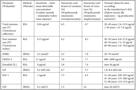
blood serum were assessed by radioimmunoassay. Sex hormone binding globulin (SHBG), luteiniz-ing hormone (LH), and growth hormone (GH) in serum were measured by immunoradiometric assay. Measurements of the selected hormone con-centrations were made using commercial reagents kits (Table 1). Based on the concentrations of the hormones in serum, the free androgen index (FAI), free testosterone, and bioavailable testoster-one were calculated according to the formulas:
– FAI = total testosterone [nmol/l] ×
100/con-centration of SHBG [nmol/l] [5],
– calculated free testosterone [ng/ml] and calcu-lated bioavailable testosterone, calcucalcu-lated with
a calculator, using the concentration of SHBG [nmol/l] and total serum testosterone [ng/ml] assuming albuminemia at 4.3 g/dl.
As the basis for this calculation, a free and bioavailable testosterone calculator was used which is described in the publication by Vermuelen, Verdonck, and Kaufman, available on the Internet [7].
Approval for the study was obtained from the Bioethics Committee at the Wroclaw Medical University (No. KB-90/2009). Persons qualified for the study gave their written consent to participate.
Statistical analysis was performed using
Student’s t test (when the condition of
homogene-ity of variance was not met, the t test was used with
a separate assessment of variance, the so-called Cochran-Cox test), the Mann-Whitney test, and the Kruskal-Wallis median test. The level of
statis-tical significance was set at p < 0.05.
Results
The men in group IA (coronary atherosclero-sis, tobacco smokers) showed significantly lower levels of free testosterone (measured by RIA) and
Table 1. Characteristics of the methods used to determine the hormonal parameters
Tabela 1. Charakterystyka metod zastosowanych w celu oznaczenia parametrów hormonalnych
Parameter
(Wskaźnik) Method (Metoda) Sensitivity – mini-mum detectable concentration (Czułość metody – minimalne wykry-wane stężenie)
Intraseries coef-ficient of variation (%)
(Współczynnik zmienności wewnątrzseryjnej)
Interseries coef-ficient of varia-tion (%) (Współczynnik zmienności międzyseryjnej)
Normal values for men – according
to the manufacturer’s kit) (Zakres normy dla mężczyzn – wg producenta zestawów)
Total testoster-one (T) (Testosteron całkowity)
RIA 0.04 ng/ml 6.5 6.7 20–49 years: 2.6–15.9 ng/ml
≥ 50 years: 1.8–7.6 ng/ml
Free testoster-one (fT) (Testosteron wolny)
RIA 0.15 pg/ml 6.2 8.5 20–39 years: 8.8–27.0 pg/ml
40–59 years: 7.2–23 pg/ml 60–80 years: 5.6–19.0 pg/ml
SHBG IRMA 0.2 nmol/l 2.2 7.8 20–70 nmol/l
DHEA-S RIA 11 ng/ml 3.8 7.7 800–5600 ng/ml
Estradiol (E2) RIA 8 pg/ml 5.8 7.4 max 44 pg/ml
LH IRMA 0.2 mIU/ml 6.7 3.7 1.8–8.4 mIU/ml
IGF-1 RIA 1 ng/ml 7.5 6.3 31–40 years: 109–293 ng/ml
41–50 years: 135–280 ng/ml 51–60 years: 114–314 ng/ml
GH IRMA 0.1 mIU/l 1.5 14 max 20 mIU/l
RIA – radioimmunoassay, IRMA – immunoradiometric assay.
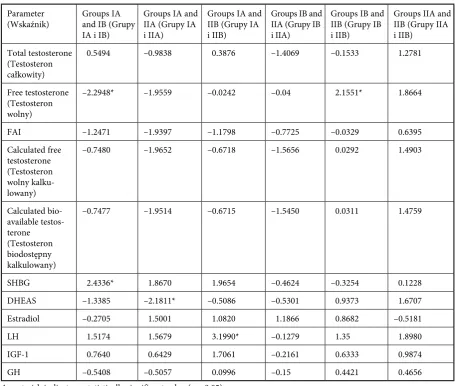
significantly higher levels of SHBG than those in group IB (smokers without changes in coronary arteries), 10.62 ± 3.5 vs. 12.77 ± 3.3 pg/ml and 44.57 ± 20.3 vs. 34.56 ± 11.5 nmol/l, respectively. Group IA also had lower levels of DHEAS (1580.29 ± ± 949.0 vs. 2163.7 ± 1079.0 ng/ml) than the healthy smokers in group IIA. The subjects in group IA also had higher LH levels than the healthy non-smokers (7.01 ± 6.1 vs. 3.8 ± 1.4 mIU/ml).
Comparing group IB (without coronary ath-erosclerosis, tobacco smokers) and the healthy smokers group (IIA), there was no significant dif-ference in any of the evaluated hormonal param-eters. Subjects of group IB had significantly higher levels of free testosterone than the healthy non-smokers of group IIB (12.77 ± 3.3 vs. 10.64 ± 2.7 pg/ml).
The comparison of all hormonal parameters in the healthy subjects from the control groups of the smokers IIA and non-smokers IIB showed no statistically significant differences. Evaluation of all four groups of men showed no differences in
the mean concentrations of total testosterone, free androgen index (FAI), calculated free testoster-one, calculated bioavailable testostertestoster-one, estradiol, growth hormone, and IGF-1 (Tables 2 and 3).
In subgroup IA (men with CHD), the relation-ship between the hormonal parameters and the progression of atherosclerosis was assessed and there was no significant link between the studied hormones and the intensity of atherogenesis, both in terms of number of considerably stenosed ves-sels and the Gensini score.
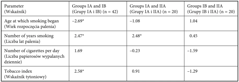
Data on smoking were different in the groups. The average age of starting smoking in group IA was 18.6 years, in group IB 21.4 years, and in group IIA 20.0 years. Patients with coronary arte-riosclerosis in group IA started smoking signifi-cantly earlier than the patients without coronary atherosclerosis in group IB.
Considering the number of years of smoking, the longest period, i.e. more than 30 years, was found in 64% of the patients with CHD. The aver-age numbers of years of smoking were 31.2 years
Table 2. Comparison of mean concentrations of selected hormones in the groups of men
Tabela 2. Porównanie średnich stężeń wybranych hormonów w badanych grupach mężczyzn
Parameter
(Wskaźnik) Group IA smokers with coronary athero-sclerosis (n = 42) (Grupa IA – osoby palące z miażdżycą naczyń wieńcowych)
Group IB smokers with-out coronary atheroscle-rosis (n = 20)
(Grupa IB – osoby palące bez miażdżycy naczyń wieńcowych)
Group IIA healthy smokers (n = 20) (Grupa IIA – ozoby zdrowe palące)
Group IIB
healthy nonsmokers (n = 20)
(Grupa IIB – zdrowi niepalący)
Total testosterone [ng/ml]
(Testosteron całkowity) 4.91 ± 1.9 4.69 ± 1.2 5.47 ± 2.1 4.75 ± 1.2 Free testosterone [pg/ml]
(Testosteron wolny) 10.62 ± 3.5 12.77 ± 3.3 12.82 ± 4.3 10.64 ± 2.7
FAI 43.75 ± 22.9 49.30 ± 11.4 52.0 ± 10.0 49.43 ± 14.2
Calculated free testoster-one [ng/ml]
(Testosteron wolny kalku-lowany)
8.99 ± 4.12 9.53 ± 1.89 10.82 ± 3.06 9.51 ± 2.31
Calculated bioavailable testosterone [ng/ml] (Testosteron biodostępny kalkulowany)
209.9 ± 96.2 223.6 ± 45.1 253.4 ± 72.2 223.1 ± 54.3
SHBG [nmol/l] 44.57 ± 20.3 34.56 ± 11.5 36.41 ± 13.1 35.88 ± 13.5
DHEAS [ng/ml] 1580.29 ± 649.0 1968.5 ± 1185.4 2163.7 ± 1079.0 1674.9 ± 679.9
Estradiol [pg/ml] 26.27 ± 6.5 27.1 ± 12.6 22.93 ± 8.6 24.24 ± 6.9
LH [mIU/ml] 7.01 ± 6.1 5.05 ± 3.8 5.19 ± 2.9 3.8 ± 1.4
IGF-1 [ng/ml] 353.91 ± 92.3 332.88 ± 102.2 339.23 ± 77.2 313.99 ± 80.3
GH [ng/ml] 0.2 ± 0.3 0.35 ± 0.4 0.38 ± 0.7 0.29 ± 0.4
in group IA, 27.2 in IB, and 26.2 in IIA. Patients in group IA smoked significantly longer than those in group IB and the healthy group IIA.
More than half of the men in the group of patients with CHD and healthy smokers smoked more than 20 cigarettes per day (60 vs. 55%). The average number of cigarettes per day was 18.6 in group IA, 14.5 in group IB, and 19.3 in group IIA. There were no significant differences in this parameter among the groups.
The tobacco index was highest in group IA of patients with CHD, lower in group IIA of healthy smokers, and lowest in group IB of patients with-out atherosclerosis (29.4 vs. 25.1 vs. 19.5). In patients with coronary arteriosclerosis (group IA), the index was higher than in the patients without CHD (group IB) (Table 4).
Assessment of the parameters characterizing smoking and the studied hormones in the men
with ischemic heart disease in group IA showed no significant relationship. In these individuals there was no correlation between the parameters related to smoking and the degree of atherosclerosis esti-mated by the number of significantly stenosed coronary arteries and the Gensini score. Analysis of the hormonal parameters in connection with tobacco addiction in subgroup IB of patients without coronary atherosclerosis showed that the men who smoked tobacco for a greater number of years had statistically significantly lower DHEAS. In subgroup IB, the higher the tobacco index, the higher the free androgen index (FAI), calculated free testosterone, and the calculated bioavailable testosterone. All these values were statistically sig-nificant. In the control group IIA of healthy male smokers, long-term smoking was associated with a significant reduction in GH concentration in blood.
Table 3. Differences between the mean concentrations of the selected hormones using the Cochran-Cox test
Tabela 3. Różnice między średnimi stężeń wybranych hormonów z użyciem testu Cochrana-Coxa
Parameter
(Wskaźnik) Groups IA and IB (Grupy IA i IB)
Groups IA and IIA (Grupy IA i IIA)
Groups IA and IIB (Grupy IA i IIB)
Groups IB and IIA (Grupy IB i IIA)
Groups IB and IIB (Grupy IB i IIB)
Groups IIA and IIB (Grupy IIA i IIB)
Total testosterone (Testosteron całkowity)
0.5494 –0.9838 0.3876 –1.4069 –0.1533 1.2781
Free testosterone (Testosteron wolny)
–2.2948* –1.9559 –0.0242 –0.04 2.1551* 1.8664
FAI –1.2471 –1.9397 –1.1798 –0.7725 –0.0329 0.6395
Calculated free testosterone (Testosteron wolny kalku-lowany)
–0.7480 –1.9652 –0.6718 –1.5656 0.0292 1.4903
Calculated bio-available testos-terone
(Testosteron biodostępny kalkulowany)
–0.7477 –1.9514 –0.6715 –1.5450 0.0311 1.4759
SHBG 2.4336* 1.8670 1.9654 –0.4624 –0.3254 0.1228
DHEAS –1.3385 –2.1811* –0.5086 –0.5301 0.9373 1.6707
Estradiol –0.2705 1.5001 1.0820 1.1866 0.8682 –0.5181
LH 1.5174 1.5679 3.1990* –0.1279 1.35 1.8980
IGF-1 0.7640 0.6429 1.7061 –0.2161 0.6333 0.9874
GH –0.5408 –0.5057 0.0996 –0.15 0.4421 0.4656
Discussion
Data on the relationship between the hor-monal status of men and coronary heart disease are unclear. This is due to a large extent to the fact that previous studies were performed in popula-tions that varied considerably in terms of the pres-ence of other coronary heart disease risk factors such as disturbances in carbohydrate mechanism. Therefore, research in men without diabetes is important, as it is commonly associated with coro-nary artery disease.
The Caerphilly Heart Disease Study, in a five-year follow-up of 2287 men aged 45–59 five-years who were selected from a group of 2512 patients after exclusion of diabetic individuals, showed lower levels of total testosterone in men with coronary artery disease compared with a healthy popula-tion [8]. In contrast, Barrett-Connor et al., in a 12-year follow-up of 1009 men aged 40–79 years, found no relationship between the level of sex hor-mones (testosterone, androstenedione, estrone, and estradiol) and the occurrence of cardiovas-cular diseases, mortality due to these diseases, ischemic heart disease, and mortality caused by it; this relationship did not change after exclud-ing factors such as hypertension, dyslipidemia, diabetes, smoking, and obesity [9]. Similarly, an analysis of the hormonal status of the participants of the Baltimore Longitudinal Study of Aging in a 34-year follow-up of 890 men aged 58.8 ± 15.8 years who were healthy at the time of enrollment did not show significant correlation between tes-tosterone levels and ischemic heart disease [10].
The present study did not reveal differenc-es in the concentrations of total tdifferenc-estosterone in the four groups. By contrast, people with coro-nary heart disease had significantly lower free
testosterone labeled by RIA and SHBG was also higher than in the patients with no change in coronary angiography. In these studies, differ-ences found were related to free testosterone levels assessed by RIA. In this respect, other methods of determining testosterone proved to be less valuable.
The anti-atherogenic and potentially benefi-cial effects of endogenous testosterone are stimu-lation of the secretion of nitric oxide (NO) by the endothelium, opening potassium channels, and intensifying the vascular response to prostaglan-din F2α. Testosterone may also act indirectly after aromatization to estradiol. Many endothelium-dependent and -inendothelium-dependent mechanisms are probably involved in this process; genomic and non-genomic actions of sex hormones are also postulated [11]. Recent studies on the hypotes-tosteronemia associated with aging showed that it is related to symptoms of metabolic syndrome: obesity, type 2 diabetes, hypertriglyceridemia, low HDL cholesterol, hypertension, and coagula-tion disorder with thrombotic predisposicoagula-tion and decrease in fibrinolytic activity [12–14].
Philips et al. examined the level of androgen dependence on coronary atherosclerosis assessed by the number of significantly stenosed large ves-sels in 154 men and found a significant negative correlation between the level of free testosterone and the intensity of atherosclerotic process [15]. Davoodi et al., in a study of 502 men, did not find an association of the levels of total and free testosterone with the degree of atherosclerosis measured with the Gensini score, and in a sub-group of patients with diabetes they also observed reduced levels of DHEAS [16]. The present study also found no significant relationship between the selected hormones and the degree of considerable
Table 4. Significance of the differences between mean parameters characterizing tobacco addiction
Tabela 4. Istotność różnic między średnimi dotyczących parametrów charakteryzujących nałóg palenia papierosów
Parameter
(Wskaźnik) Groups IA and IB (Grupy IA i IB) (n = 42) Groups IA and IIA (Grupy IA i IIA) (n = 20) Groups IB and IIA (Grupy IB i IIA) (n = 20) Age at which smoking began
(Wiek rozpoczęcia palenia) –2.69* –1.08 1.04
Number of years smoking
(Liczba lat palenia) 2.47* 2.48* 0.45
Number of cigarettes per day (Liczba papierosów wypalanych dziennie)
1.69 –0.23 –1.59
Tobacco index
(Wskaźnik tytoniowy) 2.58* 0.91 –1.29
coronary occlusion assessed by the number of sig-nificantly stenosed vessels and the Gensini score.
Data on cigarette smoking showed that the men with coronary heart disease had started smok-ing tobacco significantly earlier and had a higher tobacco index than those without atherosclerosis; this group was characterized by the highest mean number of years of smoking and it differed sig-nificantly from the group of patients with normal angiography and the group of healthy smokers. The present study, which involved an evaluation of the impact of cigarette smoking on atheroscle-rosis, did not demonstrate a link between differ-ent smoking parameters and significantly stenosed coronary arteries and the Gensini score. It seems that this result reflects the impact of many other factors besides smoking on the status of coronary atherosclerosis.
The Multiple Risk Factor Intervention Trial involved an investigation of two groups of men (284 persons) with ischemic heart disease com-pared with a 163-person control group and a posi-tive correlation between smoking and total testos-terone level was found. After four years of follow-up, total and free testosterone were elevated in the group of smokers and the control group, but this was not observed in the group with ischemic heart disease [17]. Svartberg et al. carried out a prospec-tive 6.5-year analysis of hormonal status consider-ing age, lifestyle, and chronic diseases among 1563 men aged 50–70 years. The level of total and free testosterone decreased with age in proportion to the increase in SHBG. They demonstrated a sta-tistically significant positive correlation between smoking and the levels of free and total testosterone and SHBG. Coexisting chronic diseases, including diabetes, did not change this relationship in regres-sion analysis [18]. In a study by Field et al., a group of 1241 middle-aged men was observed and higher testosterone levels as well as dihydrotestosterone (DHT), DHEAS, androstenedione, SHBG, and cortisol were found in the cigarette smokers com-pared with the non-smokers [19].
The present study revealed that male patients (tobacco smokers) with cardiac problems in whom coronary angiography was performed but who did not show changes in the coronary arter-ies had significantly higher free testosterone lev-els than healthy non-smokers. The higher the tobacco index, the higher the free androgen index (FAI), calculated free testosterone, and calculat-ed bioavailable testosterone; these relationships were statistically significant. This may mean that intensified tobacco consumption, assessed by the tobacco index, increases the concentration of cal-culated free and bioavailable testosterone and FAI. Smoking may have affected the final results,
indi-cating a reduction in free testosterone levels in the healthy non-smokers compared with the smokers who did not have coronary atherosclerosis.
Hautanen et al. suggested that smoking reduc-es the activity of 21- and 11-beta-hydroxylase in the adrenal cortex, which intensifies the synthesis of androgens. Nicotine also causes an increased response of adrenal androgens, which are precur-sors of testosterone, the adrenocorticotropic hor-mone ACTH [20].
Perhaps the absence of atherosclerosis made the group of smokers with cardiac problems simi-lar to that of healthy smokers. This is confirmed by the fact that there were no significant differences in hormonal parameters assessed between smok-ers with cardiac problems without atherosclerosis and healthy smokers.
Trummer et al. examined 1104 men with so-called idiopathic infertility and found increased levels of free and total testosterone and LH in tobacco smokers. They also made the interesting observation that men more likely to quit smok-ing had lower testosterone levels than those who continued to smoke. The question arises whether tobacco smoking causes increased secretion of LH and free and total testosterone or, conversely, men with elevated values of these hormones are more susceptible to addiction and other types of health risks [21].
The authors of the present study also found statistically significant lower levels of DHEAS in smokers with coronary atherosclerosis than in healthy smokers. In a prospective eight-year analy-sis of 119 men, Mazat et al. observed a higher rela-tive risk of death in smokers with low DHEAS levels compared with non-smokers with high levels of the hormone. Among those with high levels of DHEAS, only a slight increase in the risk of death in smokers compared with nonsmokers was reported. Perhaps a low level of DHEAS is an indicator of cardiovas-cular disease in relation to tobacco smoking, or low DHEAS may lead to disturbances in the cardio-vascular system, which further deteriorates under the influence of smoking. It is also possible that DHEAS „counteracts” the toxic effects of smok-ing. The strong correlation between smoking and DHEAS which was found suggests that this andro-gen influences mortality due to cardiovascular dis-ease or other tobacco-related disdis-eases [22].
References
[1] Ogólnopolskie i regionalne rozpowszechnienie głównych czynników ryzyka układu sercowo-naczyniowego. Wyniki wieloośrodkowego ogólnopolskiego badania stanu zdrowia ludności program WOBASZ. POLKARD 2003–2005. Kardiol Pol, Tom 63, Suplement IV.
[2] Lerner DJ, Kannel WB: Patterns of coronary heart disease morbidity and mortality in the sexes: a 26-year follow-up of the Framingham population. Am J Cardiol 1986, 111, 383–390.
[3] Mędraś M, Jankowska EA: Miażdżyca a andropauza i proces starzenia się mężczyzn. W: Andropauza. Eds.: Mędraś M, Bablok L, PZWL, Warszawa 2002, 86–89.
[4] Huxley R, Barzi F, Woodward M: Excess risk of fatal coronary heart disease associated with diabetes in men and women: meta-analysis of 37 prospective cohort studies. BMJ 2006, 332(7533), 73–78.
[5] Standardy PTK. Strona internetowa: www.pt.kardio.pl.
[6] Gensini GG: A more meaningful scoring system for determining the severity of coronary heart disease. Am J Cardiol 1983, 51, 606.
[7] Vermeulen A, Verdonck L, Kaufman JM: A critical evaluation of simple methods for the estimation of free tes-tosterone in serum. J Clin Endocrinol Metab 1999, 84, 10, 3666–3672.
[8] Lichtenstein MJ, Yarnell JW., Elwood PC, Beswick AD, Sweetnam PM, Marks V, Teale D, Riad-Fahmy D: Sex hormones, insulin, lipids, and prevalent ischemic heart disease. Am J Epidemiol. 1987, 126, 4, 647–657.
[9] Barrett-Connor E, Khaw KT: Endogenous sex hormones and cardiovascular disease in men. A prospective pop-ulation-based study. Circulation 1988, 78, 3, 539–545.
[10] Harman SM, Metter EJ, Tobin JD, Pearson J, Blackman MR: Longitudinal effects of aging on serum total and free testosterone levels in healthy men. J Clin Endocrinol Metab 2001, 86, 2, 724–731.
[11] Wu FCW, von Eckardstein A: Androgens and coronary artery disease. Endocrin Rev 2003, 24, 2, 183–217.
[12] Haffner SM, Shaten J, Stern MP, Smith GD, Kuller L: Low levels of sex hormone-binding globulin and testos-terone predict the development of non-insulin-dependent diabetes mellitus in men. MRFIT Research Group. Multiple Risk Factor Intervention Trial Am J Epidemiol 1996, 143, 9, 889–897.
[13] Khaw KT, Barrett-Connor E: Lower endogenous androgens predict central adiposity in men. Ann Epidemiol 1992, 2, 5, 675–682.
[14] Khaw KT, Barrett-Connor E: Blood pressure and endogenous testosterone in men: an inverse relationship. J Hypertens 1998, 6, 4, 329–332.
[15] Phillips GB, Pinkernell BH, Jing TY: Are major risk factors for myocardial infarction the major predicts of degree of coronary artery disease in men? Metabolism. 2004, 53, 3, 324–329.
[16] Davoodi G, Amirezadegan A, Borumand MA et al.: The relationship between level of androgenic hormones and coronary artery disease in men. Cardiovasc J Afr 2007, 18, 6, 362–366.
[17] Dai WS, Gutai JP, Kuller LH, Cauley JA: Cigarette smoking and serum sex hormones in men. Am J Epidemiol 1988, 128, 4, 796–805.
[18] Svartberg J, Midby M, Bonaa KH, Sundsfjord J, Joakimsen RM, Jorde R: The associations of age, lifestyle factors and chronic disease with testosterone in men: the Tromso Study. Eur J Endocrinol 2003, 149, 145–152.
[19] Field AE, Colditz GA, Willett WC, Longcope C, McKinlay JB: The relation of smoking, age, relative weight, and dietary intake to serum adrenal steroids, sex hormones, and sex hormone- binding globulin in middle-aged men. J Clin Endocrinol Metab 1994, 79, 5, 1310–1316.
[20] Hautanen A, Manttari M, Kupari M, Sarna S, Manninen V, Frick MH, Adlercreutz H: Cigarette smoking is associated with elevated adrenal androgen response to adrenocorticotropin. J Steroid Biochem Mol Biol 1993, 46, 2, 245–251.
[21] Trummer H, Haberman H, Haas J, Pummer K: The impact of cigarette smoking on human semen parameters and hormones. Human Reprod 2002, 17, 6, 1554–1559.
[22] Mazat L, Lafont S, Berr C, Debuire B, Tessier JF, Dartigues JF, Baulieu EE: Prospective measurements of dehy-droepiandrosterone sulfate in a cohort of elderly subjects: Relationship to gender, subjective health, smoking habits, and 10-year mortality. PNAS 2001, 98, 14, 8145–8150.
Address for correspondence:
Teresa Sicińska-Werner
Provincial Endocrinology Clinic, Provincial Medical Centre W. Witosa 26
45-418 Opole Poland
Tel.: +48 77 452 01 56 E-mail: [email protected]
Conflict of interest: None declared Received: 7.12.2009
Revised: 18.01.2010 Accepted: 7.04.2010
increases calculated free and bioavailable testos-terone and the free androgen index (FAI) in with cardiac problems smokers but without coronary atherosclerosis. There was no statistically signifi-cant relationship observed between total and free
testosterone, free and calculated bioavailable
tes-tosterone, SHBG, DHEAS, E2, IGF-1, GH, and the