The incidence of MTMC ranges from 15% to 36.1% of all TMC [2, 3] and, as some authors sug-gest, may increase [1].
The widespread use of ultrasound examina-tion of the thyroid gland, followed by ultrasound-guided fine-needle aspiration biopsy (UG-FNAB), in the general population led to an increase in the detection of very small thyroid carcinomas [4]. Over the last few decades, numerous stu dies
have described a specific type of thyroid malig-nancy, known as thyroid microcarcinoma (TMC). This small tumor very often occurs as two or more separate foci and unlike unifocal thyroid micro-carcinoma (UTMC), is defined as a multifocal tumor, i.e. multifocal thyroid microcarcinoma (MTMC) [1].
Krzysztof Kaliszewski
1, A–F, Agnieszka Zubkiewicz-Kucharska
2, C, D,
Beata Wojtczak
1, E, Marta Strutyńska-Karpińska
3, D–FMulti- and Unifocal Thyroid Microcarcinoma:
Are There Any Differences?
1 1st Department and Clinic of General, Gastroenterological and Endocrinological Surgery,
Wroclaw Medical University, Poland
2 Department of Endocrinology and Diabetology for Children and Adolescents, Wroclaw Medical University,
Poland
3 Department of Gastroenterological and General Surgery, Wroclaw Medical University, Poland
A – research concept and design; B – collection and/or assembly of data; C – data analysis and interpretation;
D – writing the article; E – critical revision of the article; F – final approval of article
Abstract
Background. Thyroid microcarcinoma (TMC) often occurs as two or more separate foci, therefore a completion of primary surgery might be necessary.
Objectives. To evaluate and compare the incidence, diagnostic accuracy, clinicopathological characteristics and surgical treatment of unifocal and multifocal thyroid microcarcinoma (UTMC vs. MTMC).
Material and Methods. We retrospectively analyzed 3,218 medical records of patients consecutively admitted and surgically treated in one center due to thyroid pathology.
Results. In the end, we evaluated 246 (7.64%) patients with thyroid malignancy. Ninety-seven of them (39.43%) were diagnosed as TMC: 84 (86.59%) UTMC and 13 (13.41%) MTMC (p < 0.0001). All MTMC were unilateral tumors. The papillary type of cancer was found in 82 (97.62%) patients with UTMC and in 12 (92.31%) with MTMC (p = 0.8661). In the UTMC group, 1 (1.19%) patient had follicular and 1 (1.19%) the medullary type of TMC. 1 (7.69%) individual in the MTMC group had tumors composed of papillary and follicular cancer. The num-ber of younger patients (age < 45) was comparable in both groups (p = 0.825). The trend was observed that ultra-sound guided fine needle aspiration biopsy (UG-FNAB) revealed malignant processes before surgery in a greater number of patients with MTMC than UTMC (84.62% vs. 58.33%, p = 0.131). In the MTMC group, the number of larger tumors (> 5 mm) was greater (84.62% vs. 65.48%), however the difference was not statistically significant. Thirteen percent of patients with UTMC presented cervical lymph node involvement, compared to nearly 8% of patients with MTMC (p = 0.298). Disease-related mortality was not observed in either group.
Conclusions. The prevalence of UTMC was significantly higher than MTMC. The majority of UTMC and MTMC were composed of the papillary type of cancer. MTMC and UTMC were equally frequent in both age groups. The accuracy of UG-FNAB was higher in patients with MTMC. The dimensions of most UTMC and MTMC was above 5 mm. The involvement of the cervical lymph node at the time of diagnosis in both groups is comparable and not infrequent (Adv Clin Exp Med 2016, 25, 3, 485–492).
Key words: cancer, thyroid, multifocal, microcarcinoma.
ORIGINAL PAPERS
Adv Clin Exp Med 2016, 25, 3, 485–492
The accuracy of preoperative ultrasound examina-tion of TMC, in the opinion of some authors, is, however, still unsatisfactory [3].
Although the prognosis for TMC is thought to be excellent, the clinical importance of this tumor remains in debate. Some authors suggest a benign course of these tumors [5], but the others describe cases with local lymph node and distant metastases at the time of diagnosis [6] as well as cancer-related deaths [7]. It is considered that multifocality may be an important predictor of recurrence [8–10]. In cases of incidental MTMC, completion of pri-mary surgery and lymph node dissection may be necessary. Moreover, the extent of surgery in uni-lateral MTMC is still not defined clearly [11, 12].
Because TMC is being diagnosed with increas-ing frequency, it seems very important to evalu-ate and compare the incidence, diagnostic accura-cy, clinicopathological characteristics and surgical treatment of unifocal and multifocal thyroid mi-crocarcinoma (UTMC vs. MTMC).
Material and Methods
Patient Cohort and Study Design
We retrospectively analyzed 3218 medical re-cords of patients consecutively admitted and surgical-ly treated due to thyroid pathology in the 1st Depart-ment of General, Gastroenterological and Endocrine Surgery, Wroclaw Medical University, between 2008 and 2013. All of the patients were staged in accor-dance with TNM staging criteria (tumor-node-metas-tasis) proposed by AJCC/UICC 2010 7th Edition [13]. Preoperative thyroid ultrasonography, UG-FNAB and cytological examinations were performed in all cases. After surgery, the final histopathological clas-sification was performed according to World Health Organization Guidelines. Serial 2 mm sections were prepared from histopathologic specimens, and sus-picious lesions were examined microscopically. Two pathologists experienced in thyroid lesions examined all of the pathology specimens to confirm the diagno-sis, features of the tumor and extent of the malignant process. Patients were diagnosed as TMC if the larg-est tumor diameter was equal to or less than 1 cm. Subsequently, patients were categorized as UTMC if only one malignant foci in the thyroid gland was found during histopathological examination, or as MTMC if there were two or more foci present.
Data Analysis
The following data was compared between the two analyzed groups (UTMC vs. MTMC): In-cidence, diagnostic accuracy (UG-FNAB),
demo-graphic information such as gender and age at the time of diagnosis, clinicopathological characteris-tics, surgical treatment and postoperative staging of tumors. For MTMCs, the dimension of the lar-gest tumor was defined as the primary tumor size.
Statistical Analysis
The statistical analysis was conducted with the use of STATISTICA (StatSoft, Inc., USA) da-ta analysis software, v. 12, based on the license owned by Wroclaw Medical University. In this tri-al, the following statistical measures were used: Ar-ithmetical mean (x) and standard deviation (SD). The Shapiro-Wilk test was used to confirm the consistency of the analyzed sample’s age tion with the normal distribution. As the distribu-tion of the analyzed samples was not significantly different from the normal distribution of statisti-cally identical variance, to assess eventual differ-ences, the t-test was used. The intergroup frequen-cy assessment was performed with the use of a c2 test. If the expected frequency was less than 5 or the total count was less than 50, Yate’s correction or Fisher’s exact test were applied.
The importance of selected independent clin-ical parameters as the predictive factors for ex-planatory variables “TC diagnosis by FNAB”, “to-tal thyroid resection” and “need of reoperation”, was tested with the use of multiple logistic regres-sion analysis.
A p-value < 0.05 was considered statistically significant, while a p value from 0.05 to < 0.10 was regarded as on the borderline of statistical signif-icance.
Results
Table 1. Demographic and clinicopathological characteristics of patients with unifocal and multifocal thyroid microcarci-noma. Descriptive data is presented as number (n), percentage (%) and mean ± standard deviation (± SD)
Parameter Unifocal thyroid microcarcinoma (n = 84)
Multifocal thyroid microcarcinoma (n = 13)
χ2 test p-value
Age (years) 48.7 ± 14.8 48.4 ± 14.0 T = 0.07 0.942 Age:
< 45
> 45 26 (30.95)58 (69.05) 5 (38.46) 8 (61.54)
0.05 0.825
Gender: female
male 78 (92.86)6 (7.14) 12 (92.31) 1 (7.69)
0.25 0.614
Prediction of thyroid cancer by FNAB: yes
no 49 (58.33)35 (41.67) 11 (84.62) 2 (15.38)
2.28 0.131
Type of resection: total
not total 50 (59.52)34 (40.48) 12 (92.31) 1 (7.69)
3.92 0.047*
Reoperation needed: no
yes 75 (89.29) 9 (10.71) 13 (100.0) 0 (0.0)
0.53 0.468
Type of cancer: papillary follicular
papillary/follicular in one patient medullary
82 (97.62) 1 (1.19) 0 (0.0) 1 (1.19)
12 (92.31) 0 (0.0) 1 (7.69) 0 (0.0)
0.29 0.864
pTNM stage: I
II III IV
83 (98.81) 0 (0.0) 1 (1.19) 0 (0.0)
13 (100.0) 0 (0.0) 0 (0.0) 0 (0.0)
1.17 0.280
pT: pT1 pT2 pT3 pT4 pTm
84 (100.0) 0 (0.0) 0 (0.0) 0 (0.0) 0 (0.0)
13 (100.0) 0 (0.0) 0 (0.0) 0 (0.0) 13 (100.0)
– –
pN: pN0 pN1a pN1b pNx
41 (48.81) 10 (11.90) 1 (1.19) 32 (38.10)
10 (76.92) 1 (7.69) 0 (0.0) 2 (15.38)
3.68 0.298
pM: pM0 pM1 pMx
43 (51.19) 1 (1.19) 40 (47.62)
11 (84.62) 0 (0.0) 2 (15.38)
5.12 0.077**
Tumor size 1–5 mm
6–10 mm 29 (34.52)55 (65.48) 2 (15.38)11 (84.62)
1.12 0.290
Table 2. Characteristics of patients with FNAB diagnosis (cancer yes/cancer no) in two groups: Unifocal and multifocal thy-roid microcarcinoma. Descriptive data is presented as number (n), percentage (%) and mean ± standard deviation (± SD)
Parameter Unifocal thyroid microcarcinoma Multifocal thyroid microcarcinoma FNAB – “cancer yes” n = 49 FNAB – “cancer no” n = 35 p-value FNAB – “cancer yes” n = 11 FNAB – “cancer no” n = 2 p-value
Age (years) 45.53 ± 15.72 51.74 ± 12.87 0.111 48.72 ± 13.9 46.50 ± 20.5 0.921 Age:
< 45
> 45 18 (36.73)31 (63.27) 8 (22.86)27 (77.14)
0.263
4 (36.36)
7 (63.64) 1 (50.0)1 (50.0)
0.670
Gender: female
male 46 (93.88) 3 (6.12) 32 (91.43) 3 (8.57)
0.669
11 (100.0)
0 (0.0) 1 (50.0)1 (50.0)
0.318
Type of resection total
not total 42 (85.71) 7 (14.29) 8 (22.86)27 (77.14)
< 0.00001
11 (100.0)
0 (0.0) 1 (50.0)1 (50.0)
0.318
Reoperation needed: no
yes 46 (93.88) 3 (6.12) 5 (82.86) 3 (17.14)
0.211
11 (100.0)
0 (95.2) 2 (100.0)0 (0.0)
–
Type of cancer: papillary follicular
papillary/follicular in one patient medullary 47 (97.91) 0 (0.0) 0 (0.0) 1 (2.09) 35 (97.22) 1 (2.78) 0 (0.0) 0 (0.0) 0.481 10 (90.91) 0 (0.0) 1 (9.09) 0 (0.0) 2 (100.0) 0 (0.0) 0 (0.0) 0 (0.0) – pTNM stage: I II III IV 48 (97.96) 0 (0.0) 1 (2.04) 0 (0.0) 35 (100.0) 0 (0.0) 0 (0.0) 0 (0.0) 0.864 11 (100.0)* 0 (0.0) 0 (0.0) 0 (0.0) 2 (100.0) 0 (0.0) 0 (0.0) 0 (0.0) – pT: pT1 pT2 pT3 pT4 pTm 49 (100.0) 0 (0.0) 0 (0.0) 0 (0.0) 0 (0.0) 35 (100.0) 0 (0.0) 0 (0.0) 0 (0.0) 0 (0.0) – 11 (100.0) 0 (0.0) 0 (0.0) 0 (0.0) 11 (100.0) 2 (100.0) 0 (0.0) 0 (0.0) 0 (0.0) 2 (100.) – pN: pN0 pN1a pN1b pNx 39 (79.59) 10 (20.41) 0 (0.0) 0 (0.0) 2 (5.71) 0 (0.0) 1 (2.86) 32 (91.43) < 0.00001* 10 (90.91) 1 (9.09) 0 (0.0) 0 (0.0) 0 (0.0) 0 (0.0) 0 (0.0) 2 (100.0) 0.001* pM: pM0 pM1 pMx 41 (83.67) 0 (0.0) 8 (16.33) 2 (5.71) 1 (1.19) 32 (91.43) < 0.00001* 11 (100.0) 0 (0.0) 0 (0.0) 0 (0.0) 0 (0.0) 2 (100.0) 0.011* Tumor size 1–5 mm
6–10 mm 7 (14.29)42 (85.71) 22 (62.86)13 (37.14)
< 0.00001*
2 (18.18)
9 (81.82) 0 (0.0)2 (100.0)
0.682
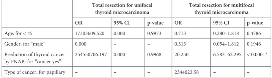
was observed that ultrasound-guided fine-nee-dle aspiration biopsy (UG-FNAB) revealed malig-nant processes before surgery in a greater number of patients with MTMC than UTMC (84.62% vs. 58.33%, p = 0.131). In the group of patients with MTMC, the number of larger tumors (6–10 mm) was higher than in the UTMC group (84.62% vs. 65.48%), however the difference was not statis-tically significant (p = 0.290). Thirteen percent of patients of the UTMC group presented cervi-cal lymph node involvement, compared to nearly 8% of patients with MTMC (p = 0.298). The char-acteristics of patients with FNAB diagnosis in the groups with unifocal and multifocal thyroid mi-crocarcinoma are presented in Table 2. Only in the group of patients with unifocal thyroid micro-carcinoma there is statistically significant differ-ence in the choice of surgical procedure between microcarcinomas confirmed by FNAB and uncon-firmed ones (p < 0.00001). Multiple logistic regres-sion analysis showed that positive results of FNAB were not dependent on the patient’s age or gender in both analyzed groups (Table 3). The results of the logistic regression analysis for the prediction of total thyroid resection in TC patients with uni-focal and multiuni-focal microcarcinoma are present-ed in Table 4. In the group of patients with multi-focal thyroid microcarcinoma, a positive result of FNAB was the only predictor of radical procedure (p < 0.0001). The risk of reoperation in patients
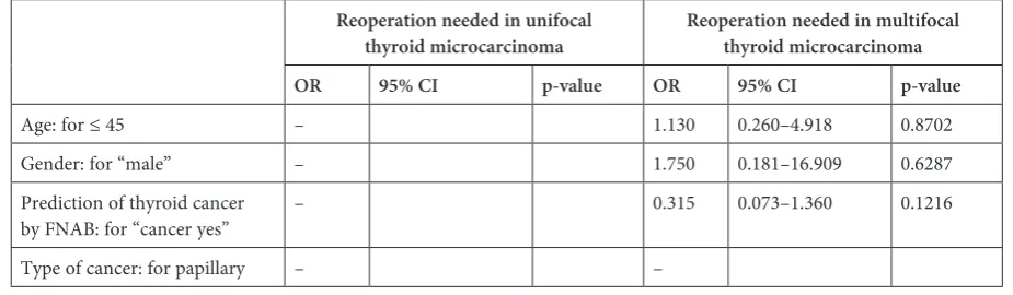
with multifocal thyroid microcarcinoma was not statistically significant for age, gender or thyroid microcarcinoma confirmation in FNAB (Table 5). Disease-related mortality was not observed in ei-ther group. Detailed results of the clinical char-acteristics of the patients with TMC diagnosis are presented in the tables (Table 1–5).
Discussion
The prevalence of UTMC and MTMC in the last decades has increased [14]. Moreover, as some authors suggest, the number of TMC in the general population will equal the incidence of occult TMC diagnosed at autopsy, reaching the prevalence rate of 36% [15].
About 47 years ago, Iida et al. [16] noticed that multifocality of thyroid cancer was a result of in-trathyroid metastases of a single primary tumor via lymphatic vessels. It was the main argument concerning multifocal lesions as very aggressive tumors. Currently, thanks to the results of stud-ies using advanced molecular techniques, MTMC is considered as a multiple synchronous prima-ry tumor arising from different cell clones [17]. In 2010 it has to be noticed however, that some relatively new findings indicate multifocality of TMC as a consequence of intraglandular metasta-ses of a primary tumor, which in such case has to
Table 3. Multiple logistic regression analysis for the prediction of TC by FNAB (cancer yes/cancer no) in the two groups of patients: with unifocal thyroid microcarcinoma and multifocal thyroid microcarcinoma
Unifocal thyroid microcarcinoma Multifocal thyroid microcarcinoma OR 95% CI p-value OR 95% CI p-value
Age: for < 45 0.571 0.028–11.849 0.7175 1.960 0.736–5.220 0.1783 Gender: for “male” 0.000 0.000 0.9966 0.696 0.132–3.669 0.6688
Table 4. Multiple logistic regression analysis for the prediction of total thyroid resection (total resection/resection not total; 1/0) in the two groups of patients: with unifocal thyroid microcarcinoma and multifocal thyroid microcarcinoma
Total resection for unifocal
thyroid microcarcinoma Total resection for multifocal thyroid microcarcinoma OR 95% CI p-value OR 95% CI p-value
Age: for < 45 17303609.520 0.000 0.9973 0.713 0.280–1.818 0.4786 Gender: for “male” 0.000 – – 0.313 0.054–1.812 0.1946 Prediction of thyroid cancer
by FNAB: for “cancer yes” 254550706.197 0.000 0.9968 20.250 6.583–62.295 < 0.0001* Type of cancer: for papillary – – – 2344023.58 – –
be considered as aggressive and therefore requires a radical treatment [18]. These contrary observa-tions justify further investigation of the clinical and pathological characteristics of TMC. What is more, as some authors recommend, a more aggressive surgical approach for MTMC (contrary to UTMC) seems to be worthwhile and useful [19]. The next important issue is the very low sensitivity of all di-agnostic tools for presurgical diagnosis of MTMC and its prognosis, which is not clearly established. Therefore, an estimation of prognostic factors of multifocality of these specific tumors is needed.
In the study by Kuo [20], as in our survey, the size of a primary tumor exceeding 5 mm in diame-ter occurred more often in MTMC. What is more, the suggestion that MTMC is more frequent in pa-tients above 45 years of age [21] can be supported by our study, however in our study the same clini-cal feature concerns UTMC patients.
UG-FNAB procedures performed in all the an-alyzed patients before surgery revealed malignancy in more than 84% of MTMC patients and only in 58% of UTMC cases. This is a completely new ob-servation as far as this aspect of presurgical diag-nostics of MTMC and UTMC.
Recent findings suggest that the incidence of recurrence or persistence was higher in patients with multifocality of TC compared to unifocal le-sions, even if they underwent more aggressive treat-ment [10]. It might be suspected that this clinical scenario may be the consequence of the fact that patients with MTMC have higher rates of lymph node metastases, soft tissue invasion and distant metastases at the time of diagnosis and treatment. Therefore, some authors recommend total aggres-sive thyroidectomy and postoperative radioiodine ablation treatment in cases of MTMC [22].
Although multifocality of thyroid carcino-ma is strongly associated with the lymph node and distant metastases in cases of MTMC [23], in our study we did not observe a higher incidence of lymph node (pN) or distant (pM) metastases at the time of diagnosis and treatment in patients
with MTMC compared to patients with UTMC (7.69% vs. 13.09%). One has to remember, though, that the number of the patients with MTMC ana-lyzed in our study is small, therefore the conclu-sions must be careful.
Some authors suggest total thyroidectomy in all cases of MTMC diagnosed preoperatively and completion of initial non-radical surgery for inci-dentally diagnosed MTMC due to the high proba-bility of contralateral tumor foci and rather poorer prognosis [3]. They add that if a partial thyroidec-tomy is performed, accurate follow-up is obligato-ry. Barczyński et al. [24] additionally suggest pro-phylactic central lymph node dissection in MTMC. Other authors suggest that subtotal thyroidec-tomy followed by radioiodine therapy without com-pletion of primary surgery and lymphadenectomy might be an acceptable option in the management of incidental MTMC [2]. They observed patients with MTMC after subtotal thyroidectomy during the 24- to 120-month follow-up period. All patients after primary surgery received an ablative dose of 131I, and during the follow-up period all of them re-mained free from relapse of the disease or metastasis. So et al. [3] concluded that multifocality of TMC localized in one lobe carried a higher chance of bilateral foci. Therefore, they recommended more extensive surgery in cases of MTMC. In our study, all analyzed patients with MTMC were clas-sified as unilateral tumors. The results of other studies showed that the clinicopathological charac-teristics did not differ between patients with mul-tifocal-unilateral and multifocal-bilateral thyroid carcinomas [10]. It was confirmed, however, that multifocality and bilaterality are both connected with aggressive pathologic features. Furthermore, no association between bilateralism and disease re-currence or persistence was found.
Other findings suggest that the factors which may influence disease recurrence are: age, sex, tu-mor dimension, extrathyroidal invasion and cervi-cal lymph node metastasis [25, 26]. In our study, we did not confirm such observations. Chow et al. [27]
Table 5. Multiple logistic regression analysis for the prediction of reoperation (yes/no; 1/0) in the two groups of patients: With unifocal thyroid microcarcinoma and multifocal thyroid microcarcinoma
Reoperation needed in unifocal
thyroid microcarcinoma Reoperation needed in multifocal thyroid microcarcinoma OR 95% CI p-value OR 95% CI p-value
Age: for ≤ 45 – 1.130 0.260–4.918 0.8702 Gender: for “male” – 1.750 0.181–16.909 0.6287 Prediction of thyroid cancer
noticed that multifocality predicted locoregional recurrence of TMC. In the current study, we did not observe distant and regional metastases, how-ever the duration of the follow-up was not long (12–36 months). For this reason, we focused on the clinicopathological characteristics of multifocal and unifocal tumors to improve their early diagno-sis and treatment. Our plan is to increase the study group and elongate the follow-up period in order to estimate the long-term influence of multifocality on disease persistence and recurrence.
The authors concluded that the prevalence of UTMC was significantly higher than MTMC. The majority of UTMC and MTMC were com-posed of papillary type of cancer. A younger age of patients was equally frequent in both groups (MTMC and UTMC). The accuracy of UG-FNAB was higher in patients with MTMC. The dimen-sions of most UTMC and MTMC were above 5 mm. The involvement of cervical lymph node at the time of diagnosis in both groups is comparable and not infrequent.
References
[1] Roti E, Rossi R, Trasforini G, Bertelli F, Ambrosio MR, Busutti L, Pearce EN, Braverman LE, Degli Uberti EC:
Clinical and histological characteristics of papillary thyroid microcarcinoma: Results of a retrospective study in 243 patients. J Clin Endocrinol Metab 2006, 91, 2171–2178.
[2] Dietlein M, Luyken WA, Schicha H, Larena-Avellaneda A: Incidental multifocal papillary microcarcinomas of the thyroid: Is subtotal thyroidectomy combined with radioiodine ablation enough? Nucl Med Commun 2005, 26, 3–8.
[3] So YK, Kim MW, Son YI: Multifocality and bilaterality of papillary thyroid microcarcinoma. Clin Exp Otorhinolaryngol 2015, 8, 174–178.
[4] Castro MR, Gharib H: Continuing controversies in the management of thyroid nodules. Ann Intern Med 2005, 142, 926–931.
[5] Ito Y, Uruno T, Nakano K, Takamura Y, Miya A, Kobayeshi K, Yokozawa T, Matsuzuka F, Kuma S, Kuma K, Miyauchi A: An observation trial without surgical treatment in patients with papillary microcarcinoma of the thyroid. Thyroid 2003, 13, 381–387.
[6] Braga M, Graf H, Ogata A, Batista J, Hakim Neto CA: Aggressive behaviour of papillary microcarcinoma in a patient with Graves’ disease initially presenting as cystic neck mass. J Endocrinol Invest 2002, 25, 250–253.
[7] Chow SM, Law SC, Au SK, Mang O, Yau S, Yuen KT, Lau WH: Changes in clinical presentation, management and outcome in 1348 patients with differentiated thyroid carcinoma: Experience in a single institute in Hong Kong, 1960–2000. Clin Oncol (R Coll Radiol) 2003, 15, 329–336.
[8] Bhanvadia VM, Sheikh SS, Santwani PM: Multifocal incidental papillary microcarcinoma of thyroid: Diagnostic pitfall in cytology. Clin Cancer Investig J 2014, 3, 570–572.
[9] Kuo SF, Chao TC, Chang HY, Hsueh C, Yang CH, Lin JD: Prognostic evaluation of patients with multicentric papillary thyroid microcarcinoma. J Form Med Assoc 2011, 110, 511–517.
[10] Kim HJ, Sohn SY, Jang HW, Kim SW, Chung JH: Multifocality, but not bilaterality, is a predictor of disease recurrence/persistence of papillary thyroid carcinoma. World J Surg 2013, 37, 376–384.
[11] Perros P, Boelaert K, Colley S, Evans C, Evans RM, Gerrard Ba G, Gilbert J, Harrison B, Johnson SJ, Giles TE, Moss L, Lewington V, Newbold K, Taylor J, Thakker RV, Watkinson J, Williams GR: Guidelines for the man-agement of thyroid cancer. Clin Endocrinol (Oxf) 2014, 81, 1–122.
[12] American Thyroid Association (ATA) Guidelines Taskforce on Thyroid Nodules and Differentiated Thyroid Cancer. Cooper DS, Doherty GM, Haugen BR, Kloos RT, Lee SL, Mandel SJ, Mazzaferri EL, McIver B, Pacini F, Schlumberger M, Sherman SI, Steward DL, Tuttle RM: Revised American Thyroid Association management guidelines for patients with thyroid nodules and differentiated thyroid cancer. Thyroid 2009, 19, 1167–1214.
[13] Edge SB, Compton CC: The American Joint Committee on Cancer: The 7th edition of the AJCC cancer staging
manual and the future of TNM. Ann Surg Oncol 2010, 17, 1471–1474.
[14] Londero SC, Krogdahl A, Bastholt L, Overgaard J, Pedersen HB, Hahn CH, Bentzen J, Schytte S, Christiansen P, Gerke O, Godballe C: Papillary thyroid carcinoma in Denmark 1996–2008: Outcome and evaluation of established prognostic scoring systems in a prospective national cohort. Thyroid 2015, 25, 78–84.
[15] Harach MR, Franssila KO, Wasenius VM: Occult papillary of the thyroid: A normal finding in Finland. A sys-tematic autopsy study. Cancer 1985, 56, 531–538.
[16] Iida F, Yonekura M, Miyakawa M: Study of intraglandular dissemination of thyroid cancer. Cancer 1969, 24, 764–771.
[17] Giannini R, Ugolini C, Lupi C, Proietti A, Elisei R, Salvatore G, Berti P, Materazzi G, Miccoli P, Santoro M, Basolo F: The heterogeneous distribution of BRAF mutation supports the independent clonal origin of distinct tumor foci in multifocal papillary thyroid carcinoma. J Clin Endocrinol Metab 2007, 92, 3511–3516.
[18] Wang W, Wang H, Teng X, Wang H, Mao C, Teng R, Zhao W, Cao J, Fathey TJ, Teng L: Clonal analysis of bilateral, recurrent, and metastatic papillary thyroid carcinomas. Hum Pathol 2010, 41, 1299–1309.
[19] Ricci JA, Alfonso AE: Multifocal micropapillary thyroid cancer: A new indication for total thyroidectomy? Am Surg 2012, 78, 1211–1214.
[21] Park SY, Park YJ, Lee YJ, Lee HS, Choi SH, Choe G, Jang HC, Park SH, Park do J, Cho BY: Analysis of differ-ential BRAF(V600E) mutational status in multifocal papillary thyroid carcinoma: Evidence of independent clonal origin in distinct tumor foci. Cancer 2006, 15, 1831–1838.
[22] Kuo SF, Lin SF, Chao TC, Hsueh C, Lin KJ, Lin JD: Prognosis of multifocal papillary thyroid carcinoma. Int J Endocrinol 2013, 2013, 809382.
[23] Mazzaferri EL, Jhiang SM: Long-term impact of initial surgical and medical therapy on papillary and follicular thyroid cancer. Am J Med 1994, 97, 418–428.
[24] Barczyński M, Konturek A, Stopa M, Nowak W: Prophylactic central neck dissection for papillary thyroid cancer. Br J Surg 2013, 100, 410–418.
[25] Leboulleux S, Rubino C, Baudin E, Caillou B, Hartl DM, Bidart JM, Travagli JP, Schlumberger M: Prognostic factors for persistent or recurrent disease of papillary thyroid carcinoma with neck lymph node metastases and/or tumor extension beyond the thyroid capsule at initial diagnosis. J Clin Endocrinol Metab 2005, 90, 5723–5729.
[26] Baek SK, Jung KY, Kang SM, Kwon SY, Woo JS, Cho SH, Chung EJ: Clinical risk factors associated with cervical lymph node recurrence in papillary thyroid carcinoma. Thyroid 2010, 20, 147–152.
[27] Chow SM, Law SC, Chan JK, Au SK, Yau S, Lau WH: Papillary microcarcinoma of the thyroid: Prognostic sig-nificance of lymph node metastasis and multifocality. Cancer 2003, 98, 31–40.
Address for correspondence:
Krzysztof Kaliszewski
1st Department and Clinic of General, Gastroenterological and Endocrinological Surgery
Wroclaw Medical University ul. Curie-Skłodowskiej 66 50-369 Wrocław
Tel: +48 71 784 21 62
E-mail address: [email protected]
Conflict of interest: None declared