© 2014 Ancharida Akaracharanya et al. This is an open access article distributed under the terms of the Creative Commons Attribution License -NonCommercial-ShareAlikeUnported License (http://creativecommons.org/licenses/by-nc-sa/3.0/).
Journal of Applied Pharmaceutical Science Vol. 4 (05), pp. 006-011, May, 2014 Available online at http://www.japsonline.com
DOI: 10.7324/JAPS.2014.40502 ISSN 2231-3354
Characterization of cellulase producing Bacillus and Paenibacillus
strains from Thai soils
Ancharida Akaracharanya
1*, Thanawan Taprig
1, Jaruwan Sitdhipol
2and Somboon Tanasupawat
31
Department of Microbiology, Faculty of Science, ChulalongkornUniversity, Bangkok 10330, Thailand. 2Bioscience Department, Thailand Institute of Scientific and Technological Research (TISTR), Pathumthani 12120, Thailand. 3 Department of Biochemistry and Microbiology, Faculty of Pharmaceutical Sciences, Chulalongkorn University, Bangkok 10330, Thailand.
ARTICLE INFO ABSTRACT
Article history:
Received on: 13/02/2014 Revised on: 11/04/2014 Accepted on: 27/04/2014 Available online: 27/05/2014
Seventeen strains of cellulase producing bacteria were isolated from soil samples collected in Nan province, Thailand. They exhibited cellulase activity as a clear zone surrounded their colonies grown on carboxymethyl cellulose (CMC) agar medium ranged from 0.63 to 2.95 cm in diameter. Their hydrolysis capacity values were 1.65 - 7.55. The bacteria isolated were divided into 2 groups and belonged to genus Bacillus and Paenibacillus
based on their phenotypic characteristics and chemotaxonomic characteristics such as meso-diaminopimelic in cell wall peptidoglycan and menaquinones of MK-7. Bacillus strains, P3-1 and P4-6 in Group I, produced maximum cellulase at 0.015 U ml-1. Their optimal pH and temperature for enzyme production and enzyme activity were 7.0 and 50 oC, respectively. The strains P3-1 and P4-6 were closely related to Bacillus velesensis
LMG 22478T (100% similarity) whereas representative strain, S10-4 in Group II, was closely related to P. cellulositrophicus KCTC 13135T(98.7% similarity) based on 16S rRNA gene sequence. On the basis of their phenotypic, chemotaxonomic characteristics and phylogenetic analysis of 16S rRNA gene sequence, the strains P3-1 and P4-6 were identified as B. velesensis and the strain S10-4 was a novel species in the genus
Paenibacillus.
Key words:
Cellulase, Bacillus, Paenibacillus, 16S rRNA gene sequence, Thai soil
INTRODUCTION
Cellulose is a linear chain of D-glucose units linked
together by β-1,4-glycosidic bond (Salmon, 1997). The β
-1,4-glycosidic linkages in the cellulose can be hydrolysed by cellulolytic enzyme, cellulase. The cellulase is a group of enzymes which composes of at least three different enzymes.
Endoglucanase (1,4-β-D-glucan -4-glucanohydrolase, E.C. 3.2.1.4)
attacks randomly internal linkages within the cellulose chain,
creating free chain ends, exoglucanase (1,4-β-D-glucan
cellobiohydrolase, E.C. 3.2.1.91) hydrolyses cellulose from the free chain ends creating mainly cellobiose as an end product, and
β-glucosidase (E.C. 3.2.1.21) hydrolyses the cellobiose to
glucose. Harmonization of exoglucanase and β-glucosidase
activities is important for glucose liberation from cellulose because accumulation of cellobiose strongly inhibits exoglucanase activity
.
* Corresponding Author
Department of Microbiology, Faculty of Science, Chulalongkorn University, Bangkok 10330, Thailand. Phone : +66-22185071 ; Fax : + 66-22527576 ; E-mail ; [email protected]
(Beguin and Aubert, 1994; Harjunpaa, 1998). During the past two decades, usage of enzyme in industrial process has significantly increased. The cellulase has been used in various industries for example; paper production, juice clarification, lignocellulosic ethanol production, extraction of valuable components from plant cells, and
nutritional improvement of animal feed, etc (Bhat, 2000; Csiszar et
al., 2001; Haki and Rakshit 2003; Lynd et al., 2002; Vielle and
Ziekus, 2001). However, difference of applications requires cellulase (s) which is different in particular properties. Several
microorganisms; bacteria, yeast and fungi; have been reported as cellulase producers. Bacterial cellulase is more thermostable than fungal cellulase. Optimal pH for fungal cellulase activity is between
4 and 6 (Zhu et al., 1982; Yazdi et al., 1990). While,
alkaline pH has been reported as optimal pH for bacterial cellulase activity (Ruttersmith and Daniel, 1993). Extracellular cellulases of
several bacteria have been studied and characterized e.g.
Clostridium, Caldocellum, and Acidothermus (Bergquist et al.,
1999), Acetovibrio (Ding et al., 1999), Ruminococcus (Aurilia et al.,
2000), Sinorhizobium (Chen et al., 2004), Cellulomonas,
In this investigation, cellulase producing bacteria were isolated from soils in Nan province, Thailand to screen for bacterial cellulase with particular property. The bacteria isolated
were also identified based on their phenotypic and
chemotaxonomic characteristics including 16S rRNA gene sequence analysis.
MATERIALS AND METHODS
Isolation and screening methods
A total of 9 soil samples were collected from Pua and Santisuk districts, Nan province, Thailand (Table 1). Cellulase producing bacteria were isolated from the soil samples not later than 24 hours after collection by an enrichment culture method. The soil sample (0.1 g) was put into a 10 ml of cellulose powder medium (CP, cellulose power 1 g, peptone 5 g, yeast extract 1 g,
K2HPO4 4 g, MgSO4.7H2O 1 g, KCl 0.2 g, FeSO4
.
7H2O 0.02 g in
1000 ml distilled water, pH 7.0) and incubated on a rotary shaker
at 200 rpm, 40oC for 2 days. One milliliter of the culture was
transferred to fresh CP medium and incubated at the same above condition for 2 more times.
The enriched cultures or their dilutions (0.1 ml) were dropped and spreaded on the CP agar medium and incubated at
40oC for 2 days. Cellulase producing capability of the cultures
was screened qualitatively by method described by Teather and Wood (1982). Their colonies grown on carboxymethyl cellulose
(CMC)-basal (CMC 5 g, (NH4)2 SO4 1 g, yeast extract 1 g in 1000
ml distilled water) agar medium at 40oC for 2 days were flooded
with 0.1% (w/v) Congo red solution for 1 min and then washed with 0.1 M NaCl. Colonies surrounded by clear zone were selected as cellulase producing isolates and then they were purified by streak plate method. Hydrolysis capacity (HC) value was calculated from clear zone diameter divided by colony diameter.
Cellulase production capability was also quantitatively determined by inoculating single colony into 10 ml of carboxymethyl cellulose medium (CMC medium, the CP medium which cellulose power was replaced by CMC) and incubated at
40oC, 200 rpm for 2 days. Three milliliters of the cultures were
transferred into 30 ml of CMC medium and incubated at the same aboved condition for 2 days. Supernatants obtained after
centrifugation of the cultures at 13,300 x g , 4o C for 15 min were
used as crude enzyme for cellulase activity assay.
Cellulase activity assay
Cellulase activity assay was done by the method described by Ghose (1987). Reaction mixture composed of 0.5 ml
of 2% (w/v) carboxymethyl cellulose in 100 mM
sodium phosphate buffer pH 7.0 , and 0.5 ml of crude
enzyme were incubated at 40oC for 30 min. The amount of
reducing sugar released was quantified by Somogyi-Nelson method (Nelson, 1944; Somogyi, 1952) using glucose as authentic sugar. The reaction stopped immediately after addition of enzyme solution was used as a reaction blank.
One unit of cellulase was defined as the amount of enzyme yielding 1 micromole of glucose within 1 min under the assay condition.
Identification methods
Cell morphology, colonial appearance, spore formation, and pigmentation of the cellulase producing bacteria isolated
grown on CMC agar medium at 37oC for 1 day were examined.
Catalase, oxidase, hydrolysis of L-arginine, aesculin, casein, gelatin, starch, tyrosine and deoxyribonuclease (DNase) activity; MR-VP, indole test, nitrate reduction, Simmon citrate test, Triple Sugar Iron agar (TSI), dihydroxyacetone from glycerol, urease activity and acid from carbohydrates were determined as described by Barrow and Feltham (1993).
Growth at different pH (5, 6, 8 and 9), in 3 and
5% NaCl and at different temperatures (10 oC, 15 oC, 20 oC, 37 oC,
45 oC, 50 oC, 55 oC and 60 oC) were investigated. All tests were
carried out by using C medium (polypeptone 5 g, yeast extract 1 g,
K2HPO4 4 g, MgSO4.7H2O 1 g, KCl 0.2 g, FeSO4.7H2O 0.02 g in
1000 ml distilled water) as a basal medium and incubated
at 37oC, except for the investigation of the effect of
temperatures. Diaminopimelic acid in the cell wall and menaquinone were determined as described by Komagata and Suzuki (1987).
DNAs were isolated from cells grown on C agar plate for 18 to 48 h, and purified by the method of Saito and Miura (1963). DNA base composition was determined by the method of Tamaoka and Komagata (1984).
The 16S rDNA gene sequence was amplified,
purified and analysed as described previously (Tanasupawat et al.,
2004). The sequences determined (1489-1545 bp) were aligned with the selected sequences obtained from the GenBank/ EMBL/DDBJ database employing CLUSTAL_X version 1.81
(Thompson et al., 1997). The alignment was manually edited to
remove gaps and ambiguous nucleotides prior to construction of phylogenetic tree. The phylogenetic tree was constructed by neighbor-joining method (Saitou and Nei, 1987) using MEGA
programme version 2.1 (Kumar et al., 2001). Confidence values
of branches of the phylogenetic tree were determined using bootstrap analyses (Felsenstein, 1985) based on 1000
resamplings.
RESULTS AND DISCUSSION
Isolation and screening
Seventeen isolates of cellulase producing bacteria which were isolated from soil samples collected in Pua and Santisuk districts, Nan province, Thailand by enrichment culture method
at 40 oC exhibited cellulase activity as a clear zone surrounded
Table. 1: Sample location, isolate no. and cellulase activity on CMC-basal agar of bacteria isolated.
District/ Province
Isolate no.
Clear zone diameter
(cm)
HC* value
Cellulase activity (Uml-1)
Identification
Pua/ Nan P1-2 2.60 2.32 0.006 Bacillus sp. P1-3 1.85 2.89 0.008 Paenibacillus sp. P1-9 1.65 5.16 0.001 Paenibacillus sp. P3-1 2.95 2.81 0.015 Bacillus sp. P4-6 2.00 5.71 0.015 Bacillus sp. P5-5 0.63 2.63 0.004 Bacillus sp. P5-7 1.63 4.29 0.002 Bacillus sp. P5-8 1.18 5.36 0.001 Bacillus sp. P6-3 1.90 5.43 0.004 Bacillus sp. P6-5 0.57 1.84 0.003 Bacillus sp. P6-6 2.82 3.92 0.007 Bacillus sp. P7-5 2.70 2.41 0.007 Bacillus sp. Santisuk/
Nan
S8-1 0.75 1.74 0.004 Paenibacillus sp.
S8-4 2.11 4.14 0.003 Paenibacillus sp. S9-2 0.70 2.33 0.001 Bacillus sp. S10-2 0.51 1.65 0.01 Bacillus sp. S10-4 2.34 7.55 0.006 Paenibacillus sp. *HC (hydrolysis capacity) was calculated from clear zone diameter divided by colony diameter.
Identification of isolates
All bacteria isolated were spore forming, Gram positive rods. They were divided into 2 groups based on their phenotypic and chemotaxonomic characteristics. All isolates were positive for catalase, oxidase, hydrolysis of L-arginine and grew at pH 5-8.
Most of the isolates grew at 15, 20, 45 and 50 o C. All were
negative for indole production, methyl red (MR), and dihydroxyacetone formation. They showed variable reaction on growth in the presence of 3-5% NaCl; DNAase, VP, citrate utilization, nitrate reduction, TSI, hydrolysis of gelatin, esculin, casein, L-tyrosine, starch, Tween 80; and on acid production from D-amygdalin, L-arabinose, D-cellobiose, D-galactose, inulin, lactose, D-maltose, D-mannitol, D-mannose, D-melibiose, salicin, sucrose, D-trehalose, and D-xylose. All did not produce acids
from gluconate, glycerol, inositol, ∞-methyl-glucoside,
D-melezitose, raffinose, L-rhamnose, D-sorbitol, and L-sorbose (Table 2).
Table. 2: Differential characteristics of Bacillus (Group I) and Paenibacillus
(Group II) isolates.
Characteristics Group I
(12)
P3-1, P4-6
Group II
(5) S10-4
Anaerobic growth - (+3) + -(+1) -
Growth at 50o C + + +(-2) -
Growth at 55o C - - -(+1) -
at pH 5-8 + + + +
at pH 9 + (-2) + + +
Growth in 5% NaCl + (-2) + + +
Hydrolysis:
Casein + (-3) + -(+2) -
Esculin + (-2) + + +
Gelatin + + +(-2) -
Starch + (-4) + -(+2) -
DNA + (-5) + - -
Tyrosine - (+2) - - -
Tween 80 - (+1) - - -
Urea + (-2) + + +
Citrate utilization - (+4) + (-1) -(+1) -
Nitrate reduction + (-5) + +(-1) +
Voges-Proskauer + (-4) + +(-1) -
Acid from:
D-Amygdalin + (-5) + +(-1) +
L-Arabinose - (+4) - + +
D-Cellobiose + (-2) + + +
D-Galactose - (+3) - +(-1) +
Inulin - (+5) + +(-1) +
Lactose + (-6) - +(-2) +
D-Maltose + (-2) - +(-2) +
D-Mannitol - (+2) + -(+1) -
D-Mannose + + +(-1) +
D-Melibiose - (+2) - +(-1) +
Salicin - (+5) - +(-2) +
Sucrose + (-1) + +(-2) +
D-Trehalose + (-4) - + +
D-Xylose + (-5) - +(-1) +
+, positive; -, negative. Numbers in parentheses indicate the number of isolates showing the reaction.
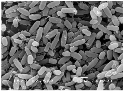
Group I contained 12 isolates. All were catalase and oxidase positive. They showed irregular, raised, entire undulate margin, smooth, dull, white colour colonies. Some strains showed circular, raised, entire margin, smooth, dull, white cream colour colonies. Isolate P4-6 produced endospore when grown on carboxymethyl
cellulose agar medium at 37 o C for 2 days (Figure 1).
Fig. 1: Scanning electron micrograph of isolate P4-6 grown on carboxymethyl cellulose agar medium at 37 o C for 2 days.
Representative isolates, P3-1 (1490 bp) and P4-6 (1489 bp), were
closely related to Bacillus velesensis LMG 22478T (100%
similarity), Bacillus polyfermenticus CJ6T (99.7% similarity) and
Bacillus subtilis DSM 10T (99.7% similarity) based on 16S rRNA
gene sequence (Figure 2). They contained meso-diaminopimelic
acid in the cell wall peptidoglycan. Therefore, they were
identified as Bacillus velezensis (Ruiz-García et al., 2005).
Group II contained 5 isolates. Colonies were circular, raised, entire margin, smooth, dull, white cream colour. Bacteria in this group showed similar characteristics, however they could be differentiated from each other. Representative isolate, S10-4,
contained meso-diaminopimelic acid in cell wall peptidoglycan
and had 53.5 mol% of DNA G+C content. This strain (1545 bp)
was closely related to P. cellulositrophicus KCTC 13135T(98.7%
similarity), P. favisporus LMG 20987T (98.6% similarity) and P.
cineris LMG 18439T (98.6% similarity) based on 16S rRNA gene
sequence (Figure 2) (Logan et al., 2004). It will be proposed as a
Paenibacillus anaericanus DSM 15890T (AJ318909)
Paenibacillus motobuensis JCM 12774T (AY741810)
Paenibacillus lautus DSM 3035T (AB073188)
Paenibacillus chibensis DSM 11731T (AB073194)
Paenibacillus cookii LMG 18419T (AJ250317)
Paenibacillus azoreducens DSM 13822T (AJ272249)
Paenibacillus rhizosphaerae LMG 21955T (AY751754)
Paenibacillus cineris LMG 18439T (AJ575658)
S10-4
Paenibacillus cellulositrophicus KCTC 13135T (FJ178001)
Paenibacillus favisporus LMG 20987T (AY208751)
86
83
59 100
56
Bacillus amyloliquefaciens ATCC 23350T (X60605)
Bacillus polyfermenticus CJ6 T (FJ436014)
Bacillus subtilis DSM10T (AJ276351)
Bacillus vallismortis DSM11031T (AB021198)
P4-6 P3-1
Bacillus velezensis LMG 22478T (AY603658) 62
0.01
Fig. 2: Phylogenetic tree based on 16S rRNA gene sequence, showing relationship between isolates P3-1, P4-6, S10-4 and related Bacillus and Paenibacillus
species.
(A) (B)
(C) (D)
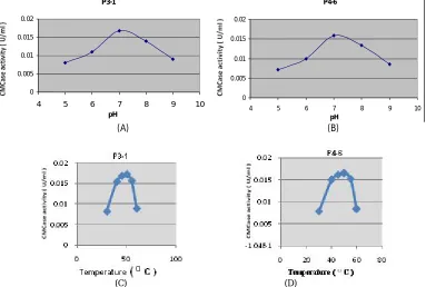
Fig. 3: Effect of pH and temperature on cellulase production of the isolates, P3-1 (A, C) and P4-6 (B, D).
0 0.005 0.01 0.015 0.02
4 5 6 7 8 9 10
pH P3-1
0 0.005 0.01 0.015 0.02
4 5 6 7 8 9 10
Cellulase activity
The isolates P3-1 and P4-6 gave the highest cellulase
activity (0.015 U ml-1 ) when grown in carboxymethyl cellulose
(CMC) broth. Their optimal pH and temperature for cellulase
production were at 7 and 50 oC, respectively.
Several strains of Bacillus including B. brevis, B. firmus,
B. polymyxa, B. pumilus, B. subtilis, B. circulans were reported as
cellulase producing bacteria (Priest, 1977; Hakamada et al., 2002;
Sa-Pereira et al., 2002). Studies of Bacillus cellulase were lagged
far behind that of fungal cellulase due to the fact that most Bacillus
cellulase hydrolyses synthetic carboxymethyl cellulose (CMC) but barely hydrolyses crystalline form of cellulose. Ito (1989) isolated
alkaline cellulase producing Bacillus and showed that this bacterial
cellulase was an effective additive of laundry detergents. Other
bacterial alkaline cellulase from Bacillus sp. (Eudo et al., 2001),
B. circulans (Hakamada et al., 2002) and Paenibacillus sp.
(Ogawa et al., 2007) were reported. In addition, Kawai et al.
(1988) reported that neutrophilic Bacillus produced alkaline
cellulase. By this study, Bacillus and Paenibacillus strains
isolated were found to be different from previous report based on
their phenotypic characteristics (Table 2). Ten Bacillus and 2
Paenibacillus strains were isolated from Pua district, while 2
Bacillus strains and 3 Paenibacillus strains were isolated from
Santisuk district. Paenibacillus sp. S10-4 isolated from Santisuk
district showed high cellulase activity on CMC agar plate but low
cellulase activity in CMC broth compared to Bacillus strains P3-1
and P4-6 that isolated from Pua district.
CONCLUSION
Seventeen cellulase producing bacteria were isolated from soil samples collected in Nan provice, Thailand by
enrichment culture method at 40oC. They were all spore forming,
Gram-positive rods. Based on their morphological, cultural,
physiological and biochemical characterictics including
chemotaxonomic characterictics, they were divided into 2 groups. Representative strains of each group were selected and
characterized by 16S rRNA gene sequence analysis.
Phylogenetic tree constructed of the representative strains revealed
that they were Bacillus strains and a novel species Paenibacillus.
Highest cellulase producing isolates (0.015 U ml-1) were belonged
to genus Bacillus. Their cellulase had an optimal pH and
temperature at 7 and 50oC, respectively. Isolate with the highest
hydrolysis capacity (7.55) belonged to genus Paenibacillus.
ACKNOWLEDGEMENT
This study was supported in part by Government Research Grant, Chulalongkorn University (2006).
REFERENCES
Aurilia V, Ding SY, Rincon MT, Lamed R, Martin JC, McCrae SI, Shoham Y, Bayer EA, Flint HJ. Cellulosomal scaffoldin-like proteins from Ruminococcus flavefaciens. J Bacteriol,2000; 183(6): 1945-1953.
Barrow GH, Feltham RKA. Cowan and Steel's Manual for Identification of Medical Bacteria. 3rded., Cambridge: Cambridge University Press, 1993; 331pp.
Beguin P, Aubert JP. The biological degradation of cellulose. FEMS Microbiol Rev, 1994; 13: 25-58.
Bhat MK. Cellulases and related enzymes in biotechnology. Biotechnol. Adv., 2000; 18: 355-383.
Chen P, Wei T, Chang Y, Lin L. Purification and characterization of carboxymethyl cellulase from Sinorhizobium fredii. Bot Bull Acad Sin, 2004; 45: 111-118.
Csiszar E, Urbanszki K, Szakacs G. Biotreatment of desized cotton fabric by commercial cellulase and xylanase enzymes. J Mol Catal B: Enzyme,2001; 11: 1065-1072.
Ding SY, Bayer EA, Steiner D, Shoham Y, Lamed R. A Novel cellulosomal scaffoldin from Acetivibrio cellulolyticus that contains a family 9 glycosyl hydrolase. J Bacteriol,1999; 181: 6720-6729.
Eudo K, Hakamada Y, Takizawa S, Kubota H. A novel alkaline endoglucanase from an alkaliphilic Bacillus isolate: enzymatic properties, and nucleotide and deduced amino acid sequences. Appl Microbiol Biotechnol, 2001; 57: 109-116.
Felsenstein J. Confidence limits on phylogenies: An approach using the bootstrap. Evolution, 1985; 39: 783-791.
Ghose TK. Measurement of cellulase activities. Pure Appl Chem,1987; 59:257-268.
Hakamada Y, Endo K, Takizawa S, Kobayashi T, Shirai T, Yamane T, Ito S. Enzymatic properties, crystallization, and deduced amino acid sequence of an alkaline endoglucanase from Bacillus circulans. Biochim Biophys Acta, 2002; 1570: 174-180.
Haki GD, Rakshit SK. Developments in industrially important thermostable enzymes. Biores Technol, 2003; 89: 17-34.
Harjunpaa V. Enzymes hydrolyzing wood polysaccharides. Technical Research Centre of Finland. VTT Publications, 1998; 372: 1-76.
Immanuel G, Dhanusa R, Prema P, Palavesam A. Effect of different growth parameters on endoglucanase enzyme activity by bacteria isolated from coir retting effluents of estuarine environment.Int J Environ Sci Tech, 2006; 3(1): 25-34.
Ito S, Shikata S, Ozaki K, Kawai S, Okamoto K, Inque S, Takel A, Ohta YI, Satoh T. Alkaline cellulase for laundry detergents: production by Bacillus sp. KSM-635 and enzymatic properties. Agric Biol Chem, 1989; 53:1275-1281.
Kawai S, Okoshi H, Ozaki K, Shikata S, Ara K, Ito S. Neurophilic Bacillus strain, KSM-522, that produces an alkaline carboxymethyl cellulase. Agric Biol Chem, 1988; 52:
1425-1431.
Komagata K, Suzuki K. Lipid and cell-wall analysis in bacterial systematics. Methods Microbiol, 1987; 19: 161-207.
Kumar S, Tamura K, Jakobson IB, Nei M. MEGA 2: Molecular evolution analysis software. Bioinformatics, 2001; 17: 1244-1245.
Logan NA, Clerck ED, Lebbe L, Verhelst A, Goris J, Forsyth G, Rodriguez-Diaz M, Heyndrickx M, Vo PD. Paenibacillus cineris sp. nov. and Paenibacillus cookii sp. nov., from Antarctic volcanic soils and a gelatin-processing plant. Int J Syst Evol Microbiol, 2004; 54:1071-1076.
Lynd LR, Weimer PJ, Zyl WHV, Isak SP. Microbial Cellulose Utilization: Fundamentals and Biotechnology. Microbiol Mol Biol Rev, 2002; 66: 506-577.
Nelson N. A photometric adaption of the Somogyi method for the determination of glucose. Biol Chem, 1944; 153: 375-380.
Ogawa A, Suzumatsu A, Takizawa S, Kuboto H, Sawada K, Hakamada Y, Kawai S, Kobayashi T, Ito S. Endoglucanases from
Paenibacillus spp. from a new clan in glycoside hydrolase family 5. J Biotechnol, 2007;129(3): 406-414.
Priest FG. Extracellular enzyme synthesis in the genus Bacillus. Bacteriol Rev, 1977; 41: 711-753.
Ruttersmith LD, Daniel RM. Thermostable â-glucosidase and á-xylosidase from Thermotoga sp. From strain FjSS3B 1. Biochim Biophys Acta, 1993; 1156: 167-172.
Saito H, Miura K. Preparation of transforming DNA by phenol treatment. Biochem Biophys Acta, 1963; 72: 619-629.
Saitou N, Nei M. The neighboring-joining method: a new method for reconstructing phylogenetic trees. Mol Biol Evol, 1987; 4: 406-425.
Sa-Pereira P, Mesquita A, Duarte JC, Barros MRA, Costa-Ferreira M. Rapid production of thermostable cellulase-free xylanase by a strain of Bacillus subtilis and its properties. Enzyme Microbial Technol, 2002; 30: 924-933.
Salmon L, Sahlberg U, Oscarsson A. The fibrillar orientation in the S2-layer of wood fibres as determined by x-ray diffraction analysis. Wood Sci Technol, 1997; 31: 77-86.
Somogyi M. Notes on sugar determination. Biol Chem, 1952; 195: 19-23.
Tamaoka K, Komagata K. Determination of DNA base comparision by reversed-phase high-performance liquid chromatography.
FEMS Microbiol Lett, 1984; 25: 125-128.
Teather RM, Wood PJ. Use of congo red-polysaccharide interactions in enumeration and characterization of cellulolytic bacteria from bovine rumen. ApplEnviron Microbiol, 1982; 43: 777-780.
Thompson JD, Gibson TJ, Plewniak F, Jeanmougin F, Higgins DG. The CLUSTAL_X windows interface: flexible strategies for multiple sequence alignment aided by quality analysis tools. Nucleic Acids Res, 1997; 25: 4876-4882.
Tanasupawat S, Thawai C, Yukphan P, Moonmangnee D, Itoh T, Adachi O, Yamada Y. Gluconobacter thailandicus sp. nov., an acetic acid bacterium in the -proteobacteria. J Gen Appl Microbiol, 2004; 50: 159-167.
Vielle C, Zeikus GJ. Hyperthermophilic enzymes: sources, uses, and molecular mechanisms for thermostability. MicrobiolMol Biol Rev, 2001; 65(1): 1-34.
Yazdi MT, Woodward JR, Radford A. The cellulase of
Neurospora crassa: activity, stability and release. J Gen Microbiol, 1990; 136: 1313-1319.
Zhu YS., Wu YQ, Chen W, Tan C, Gao JH, Fei JX, Shih CN. Induction and regulation of cellulase synthesis in Trichoderma pseudokoningii mutants EA3-867 and N2-78. Enzyme Microbial Technol,
1982; 4: 3-12.
How to cite this article: