Cite as
Plesiński K, Adamczyk P, Świętochowska E, et al. Evalua-tion of liver-type fatty acid binding protein (L-FABP) and inter leukin 6 in children with renal cysts. Adv Clin Exp Med. 2019;28(12):1675–1682. doi:10.17219/acem/110312
DOI
10.17219/acem/110312
Copyright
© 2019 by Wroclaw Medical University This is an article distributed under the terms of the Creative Commons Attribution 3.0 Unported (CC BY 3.0) (https://creativecommons.org/licenses/by/3.0/)
Address for correspondence
Maria Szczepańska
E-mail: [email protected]
Funding sources
Grant KNW-102/N/6/K from the Medical University of Silesia in Katowice, Poland.
Conflict of interest
None declared
Received on March 26, 2019 Reviewed on May 29, 2019 Accepted on June 27, 2019
Published online on November 28, 2019
Abstract
Background. Renal cysts, according to their etiology, can be divided into genetic and acquired cysts. This is of great importance in patients with cystic kidney disease with a possible poor prognosis to identify markers of early kidney damage.
Objectives. The objective of this study was to evaluate the concentration of serum and urine liver-type fatty acid binding protein (L-FABP) and interleukin 6 (IL-6) in children with kidney cysts.
Material and methods. The study was conducted on a group of 39 children with kidney cysts including 20 subjects with autosomal dominant polycystic kidney disease (ADPKD).
Results. Serum and urine L-FABP concentration in children with renal cysts was significantly higher compared to the controls, regardless of the underlying type of cystic degeneration, number of cysts and gender. Also, serum and urinary IL-6 concentration was significantly higher than in the control group. There was a significant negative correlation between serum L-FABP concentration and standard deviation score (SDS) for diastolic blood pressure (DBP). A significant negative correlation was found between serum IL-6 concentration and systolic blood pressure (SBP), DBP and mean arterial pressure (MAP) values as well as SDS for SBP and DBP. In addition, a significant positive correlation was found between urinary IL-6 concentration and estimated glomerular filtration rate (eGFR).
Conclusions. Higher concentration of L-FABP in serum and urine in children with kidney cysts indicates the early damage to the renal parenchyma, detectable before the onset of hypertension and other organ damage. Significantly higher serum and urinary IL-6 levels in children with cystic kidney disease compared to healthy children may suggest the role of this cytokine in chronic kidney disease development.
Key words: children, interleukin 6, autosomal dominant polycystic kidney disease, liver-type fatty acid binding protein, kidney cysts
Evaluation of liver-type fatty acid binding protein (L-FABP)
and interleukin 6 in children with renal cysts
Krzysztof Plesiński
1,A–D, Piotr Adamczyk
2,A,D,E, Elżbieta Świętochowska
3,C,E, Aurelia Morawiec-Knysak
4,C,F,
Aleksandra Gliwińska
4,B,E, Wojciech Korlacki
5,B,E,F, Maria Szczepańska
2,A,E,F1 Cardiological Outpatient Center “Medicor”, Myszków, Poland
2 Department of Pediatrics, School of Medicine with the Division of Dentistry in Zabrze, Medical University of Silesia in Katowice, Poland
3 Chair and Department of Medical and Molecular Biology, School of Medicine with the Division of Dentistry in Zabrze, Medical University of Silesia in Katowice, Poland 4 Pediatric Nephrology Ward, Public Clinical Hospital No. 1 in Zabrze, Poland
5 Department of Children’s Developmental Defects Surgery and Traumatology, School of Medicine with the Division of Dentistry in Zabrze, Medical University of Silesia
in Katowice, Poland
A – research concept and design; B – collection and/or assembly of data; C – data analysis and interpretation; D – writing the article; E – critical revision of the article; F – final approval of the article
Introduction
The kidney cyst is defined as a space filled with fluid of non-uniform size, which arises in various locations as a result of widening and changing of the structure of re-nal tubules. The most common are simple kidney cysts that can appear in kidneys not affected with any defined disease, and their number often increases with age. They usually do not cause clinical symptoms and, therefore, are detected accidentally during an ultrasound examina-tion. In 1964, Osathanondh et al. classified renal cysts into 4 categories. Type I was identified with autosomal recessive polycystic kidney disease (ARPKD), type II was described as polycystic renal dysplasia, type III referred to autosomal dominant polycystic kidney disease (ADPKD), while type IV included pathologies associated with impaired urine outflow and the presence of hydronephrosis.1
Currently, the classification of diseases related to the presence of cysts in the kidneys is of an etiologi-cal basis and includes, apart from the family history, also the clinical picture, the location and morphology of the cysts as well as the presence of extrarenal symp-toms.2 Genetically caused cystic kidney diseases were
systematized according to a new classification developed by Kim et al.2 Of these, ADPKD is the most common.3–6
Diagnosis of ADPKD due to the typical symptoms, initially before confirmation by genetic testing, is based on fam-ily history and imaging.6–8 For the diagnosis of ADPKD,
the ultrasound criteria according to Ravine are currently used.6 The ADPKD is the most common genetically
de-termined cause of end-stage renal failure in adults.3,5,9
The reason for the development of ADPKD in 80–85% of cases is the mutation of the PKD1 gene, which encodes the polycystine-1 protein, while in the remaining 15–20% of patients it is the PKD2 gene encoding polycystin-2.10
Incorrectly encoded proteins that arise as a result of muta-tions lead to the growth and proliferation of kidney tubules cells with the formation of cysts, then to their enlargement and secretion and collection of fluid inside them.11
In pa-tients with ADPKD, hypertension occurs much earlier than in the general population and often in childhood, which leads to left ventricular hypertrophy, followed by diastolic and later on systolic heart dysfunction.12,15,16
In about 30–50% of patients with ADPKD, hematuria may occur. In children proteinuria or albuminuria may be present as well. In the course of ADPKD, concomitant cysts in the liver and other organs could be present as well as intracerebral aneurysms, which carry the risk of sub-arachnoid hemorrhage.13,17
Fatty acid binding proteins (FABPs) are low molecular weight cytoplasmic proteins of 14–15 kDa. The physiologi-cal role of these proteins is to bind free long chain fatty acids and transfer them to intracellular sites of utilization or storage in the cytoplasmic reticulum and cytoplasm.18,19
We currently know 9 types of these proteins with their names derived from the organ or tissue in which they
were identified for the first time. Liver fatty acid binding protein (L-FABP) was first detected in hepatocytes, but it is also synthesized in the small and large intestine, lungs and kidneys.19–21 It binds fatty acids and transports them
to mitochondria and peroxisomes, where they are metabo-lized by beta-oxidation and in that manner provide energy to the epithelial cells.19,21,22 It has a high affinity to fatty
acid peroxidation products, participating in the urinary excretion process, which makes L-FABP an endogenous antioxidant.19 In recent years, attention has been paid
to the prognostic value of L-FABP in kidney disease. Liver fatty acid binding proteins circulating in the blood are fil-tered in the glomeruli and then reabsorbed at the proximal renal tubules, which explains the increase in their concen-tration in the urine in the case of damage to the proximal tubules.19,22,23
Interleukin 6 (IL-6) is one of the main factors regulating defense mechanisms of the body. Formerly, IL-6 was deter-mined as factor 2 stimulating B lymphocytes, β2 interferon or the growth and differentiation factor of T cytotoxic lymphocytes. It is secreted primarily by macrophages and monocytes, but also by endothelial cells, fibroblasts, chon-drocytes, and T and B lymphocytes.24 The most important
role of IL-6 is its participation in the immune response and inflammatory processes by stimulating the differentiation of B lymphocytes into plasma cells, production of acute phase proteins and activation of T lymphocytes.24–26
In-terleukin 6 also participates in hematopoiesis by stimulat-ing hematopoietic stem cells and inducby stimulat-ing differentiation of megakaryocytes into platelets. Interleukin 6 produced in excess leads to the development of chronic inflamma-tory reaction, conducive to the development of autoreactive immune response, leading to the destruction of tissues and organs. Elevated levels of IL-6 have been confirmed in mul-tiple myeloma, chronic lymphocytic leukemia, Alzheimer’s disease, systemic lupus erythematosus, rheumatoid arthri-tis, Castelman’s disease, connective tissue diseases, as well as hypertension.25–27 Chronic inflammatory process,
me-diated by IL-6 among others, contributes to the increase in blood pressure (BP). Interleukin 6 also affects the growth and development of hypertension by stimulating the sym-pathetic nervous system and controlling the expression of angiotensinogen, resulting in an increase in angioten-sin II. In addition to the direct effect on BP, the increase in IL-6 concentration is associated with obesity, coronary heart disease and diabetes, the last of which also contrib-utes to the development of hypertension and enhances the progression of chronic kidney disease (CKD).28
Recently, new markers have been proposed for the diag-nosis of early kidney damage in the course of cystic kidney disease. This could help to identify children whose disease progresses faster, with a higher risk of developing organ complications and end-stage renal failure in adolescence or young adulthood.
acids of liver origin (L-FABP) and IL-6. The aim of the study was to assess the usefulness of determining protein-FABP and IL-6 concentrations in serum and urine in children with kidney cysts of various etiologies and their relation-ship to the underlying cystic kidney disease, the number of cysts found, gender, and hypertension.
Material and methods
The study group consisted of 39 children and adoles-cents (23 girls and 16 boys) between 1.9 and 20 years of age (mean age 10.9 ±5.0 years) with renal cysts. In all children, we confirmed the presence of cysts in the renal parenchyma on the basis of an ultrasound examination. After conducting family history and evaluation of ul-trasounds according to Ravine’s criteria,7 we identified
in the study group the following subgroups of children: with ADPKD (20 children – 51%) and without diagno-sis of this disease – non-ADPKD (19 children – 49%). Analyzing the number of cysts found on the basis of ul-trasound, we identified children with single cysts – less than 10 cysts (29 children – 74%) and multiple cysts (10 children – 26%) in the study group. Most of the chil-dren in the study group did not take any medications. Only 4 children with hypertension were successfully treated with antihypertensive drugs. In 15 children from the study group, we showed a positive family history of ADPKD. Body mass, height, BP measurement and rou-tine biochemical tests were performed in the study group – assessment of sodium, potassium, creatinine, urea, and uric acid serum concentration. Blood pressure measure-ments were made using an oscillometric pressure device, with a cuff placed on the arm. The tests were performed 3 times, in the sitting position, after 5–10 min rest, ac-cording to the recommended standards, and the aver-age value of the measurements was calculated. The size of the cuff of the oscillometric pressure device was ap-propriately matched to the circumference and length of the child’s arm in accordance with the recommended measurement principles. Hypertension was diagnosed if the mean of measured systolic blood pressure (SBP) or diastolic blood pressure (DBP) was equal to or exceed-ing 95.cc for age, sex and height accordto or exceed-ing to the Polish pediatric population.29–31 Estimated glomerular filtration
rate (eGFR) was calculated using the Schwartz formula [mL/min/1.73 m²].14
The control group included 20 healthy children (10 girls and 10 boys). These were ambulatory patients diagnosed due to bedwetting or qualified for “one-day” surgical proce-dures, who additionally agreed to participate in the study. All subjects were in good clinical condition and had no symptoms of acute infection.
We obtained the consent of the Bioethics Commit-tee of the Silesian Medical University in Katowice, Po-land (resolution No. KNW/0022/KB1/21/15). Prior
to the examination, written consent was obtained for par-ticipation in the project from parents or legal guardians and from children over 16 years of age.
Laboratory tests
Blood samples (3–5 mL) for laboratory tests were collect-ed in the morning (8.00–9.00 am) into Eppendorf tubes, during routine tests related to periodic outpatient supervi-sion. After centrifugation at 1,000 × g for 15 min at 4°C, the serum was stored at −20°C until laboratory tests were performed. Urine samples (50–100 mL) were also collected in the morning and stored at −20°C until testing. The stan-dard BioVendor kit (BioVendor, Brno, Czech Republic) was used to determine the concentration of L-FABP in serum and urine according to the manufacturer’s protocol (sen-sitivity 0.08 ng/mL). The concentration of IL-6 in serum and urine was tested with enzyme-linked immunosorbent assay (ELISA) using a standard kit from Diaclone (Besan-çon, France) according to the manufacturer’s protocol (sensitivity 0.81 pg/mL).
Statistical analysis
The Excel spreadsheet from Microsoft Office package v. 16.0 (Microsoft Corp., Armonk, USA) was used to pre-pare the database. The calculations were performed using STATISTICA v. 12.0 software (StatSoft Inc., Tulsa, USA). In statistical calculations, the level of significance was p < 0.05. As parameters of descriptive statistics, the fol-lowing were assumed: arithmetic mean, median, mini-mum and maximini-mum value, lower and upper quartiles, and standard deviation (SD). For all parameters, their distribution was checked for compatibility with the nor-mal distribution using the Shapiro–Wilk test. We tested the homogeneity of variance using Levene’s test. For com-parison of variables with a normal distribution, paramet-ric tests with a separate variance assessment were used. For comparative analysis of variables with distribution deviated from the normal distribution, nonparametric Mann–Whitney U test was used. We performed the corre-lation analysis using the Pearson’s test or Spearman’s rank correlation test – according to the distribution of the vari-ables studied.
Results
SDS in a subgroup of children with multiple cysts com-pared to subgroups of subjects with single cysts. The re-sults of laboratory tests in the study group and in separate subgroups are presented in Table 2.
The mean values of basic laboratory tests in the group of ADPKD and non-ADPKD children as well as single and multiple cysts did not differ significantly. The mean value of eGFR in the study group was 119.8 ±32.7 mL/min/1.73 m2;
in 5 children in the group with cystic kidney disease
the value of eGFR was reduced. Moreover, in 4 children in the study group, we diagnosed hypertension (all of them belonged to the ADPKD subgroup).
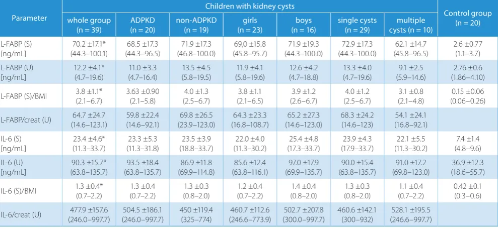
The concentrations of the tested markers (L-FABP, IL-6) in serum and urine in children from the study and con-trol groups are presented in Table 3. The concentration of L-FABP and IL-6 in serum and urine was significantly higher in children with cystic kidney disease compared to the healthy children. In contrast, serum and urinary
Table 1. Characteristics of examined children with renal cysts and controls
Parameter
Children with kidney cysts Control group (n = 20) whole group
(n = 39) ADPKD (n = 20 non-ADPKD (n = 19) single cysts (n = 29) cysts (n = 10)multiple (n = 23)girls (n = 16)boys
Age [years] 10.9 ±4.9 (1.9–19.8)
10.9 ±5.1 (3.4–19.8)
11.0 ±5.0 (1.9–18.7)
10.5 ±4.9 (1.9–19.8)
12.3 ±5.2 (5.6–18.9)
10.8 ±4.6 (3.4–18.7)
11.1 ±5.6 (1.9–19.8)
8.8 ±3.9 (1.8–17.2)
Height [cm] (80.0–184.5)142.3 ±24.9 (97.0–184.5)141.7 ±26.0 142.9 ±24.3 (80–176) (80.0–184.5)140.0 ±25.7 148.8 ±22.2 (115–176) 142.1 ±22.5 (97–173) (80.0–184.5)142.5 ±28.7 130.8 ±22.7 (82–172)
SDS for height (−2.97–2.24)−0.03 ±1.12 −0.05 ±1.00 (−2.97–1.80) –0.02 ±1.2 (−1.9–2.2) (−2.97–1.80)−0.16 ±1.20 (−1.0–2.2)0.3 ±1.0 (−2.97–1.81)0.2 ±1.2 −0.3 ±1.0 (−1.9–2.2) −0.4 ±0.9 (−1.9–2.0)
Body weight [kg] 40.7 ±17.8 (11–78) 41.7 ±19.8 (13–78) 39.6 ±16.0 (11–69) 38.5 ±17.3 (11–70) 47.2 ±18.7 (21–78) 40.5 ±17.6 (13–78) 41.0 ±18.8 (11–70) 33.0 ±16.1 (9.7–63.0)
SDS for BW (−3.20–2.07)0.13 ±1.09 (−3.2–1.9)0.2 ±1.2 (−1.3–2.1)0.05 ±1.0 −0.04 ±1.00 (−3.2–1.7) (−1.4–2.1)0.6 ±1.1 (−3.20–1.91)0.2 ±1.2 (−1.30–2.07)−0.06 ±1.0 −0.003 ±1.000 (−1.6–1.8)
BMI [kg/m²] 18.90 ±3.29 (13.5–28.7)
19.3 ±4.0 (13.5–28.7)
18.4 ±2.4 (14.4–22.3)
18.3 ±2.9 (13.5–25.0)
20.4 ±4.0 (13.9–28.7)
18.9 ±3.7 (13.5–28.7)
18.9 ±2.7 (14.8–23.4)
18.0 ±3.4 (14.2–26.6)
SDS for BMI (−1.38–2.02)0.23 ±0.93 (−1.40–2.02)0.3 ±1.1 (−1.3–1.7)0.2 ±0.8 (−1.4–1.8)0.1 ±0.8 (−1.2–2.0)0.6 ±1.2 (−1.38–2.02)0.3 ±1.0 (−1.3–1.7)0.2 ±0.9 (−1.5–8.8)0.7 ±2.1
SBP 109.8 ±13.3 (90–137) 111.6 ±14.0 (90–137) 107.9 ±12.6 (90–130) 106.4 ±12.0(90–130)a 119.5 ±12.7 (90–137) 111.4 ±13.3 (90–137) 107.4 ±13.4 (90–130)
DBP 64.9 ±8.9 (45–84) 66.7 ±10.5 (45–84) 63.1 ±6.8 (55–75) 62.3 ±7.1(45–75)a 72.7 ±9.6 (55–84) 66.5 ±9.2 (50–84) 62.8 ±8.3 (45–77)
MAP (61.7–98.3)79.9 ±9.8 (61.7–98.3)81.7 ±11.1 (66.7–93.3)78.0 ±8.2 (61.7–93.3)77.0 ±8.1a (73.3–98.3)88.3 ±9.8 (63.3–98.3)81.5 ±10.0 (61.7–93.3)77.7 ±9.3
SDS for SBP 0.58 ±1.00 (−1.22–2.63)
0.8 ±1.1 (−0.8–2.6)
0.4 ±0.9 (−1.2–1.6)
0.34 ±0.9a
(−1.2–2.6)
1.2 ±09 (−0.6–2.2)
0.8 ±1.0 (−0.84–2.63)
0.3 ±0.9 (−1.2–1.8)
SDS for DBP (−0.95–2.07)0.39 ±0.70 (−0.9–2.07)0.5 ±0.8 (−0.8–2.0)0.2 ±0.7 (−0.9–2.0)0.2 ±0.7a (−0.2–2.1)0.9 ±0.6 (−0.6–2.07)0.5 ±0.7 (−0.9–2.0)0.2 ±0.8
Data is presented as: mean ± standard deviation (SD) (minimum–maximum); a p < 0.05 children with single cysts vs children with multiple cysts;
BW SDS – SDS body weight, BMI – body mass index; SBP – systolic blood pressure, DBP – diastolic blood pressure, MAP – mean arterial pressure; ADPKD – autosomal dominant polycystic kidney disease.
Table 2. Laboratory tests and eGFR value in children with renal cysts
Parameter Whole group (n = 39) (n = 20)ADPKD Non–ADPKD(n = 19) Single cysts (n = 29) Multiple cysts (n = 10) (n = 23)Girls (n = 16)Boys
Na
[mmol/L] 140.1 ±2.2 (134–143) 140.0 ±2.5 (134–143) 140.2 ±1.7 (136–143) 140.2 ±1.8 (136–143) 139.9 ±3.1 (134–143) 140.3 ±1.7 (136–143) 139.8 ±2.8 (134–143) Creatinine
[umol/L]
48.0 ±15.6 (22–77)
48.3 ±17.5 (22–77)
47.8 ±13.7 (29–73)
47.6 ±14.9 (22–77)
49.4 ±18.1 (24–76)
45.8 ±13.7 (22–69)
51.25 ±17.90 (27–77) Uric acid
[umol/L] 266.2 ±67.8 (134–370) 267.4 ±66.5 (150–368) 264.9 ±71.0 (134–370) 276.6 ±69.0 (134–370) 235.7 ±56.7 (150–347) 253.4 ±65.0 (134–351) 284.4 ±69.7 (151–370) Urea
[mmol/L]
4.3 ±1.3 (2.4–6.8)
4.4 ±1.4 (2.4–6.8)
4.2 ±1.2 (2.5–6.1)
4.4 ±1.2 (2.4–6.8)
4.0 ±1.4 (2.5–6.6)
4.16 ±1.25 (2.4–6.6)
4.4 ±1.3 (2.5–6.8) eGFR
[mL/min/1.73 m2] (38.2–179.3)119.8 ±32.7 (38.2–176.0)121.5 ±35.0 (47.8–179.3)117.9 ±30.8 (38.2–179.3)115.1 ±35.2 133.3 ±19.9 (103–162) 115.90 ±28.25 (38.2–156.2) 125.3 ±38.5 (47.8–179.3)
concentrations of L-FABP and IL-6 in individual subgroups of children with renal cysts were comparable.
When analyzing the correlation of L-FABP and IL-6 con-centrations in both serum and urine, and anthropometric measurements, we did not show any significant relation-ships in the whole group of children with renal cysts.
We showed a significant negative correlation between se-rum L-FABP concentration and SDS for DBP in the whole study group (Table 4). In children with cystic kidney dis-ease, we observed a significant negative correlation be-tween IL-6 serum concentration and absolute values and SDS for SBP, DBP and MAP. There was also a significant negative correlation between urinary IL-6 concentration and SDS for SBP and DBP. In addition, a significant positive correlation was found between urinary IL-6 concentration and eGFR. Similar correlations were observed in the study
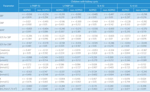
group by analyzing the correlation of IL-6 with body mass index (BMI)(data not shown). However, in the whole group with cystic renal changes, we found a negative correlation between L-FABP/BMI ratio and SBP (r = −0.460, p < 0.01), DBP (r = −0.378, p < 0.05), MAP (r = −437, p < 0.01), SDS for SBP (r = −0.373, p < 0.05), and eGFR (r = −0.398, p < 0.05), and positive correlation between L-FABP/BMI ratio and serum urea concentration (r = 0.343, p < 0.05). We also analyzed correlations occurring in subgroups, divided into ADPKD and non-ADPKD (Table 5). We have dem-onstrated in children with ADPKD a positive correlation between serum L-FABP concentration and serum creati-nine concentration and serum uric acid concentration. In addition, in children with ADPKD, we found a positive correlation between the excretion of L-FABP in the urine and the concentration of serum uric acid. In children from the non-ADPKD subgroup, we found a negative correla-tion between serum L-FABP concentracorrela-tion, SDS for BMI and SDS for DBP and DBP absolute values. In the whole study group with renal cysts, we found a negative cor-relation between L-FABP/BMI ratio and SBP (r = −0.460, p < 0.005), DBP (r = −0.378, p < 0.02), MAP (r = −0.437, p < 0.005), SDS for SBP (r = −0.377, p < 0.02), and eGFR (r = −0.398, p < 0.02), and a positive correlation between L-FABP/BMI ratio and serum urea (r = 0.343, p < 0.05). However, after normalizing the concentration of L-FABP in urine to the concentration of creatinine, we did not observe the above relationships.
When analyzing the correlation of serum and urine IL-6 concentrations with the results of anthropometric measurements and the IL-6/BMI ratio with BP values, we demonstrated a similar relationship in ADPKD chil-dren as in the whole study group, and a significant positive
Table 3. Concentration of the examined parameters in serum and in urine in children with kidney cysts (divided into subgroups) and in controls
Parameter
Children with kidney cysts
Control group (n = 20) whole group
(n = 39) (n = 20)ADPKD non-ADPKD (n = 19) (n = 23)girls (n = 16)boys single cysts (n = 29) cysts (n = 10)multiple L-FABP (S)
[ng/mL]
70.2 ±17.1* (44.3–100.1)
68.5 ±17.3 (44.3–96.5)
71.9 ±17.3 (46.8–100.0)
69,0 ±15.8 (45.8–95.7)
71.9 ±19.3 (44.3–100.0)
72.9 ±17.3 (44.3–100.0)
62.1 ±14.7 (45.8–96.5)
2.6 ±0.77 (1.1–3.7) L-FABP (U)
[ng/mL] 12.2 ±4.1*(4.7–19.6) (4.7–16.4)11.0 ±3.3 (5.8–19.5)13.5 ±4.5 (5.8–19.6)11.9 ±4.1 (4.7–18.8)12.6 ±4.2 13.3 ±4.0 (4.7–19.6) (5.9–14.6)9.1 ±2.5 (1.86–4.10)2.76 ±0.6
L-FABP (S)/BMI 3.8 ±1.1*(2.1–6.7) 3.63 ±0.90(2.1–5.8) (2.5–6.7)4.0 ±1.3 (2.1–6.5)3.8 ±1.1 (2.6–6.7)3.9 ±1.2 (2.5–6.7)4.0 ±1.2 (2.1–4.8)3.1 ±0.8 (0.06–0.26)0.15 ±0.06
L-FABP/creat (U) (14.6–123.1)64.7 ±24.7 (14.6–92.1)59.8 ±22.4 (23.9–123.0)69.8 ±26.5 (16.8–108.7)64.3 ±23.3 (14.6–123.0)65.2 ±27.3 68.3 ±24.2(14.6–123) (16.8–92.1)54.1 ±24.1
IL-6 (S)
[ng/mL] 23.4 ±4.6*(11.3–33.7) (11.3–31.8)23.3 ±5.3 (18.8–33.7)23.5 ±3.9 (11.3–30.2)22.0 ±4.0 (17.3–33.7)25.4 ±4.8 (17.9–33.7)23.9 ±4.3 (11.3–30.2)22.1 ±5.5 (4.8–9.6)7.4 ±1.4 IL-6 (U)
[ng/mL]
90.3 ±15.7* (63.8–135.7)
93.5 ±18.4 (63.8–135.7)
86.9 ±11.8 (69.9–114.8)
85.6 ±12.4 (63.8–116.1)
97.0 ±17.9 (69.9–135.7)
90.0 ±15.4 (63.8–135.7)
91.0 ±17.2 (69.8–123.0)
36.9 ±12.3 (18.6–55.7)
IL-6 (S)/BMI 1.3 ±0.4*(0.7–2.2) (0.7–2.2)1.3 ±0.4 (0.8–2.0)1.3 ±0.3 (0.7–2.2)1.2 ±0.4 (0.8–2.0)1.4 ±0.4 (0.8–2.0)1.3 ±0.3 (0.7–2.2)1.1 ±0.4 0.42 ±0.1(0.3–0.6)
IL-6/creat (U) (246.0–997.7)477.9 ±157.6 (246.0–997.7)504.5 ±186.1 450 ±119.4(325–774) (246.6–773.9)460.7 ±112.6 (300.0–997.7)502.7 ±207.8 460.6 ±142.1(300–932) (246.6–997.7)528.1 ±195.5
Data is presented as: mean ± standard deviation (SD) (minimum–maximum); p *, p < 0.05 the whole group of children with kidney cystic disease vs control group; L-FABP – liver fatty acid binding protein; IL-6 – interleukin 6; S – serum; U – urine; ADPKD – autosomal dominant polycystic kidney disease; BMI – body mass index.
Table 4. Analysis of correlation between blood pressure, eGFR values and the tested markers concentration
Parameter Children with kidney cysts – whole group SBP [mm Hg] serum IL-6 r = –0.485, p < 0.01 DBP [mm Hg] serum IL-6 r = –0.425, p < 0.01 MAP [mm Hg] serum IL-6 r = –0.477, p < 0.01
SBP SDS serum IL-6 r = –0.565, p < 0.0001urine IL-6 r = –0.452, p < 0.01
DBP SDS serum L-FABP r = –0.356, p < 0.05serum IL-6 r = –0.417, p < 0.01 urine IL-6 r = –0.379, p < 0.05 eGFR [mL/min/1.73 m²] urine IL-6 r = 0.400, p < 0.05
correlation between serum IL-6 concentration and serum potassium concentration. Also in children with ADPKD, we found a negative correlation between urinary IL-6 ex-cretion and SDS for SBP and SDS for DBP and DBP ab-solute values. In the subgroup with non-ADPKD, we ob-served only a negative correlation between serum IL-6 concentration and SBP values and SDS for SBP.
In the subgroup of children with single cysts, we ob-served a significant negative correlation between IL-6 se-rum concentration and SBP (r = −0.404, p < 0.05), MAP (r = −0.368, p < 0.05), and SDS for SBP and SDS for DBP (r = −0.633, p < 0.0001, r = −0.381, p < 0.05, respectively). Also, in children with single cysts, we showed a negative correlation between urinary IL-6 excretion and SDS for SBP and SDS for DBP (r = −0.528, p < 0.01, r = −0.523, p < 0.01), and a positive correlation between IL-6 excretion in the urine and eGFR (r = 0.451, p < 0.05). Similar correla-tions were observed by analyzing the correlation between IL-6 and BMI(data not shown). However, we observed a sig-nificant positive correlation between IL-6/creatinine ratio and the age of the examined children (r = 0.388, p < 0.05), height (r = 0.391, p < 0.05) and body weight (r = 0.387, p < 0.05). In a subgroup with multiple cysts, we observed a significant negative correlation between L-FABP/BMI ra-tio and SDS for SBP (r = −0.769, p < 0.01). However, we did not observe similar correlations in the subgroup of chil-dren with single cysts. In the subgroup of chilof chil-dren with
multiple cysts, we observed a negative correlation between serum IL-6 concentration and MAP (r = −0.642, p < 0.05). We also recorded a significant negative correlation be-tween IL-6 and BMI and SBP (r = −0.873, p < 0.01), MAP (r = −0.758, p < 0.05), and serum creatinine (r = −0.708, p < 0.05).
Discussion
Numerous clinical studies have confirmed the increased urinary excretion of L-FABP in kidney diseases in both children and adults. Many studies indicate the usefulness of the L-FABP assay as a biomarker for kidney disease, and L-FABP has also been shown to be able to relieve the occurrence of kidney damage.19,22,32 Oxidative stress
and damage to the proximal renal tubules due to ischemia lead to increased urinary excretion of L-FABP, as shown by Małyszko et al.21 This was also confirmed
by a meta-analysis carried out by Susantitaphong et al., which con-cluded that L-FABP provides adequate data to diagnose AKI and predict the necessity of renal replacement therapy and acute mortality.23
Khatir et al. evaluated the concentration of L-FABP in the group of adult patients with CKD stage 3–4, from which, however, patients with polycystic kidney disease were excluded. These authors showed that elevated L-FABP
Table 5. Evaluation of the correlation between the tested markers and biochemical parameters and blood pressure values
Parameter
Children with kidney cysts
L-FABP (S) L-FABP (U) IL-6 (S) IL-6 (U)
ADPKD non-ADPKD ADPKD non-ADPKD ADPKD non-ADPKD ADPKD non-ADPKD
SBP r = −0.038 p = 0.874 r = −0.254p = 0.294 r = −0.293 p = 0.210 r = −0.092p = 0.709 r = −0.485 p < 0.05 r = −0.497p < 0.05 r = −0.245 p = 0.297 r = −0.298p = 0.216
DBP r = 0.021
p = 0.930
r = −0.493 p < 0.05
r = −0.186 p = 0.433
r = −0.300 p = 0.212
r = −0.469 p < 0.05
r = −0.350 p = 0.142
r = −0.228 p = 0.333
r = −0.392 p = 0.097
MAP r = −0.003 p = 0.991 r = −0.404p = 0.086 r = −0.240 p = 0.307 r = −0.213p = 0.381 r = −0.500 p < 0.05 r = −0.450 p = 0.053 r = −0.247 p = 0.293 r = −0.370p = 0.119
SDS for SBP r = −0.206 p = 0.384 r = −0.393p = 0.096 r = −0.221 p = 0.349 r = −0.128p = 0.600 r = −0.556 p < 0.05 r = −0.603p < 0.01 r = −0.572 p < 0.01 r = −0.417p = 0.076
SDS for DBP r = −0.167 p = 0.481 r = −0.570p < 0.05 r = −0.158 p = 0.505 r = −0.316p = 0.187 r = −0.505 p < 0.05 r = −0.286p = 0.235 r = −0.453 p < 0.05 r = −0.445 p = 0.056
Creatinine
[umol/L] r = 0.497 p < 0.05 r = −0.131p = 0.592 p = 0.314r = 0.237 p = 0.939r = 0.019 r = −0.053 p = 0.824 r = −0.324p = 0.176 p = 0.844r = 0.047 r = −0.367p = 0.122 Urea
[umol/L]
r = −0.088 p = 0.712
r = 0.340 p = 0.154
r = 0.022 p = 0.925
r = 0.377 p = 0.112
r = 0.259 p = 0.270
r = 0.300 p = 0.212
r = −0.222 p = 0.346
r = 0.132 p = 0.589 Uric acid
[mmol/L] r = 0.572 p < 0.01 r = −0.120p = 0.624 r = 0.586 p < 0.01 p = 0.727r = 0.086 p = 0.907r = 0.028 p = 0.403r = 0.203 p = 0.725r = 0.084 p = 0.535r = 0.152 Na
[mmol/L] r = −0.163 p = 0.493 p = 0.548r = 0.147 r = −0.331 p = 0.154 p = 0.112r = 0.376 p = 0.602r = 0.124 r = −0.020p = 0.934 r = −0.041 p = 0.865 r = −0.243p = 0.315 K
[mmol/L]
r = −0.100 p = 0.676
r = 0.431 p = 0.065
r = −0.004 p = 0.986
r = 0.435 p = 0.063
r = 0.457 p < 0.05
r = 0.025 p = 0.920
r = 0.078 p = 0.744
r = 0.024 p = 0.923 eGFR
[mL/min/1.73 m²] r = −0.060 p = 0.802 p = 0.846r = 0.047 r = −0.284 p = 0.224 r = −0.177p = 0.469 p = 0.463r = 0.174 p = 0.321r = 0.240 p = 0.077r = 0.404 p = 0.091r = 0.398
concentration in the urine correlates with the loss of renal function only in the absence of significant albuminuria.33
Matsui et al. followed 244 adult CKD patients for 3 years and confirmed that elevated urinary L-FABP concentration was associated with progression to ESRD and the incidence of severe cardiovascular events in these patients.34
In lit-erature, no one before conducted research on the evalua-tion of L-FABP in a selected group of children with cystic kidney disease, nor did anyone make comparisons between groups of children with single and multiple renal cysts.
In our study, we showed a higher L-FABP concentration both in serum and in urine in children with cystic kidney disease, regardless of the underlying cysts etiology, which may indicate early damage of the proximal tubules even before the occurrence of renal impairment. McMahon et al. reviewed the usefulness of biomarkers in the diagno-sis of kidney disease and indicated that increased urinary L-FABP excretion in patients with CKD correlated with the severity of proteinuria.22 When analyzing
the correla-tion of serum and urine L-FABP concentrathe correla-tions by sub-group division, we showed a positive correlation between serum L-FABP concentration and serum creatinine and uric acid concentration in ADPKD children, which may indicate that L-FABP may be an early marker of CKD development.
Ichikawa et al. performed a study on experimental animals of the RAS-activated mouse model to evalu-ate the renoprotective effect of renal hL-FABP (human L-FABP). This study suggested that the increased expres-sion of renal hL-FABP, known an antioxidant, together with the suppression of angiotensin II type 1 receptor ap-pearance, blocked the production of pro-inflammatory cytokines and diminished the tubulointerstitial damage.35
In our previous work in the same group of children with renal cysts, we also demonstrated the activation of intra-renal RAAS.36 Ichikawa et al. concluded that agents that
amplify the expression of renal L-FABP in the proximal tubules might be useful as therapeutic agents in future.37
In our study, a group of children with cystic kidney dis-ease showed a significant negative correlation between serum L-FABP concentration and SDS for DBP. The above results may indicate the beneficial effect of L-FABP and the contribution of this antioxidant in inhibiting the de-velopment of hypertension in children with renal cystic changes. The pilot studies conducted by us also indicate the need for further research in a larger group of children with kidney cysts.
Many clinical trials in adults and children have con-firmed the association of elevated IL-6 levels with kidney disease, especially CKD progression. It is believed that the cause of this phenomenon is chronic inflammation in the kidney parenchyma, which is a common feature, characteristic for kidney damage due to various reasons. Determination of serum IL-6 concentration may be use-ful in predicting the risk of end-stage renal disease.38
Nakamura et al., similarly to our results, found elevated
IL-6 concentration in ADPKD adults, where the mean age of the study group was 57 years. In addition, these authors confirmed that IL-6 levels decreased as a result of the reno-protective treatment with angiotensin converting enzyme inhibitor and/or angiotensin receptor blockade.39 In our
study, we found in children with cystic renal changes sig-nificantly higher IL-6 concentration in serum and urine, both in subgroups with ADPKD and non-ADPKD as well as with the division due to the number of cysts and gender, in comparison with healthy children, with no difference between individual subgroups. In addition, we have dem-onstrated a significant positive correlation between urinary IL-6 concentration and eGFR, which indicates IL-6 contri-bution in the deterioration of renal function. Menon et al. also showed increased levels of IL-6 in the serum of adoles-cents and adult patients with ADPKD. In this study, they divided the group of patients into 3 subgroups: ADPKD with normal BP and normal eGFR, ADPKD with sion but with normal eGFR and ADPKD with hyperten-sion and reduced eGFR. The highest IL-6 concentrations occurred in the subgroup with ADPKD with hypertension and depressed eGFR.40 In contrast, Soleimani et al. showed
higher levels of L-FABP in urine in patients with ADPKD, but not in serum, compared to the healthy group, which would suggest only the local presence of inflammation in the renal parenchyma.41 Ma et al. confirmed
the associa-tion of 572C> GG IL-6 polymorphism with the develop-ment of hypertension in the Asian population.28 However,
in our study, we showed a significant negative correlation between the serum IL-6 concentration and absolute values and SDS for SBP and DBP, and the value of MAP. There was also a significant negative correlation between urinary IL-6 concentration and SDS for SBP and DBP. Perhaps this is due to the other polymorphisms of the IL-6 gene in the Polish population. Also, the mechanism of activation via binding of IL-6 to its membrane-bound receptor IL-6R, which is named classic signaling (anti-inflammatory ac-tion) or binding via the trans-signaling with pro-inflam-matory features, could play a role.17 These results indicate
the need for further studies on a larger group of children with renal cysts to more accurately assess the role of this cytokine in the course of cystic changes in children and its contribution to the pathogenesis of hypertension.
ORCID iDs
Krzysztof Plesiński https://orcid.org/0000-0002-0783-5620 Piotr Adamczyk https://orcid.org/0000-0001-9557-221X Elżbieta Świętochowska https://orcid.org/0000-0001-5787-7880 Aurelia Morawiec-Knysak https://orcid.org/0000-0003-4914-6513 Aleksandra Gliwińska https://orcid.org/0000-0002-4473-6840 Wojciech Korlacki https://orcid.org/0000-0002-2632-3567 Maria Szczepańska https://orcid.org/0000-0002-6772-1983
References
1. Osathanondh V, Potter EL. Pathogenesis of polycystic kidneys: Sur-vey of results of microdissection. Arch Pathol. 1964;77:510–512. 2. Kim B, King BF, Vrtiska TJ, Irazabal MV, Torres VE, Harris PC. Inherited
renal cystic diseases. Abdom Radiol (NY). 2016;41(6):1035–1051. 3. Audrezet MP, Corbiere C, Lebbah S, et al. Comprehensive PKD1 and
PKD2 mutation analysis in prenatal autosomal dominant polycystic kidney disease. J Am Soc Nephrol. 2016;27(3):722–729.
4. Norman J. Fibrosis and progression of autosomal dominant poly-cystic kidney disease (ADPKD). Biochim Biophys Acta. 2011;1812(10): 1327–1336.
5. Nowak KL, Chonchol M, You Z, Gupta M, Gitomer B. Affected parent sex and severity of autosomal dominant polycystic kidney disease: A retrospective cohort study. Clin Nephrol. 2018;89(3):196–204. 6. Ravine D, Gibson RN, Donlan J, Sheffield LJ. An ultrasound renal cyst
prevalence survey: Specificity data for inherited renal cystic diseases. Am J Kidney Dis. 1993;22(6):803–807.
7. Turco D, Severi S, Mignani R, Aiello V, Magistroni R, Corsi C. Reliability of total renal volume computation in polycystic kidney disease from magnetic resonance imaging. Acad Radiol. 2015;22(11):1376–1384. 8. Ravine D, Gibson RN, Walker RG, Sheffield LJ, Kincaid-Smith P,
Danks DM. Evaluation of ultrasonographic diagnostics criteria for auto-somal dominant polycystic kidney disease 1. Lancet. 1994;343(8903): 824–827.
9. Müller RU, Benzing T. Cystic kidney diseases from the adult nephrol-ogist’s point of view. Front Pediatr. 2018;6:1–8.
10. Simms RJ. Autosomal dominant polycystic kidney disease. BMJ. 2016; 352:1–10.
11. Pinto CS, Raman A, Reif GA, et al. Phosphodiesterase isoform regula-tion of cell proliferaregula-tion and fluid secreregula-tion in autosomal dominant polycystic kidney disease. J Am Soc Nephrol. 2016;27(4):1124–1134. 12. Niemczyk M, Pilecki T, Gradzik M, Bujko M, Niemczyk S, Pączek L.
Blood pressure and intracranial aneurysms in autosomal dominant polycystic kidney disease. Kidney Blood Press Res. 2014;39(6):630–635. 13. Dembowska M, Nieszporek T, Więcek A. Zwyrodnienie wielotorbie-lowate nerek jako przyczyna nadciśnienia tętniczego. Terapia. 2011; 19(7–8):14–18.
14. Schwartz GJ, Munoz A, Schneider MF, et al. New equations to esti-mate GFR in children with CKD. J Am Soc Nephrol. 2009;20(3):629–637. 15. Cornec-Le Gall E, Audrezet MP, Rousseau A, et al. The PROPKD Score:
A new algorithm to predict renal survival in autosomal dominant polycystic kidney disease. J Am Soc Nephrol. 2016;27(3):942–951. 16. Marlais M, Cuthell O, Langan D, Dudley J, Sinha MD, Winyard PJ.
Hyper-tension in autosomal dominant polycystic kidney disease: A meta-analysis. Arch Dis Child. 2016;101(12):1142–1147.
17. Hogan MC, Abebe K, Torres VE, et al. Liver involvement in early auto-somal-dominant polycystic kidney disease. Clin Gastroenterol Hepa-tol. 2015;13(1):155–164.
18. Glatz JF, Van der Vusse GJ. Cellular fatty acid-binding proteins: Their function and physiological significance. Prog Lipid Res. 1996;35(3): 243–282.
19. Xu Y, Xie Y, Shao X, Ni Z, Mou S. L-FABP: A novel biomarker of kidney disease. Clin Chim Acta. 2015;445:85–90.
20. Smathers RL, Petersen DR. The human fatty acid-binding protein family: Divergences and functions. Hum Genom. 2011;5(3):170–191.
21. Małyszko J, Bachorzewska-Gajewska H, Dobrzycki S. Biomarkers of contrast-induced nephropathy: Which ones and what is their clini-cal relevance? Interv Cardiol Clin. 2014;3(3):379–391.
22. McMahon GM, Waikar SS. Biomarkers in nephrology: Core Curricu-lum 2013. Am J Kidney Dis. 2013;62(1):165–178.
23. Susantitaphong P, Siribamrungwong M, Doi K, Noiri E, Terrin N, Jaber BL. Performance of urinary liver-type fatty acid-binding protein in acute kidney injury: A meta-analysis. Am J Kidney Dis. 2013;61(3): 430–439.
24. Schaper F, Rose-John S. Interleukin 6: Biology, signaling and strate-gies of blockade. Cytokine Growth Factor Rev. 2015;26(5):475–487. 25. Wolf J, Rose-John S, Garbers C. Interleukin-6 and its receptors:
A high-ly regulated and dynamic system. Cytokine. 2014;70(1):11–20. 26. Tanaka T, Kishimoto T. The biology and medical implications
of inter-leukin 6. Cancer Immunol Res. 2014;2(4):288–294.
27. Chiesa C, Pacifico L, Natale F, Hofer N, Osborn JF, Resch B. Fetal and early neonatal interleukin 6 response. Cytokine. 2015;76(1):1–12. 28. Ma H, Sun G, Wang W, et al. Association between interleukin-6 -572
C> G and -174- G> C polymorphisms and hypertension: A meta-anal-ysis of case-control studies. Medicine. 2016;95(2):e2416. doi: 10.1097/ MD.0000000000002416
29. Litwin M. Nadciśnienie tętnicze pierwotne u dzieci i młodzieży – pato-fizjologia. [In:]. Litwin M, Januszewicz A, Prejbisz A. ed. Nadciś nienie tętnicze u młodzieży i młodych dorosłych. Zapobieganie, diagnostyka i leczenie. Kraków, Poland: Medycyna Praktyczna; 2011:289–315. 30. Kułaga Z, Litwin M, Grajda A, Gurzkowska B, Napieralska E, Kułaga K.
Rozkłady wartości ciśnienia krwi w populacji referencyjnej dzieci i młodzieży w wieku szkolnym. Standardy medyczne. Pediatria. 2010; 7:100–111.
31. Kułaga Z, Rożdżyńska A, Palczewska I, Grajda A, Gurzkowska B, Napie-ralska E. Siatki centylowe wysokości, masy ciała i wskaźnika masy ciała dzieci i młodzieży w Polsce – wyniki badania OLAF. Standardy medyczne. Pediatria. 2010;7:690–700.
32. Sato R, Suzuki Y, Takahashi G, Kojika M, Inoue Y, Endo S. A newly developed kit for the measurement of urinary liver-type fatty acid-binding protein as a biomarker for acute kidney injury in patients with critical care. J Infect Chemother. 2015;21(3):165–169.
33. Khatir DS, Bendtsen MD, Birn H, et al. Urine liver fatty acid binding protein and chronic kidney disease progression. Scand J Clin Lab Invest. 2017;77(7):549–554.
34. Matsui K, Kamijo-Ikemori A, Imai N, et al. Clinical significance of uri-nary liver-type fatty acid-binding protein as a predictor of ESRD and CVD in patients with CKD. Clin Exp Nephrol. 2016;20(2):195–203. 35. Ichikawa D, Kamijo-Ikemori A, Sugaya T, et al. Renoprotective effect
of renal liver-type fatty acid binding protein and angiotensin II type 1a receptor loss in renal injury caused by RAS activation. Am J Physiol Renal Physiol. 2014;306(6):F655–F663.
36. Plesiński K, Adamczyk P, Świętochowska E, et al. Angiotensinogen and interleukin 18 in serum and urine of children with kidney cysts. J Renin Angiotensin Aldosterone Syst. 2019;20(3):1470320319862662. doi:10.1177/1470320319862662
37. Ichikawa D, Kamijo-Ikemori A, Sugaya T, et al. Renal liver-type fatty acid binding protein attenuates angiotensin II-induced renal injury. Hypertension. 2012;60(4):973–980.
38. Jones SA, Fraser DJ, Fielding CA, Jones GW. Interleukin 6 in renal dis-ease and therapy. Nephrol Dial Transplant. 2015;30(4):564–574. 39. Nakamura T, Sato E, Fujiwara N, et al. Changes in urinary albumin
excre-tion, inflammatory and oxidative stress markers in ADPKD patients with hypertension. Am J Med Sci. 2012;343(1):46–51.
40. Menon V, Rudym D, Chandra P, Miskulin D, Perrone R, Sarnak M. Inflam-mation, oxidative stress, and insulin resistance in polycystic kidney disease. Clin J Am Soc Nephrol. 2011;6(1):7–13.