R E S E A R C H
Open Access
Hox
gene expression during postlarval
development of the polychaete
Alitta virens
Nadezhda I Bakalenko
*†, Elena L Novikova
†, Alexander Y Nesterenko and Milana A Kulakova
Abstract
Background:Hoxgenes are the family of transcription factors that play a key role in the patterning of the anterior-posterior axis of all bilaterian animals. These genes display clustered organization and colinear expression.
Expression boundaries of individualHoxgenes usually correspond with morphological boundaries of the body. Previously, we studiedHoxgene expression during larval development of the polychaeteAlitta virens(formerly
Nereis virens) and discovered thatHoxgenes are expressed in nereid larva according to the spatial colinearity principle. AdultAlitta virensconsist of multiple morphologically similar segments, which are formed sequentially in the growth zone. Since the worm grows for most of its life, postlarval segments constantly change their position along the anterior-posterior axis.
Results:We studied the expression dynamics of theHoxcluster during postlarval development of the nereidAlitta virensand found that 8 out of 11Hoxgenes are transcribed as wide gene-specific gradients in the ventral nerve cord, ectoderm, and mesoderm. The expression domains constantly shift in accordance with the changing proportions of the growing worm, so expression domains of mostHoxgenes do not have stable anterior or/and posterior boundaries.
In the course of our study, we revealed long antisense RNA (asRNA) for someHoxgenes. Expression patterns of two of these genes were analyzed using whole-mountin-situhybridization. This is the first discovery of antisense RNA forHoxgenes in Lophotrochozoa.
Conclusion:Hoxgene expression in juvenileA. virensdiffers significantly fromHoxgene expression patterns both inA. virenslarva and in other Bilateria.
We suppose that the postlarval function of the Hox genes in this polychaete is to establish and maintain positional coordinates in a constantly growing body, as opposed to creating morphological difference between segments.
Keywords:Hox genes, ncRNA, Polychaete, Positional information
Background
The amazing diversity of body plans of bilateral ani-mals is a result of the structural and regulatory evolution of genes directly connected to morpho-genetic processes. Among these, Hox genes, which play a crucial role in regionalization of the anterior-posterior (AP) axis in bilateral animals, are particu-larly interesting [1,2]. These genes display clustered
organization and colinear expression, and are highly conserved.
Current knowledge of the function ofHox genes, and even of their expression patterns, is nonuniform across different clades of Bilateria. While in Deuterostomia and Ecdysozoa Hox gene functions are well studied, at least for vertebrates and arthropods, research in Lophotrochozoa is still at the initial stage. There are only a few studies of this animal group that describe expression patterns of all Hox genes in the cluster [3-5]. However, the Lophotrochozoa group includes an unsurpassed amount of diverse body plans and is very promising for the study of mechanisms of mor-phogenetic evolution.
* Correspondence:[email protected] †Equal contributors
Department of Embryology, Laboratory of Experimental Embryology, SaintPetersburg State University, Oranienbaumskoe sh., 2. St. Peterhof, Saint Petersburg, Russia
One of the major phyla among Lophotrochozoa is An-nelida. Polychaeta is a basal class of Annelida [6]. Many species in this group have indirect development that in-cludes stages of an unsegmented trochophore larva (a trait shared with many other Lophotrochozoa phyla) and a segmented larva, nectochaete. In the postlarval stage, polychaetes generate segments through a subterminal growth zone (GZ).
The diversity of polychaetes is manifest in various seg-ment morphologies. There are species with morpho-logically similar segments, like representatives of the Nereididae family, as well as heteronomously segmented species that have different segments grouped into tag-mata. To date, the expression of Hox genes has been studied in two heteronomously segmented polychaetes,
Chaetopterus and Capitella. As in most bilateral ani-mals, the expression boundaries of individual Hoxgenes in these species correspond with morphological bound-aries of the body [5,7]. These expression patterns are consistent with the possible role of Hox genes in establishing morphological identity along the AP axis.
Our model object is a nereid polychaete Alitta (Nereis) virens. This is an errant homonomously segmented worm. The ontogenesis of this polychaete includes a lecithotrophic trochophore, a nectochaete with three seta-bearing seg-ments and a multi-segmented worm that grows throughout most of its life. This body plan is likely to be basal among Polychaeta [8]. Previously, we completed a study of larval Hox gene expression in nereids Alitta virens
andPlatynereis dumerilii[4]. The genomes of these spe-cies contain the whole complement of Hox genes spe-cific to Lophotrochozoa:Hox1(PG1),Hox2(PG2),Hox3
(PG3), Hox4(PG4), Hox5(PG5), Lox5(PG6–8), Hox7
(PG6–8), Lox4(PG6-8), Lox2(PG6-8), Post2(PG9+) and
Post1(PG9+) [4]. The Hox genes in a segmented larva seem to define its body plan according to the principle of spatial colinearity, as they do in most bilaterian animals.
During postlarval development, A. virens continues to form new segments in the GZ for almost its whole life.
Alitta’s body does not have any apparent morphological
boundaries. This raises a question about the role of the
Hoxgenes in such an animal.
Apart from morphological heteronomy of the seg-ments, most polychaetes display primary segmental heteronomy, which is based on differences between lar-val and postlarlar-val segments. This is characteristic of both heteronomously and homonomously segmented polychaetes. Many researchers point out significant dis-similarities in formation, structure and ability to regener-ate larval and postlarval segments [9-11]. In particular, the larval segments form almost simultaneously by split-ting the single somatic plate into metameres; they do not produce reproductive products, and do not have metanephridia (only protonephridia are present) [4,10,12].
Postlarval segments, on the other hand, are formed se-quentially in the posterior GZ, have metanephridia, and can produce reproductive products. There are some groups of polychaetes that use only one type of segmenta-tion. For example, Polygordiidae lack a segmented larva, and a juvenile worm is formed right after trochophore metamorphosis. Dinophilidae (Archiannelida), on the other hand, have only‘larval’segments. Sometimes within a single family (such as Nereididae), there are species that have all stages of development from trochophore to an adult seg-mented worm (Platynereis dumerilii), as well as species with direct development (Neanthes arenaceodentata) [10,13,14]. Finally, different individuals of the same species can have direct development as well as development through a larval stage; moreover, the larvae can be of differ-ent types (planktonic and bdiffer-enthic) [15,16].
The evolutionary and ontogenetic flexibility of poly-chaetes suggests that stages of their development are inde-pendent ‘modules’, controlled by different morphogenetic programs. In this case, we can expect thatHoxgene ex-pression patterns in larval and postlarval development are significantly different.
In this study, we describe detailed Hox gene expression patterns using whole-mountin-situhybridization (WMISH) in polychaete Alitta virens during postlarval stages. We address three main questions. First, areHoxgenes involved in patterning of postlarval segments? Second, are their expression patterns during postlarval development consist-ent with a conservedHoxgene function to convey morpho-logical segment identity? Third, does A. virens have considerable differences between larval and postlarval Hox
gene expression?
Methods
Animals
Adult Alitta virens were collected near the Kartesh Marine Biological Station of the Zoological Institute (RAS), at the White Sea, Chupa Inlet. Mature animals were caught with a hand net at the water surface during their spawning period (June and July). Artificial fertilization and cultivation of the embryos were carried out at 10.5°C [17]. A culture of postlarval animals was kept in the Laboratory of Experimental Embryology (Peterhof, Russia) under the following conditions: temperature−18°C, salinity−23‰, artificial seawater (Red Sea salt). The size of nechtochaetes is about 0.8 mm; 4 to 6 segment worms, 1 mm; 10 to 12 segment worms, 2 mm; 15 to 20 segment worms, 3 to 4 mm; worms with more than 20 segments, up to 6 mm.
Cloning ofA. virens Hoxgenes
were inserted into pGEMW-T Easy Vector (Promega). Nvi-Hox3 was inserted into pBluescript II SK+ (Fermentas). The vector sequence allows sense and antisense probes to be obtained from different promoters (T7 and Sp6). Ribo-probes were generated from fragments of the following lengths: 548bp for Nvi-Hox1, 580bp for Nvi-Hox2, 550bp forNvi-Hox3, 453bp forNvi-Hox4, 1010bp forNvi-Hox5, 573bp forNvi-Lox5, 522bp forNvi-Hox7, 302bp for Nvi-Lox4, 498bp forNvi-Lox2and 380bp forNvi-Post2.
Whole-mountin-situhybridization (WMISH)
Whole-mount in-situ hybridization was performed for
Alittaas described previously [4] with the following modi-fications. Hybridization was carried out at 65°C, and washings from probes at 67°C. Collagenase treatment (col-lagenase (Sigma) 100 γ/ml, 2.5 mM dithiothreitol (DTT); 1 mM CaCl2) was performed for 5 to 10 min, proteinase K (Sigma) treatment was performed for 10 to 20 min (10 γ/ml). Washings from the probes were performed as follows: 100% Hybe 2 × 60 min, 80% prehybe/20% PTw 2 × 20 min, 50% prehybe/50% PTw 4 × 30 min, 20% prehybe/80% PTw 2 × 20 min, 100% PTw 2 × 20 min at 67°C. Washings from antibodies were carried out for 10 × 20 min in PTw on the shaker. The detailed protocol is available on request. Between 8 and 20 worms were used for each stage. BM-purple (Roche) was used as a
chromogenic substrate to localize the hybridized probe. The time of incubation in substrate was 12 h for sense transcripts and 24 to 48 h for antisense transcripts and sense transcript ofNvi-Lox4. The worms were mounted in clove oil before microscopic analysis. We used the follow-ing negative control: animals taken through the entire in-situprotocol, but with no probe (see Additional file 1). The results were imaged on a DMRXA microscope (Leica) with a Leica DC500 digital camera with Nomarski optics. Optical sections were assembled with Helicon Focus soft-ware. Brightness, contrast, and color values were corrected in all images using Adobe Photoshop CS5 image process-ing software.
Results
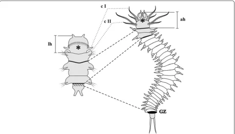
Brief description ofAlitta virenspostlarval development The main stages ofAlitta virenslarval development were described previously [4]. The postlarval growth begins with origination of the fourth segment anlage (this is the first postlarval segment) from posterior GZ (Figure 1). Postlarval segments form sequentially, one by one, in the GZ. When a worm has 6 to 8 segments, its first chaete-bearing segment (the first larval segment) changes signifi-cantly. Its parapodia are considerably reduced, they lose their chaetae, their cirri grow and transform into a second pair of peristomial cirri. Then, the first larval segment
becomes part of the peristome of the adult head. After this stage, the second larval segment becomes the first body segment of a juvenile worm (Figure 1). Formation of new segments continues for most of Alitta’s life and stops
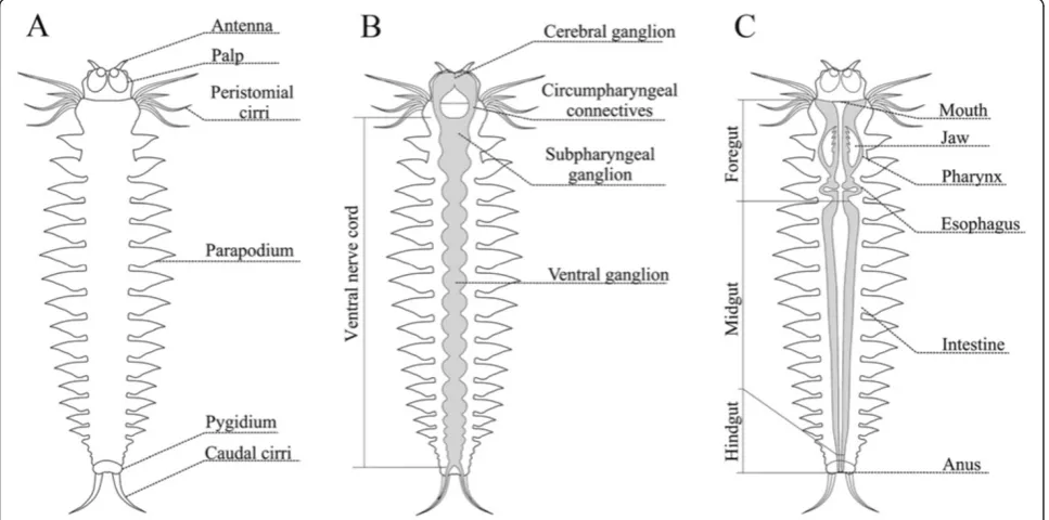
when the worm has about 200 segments. The morphology of the juvenile worm is shown in Figure 2.
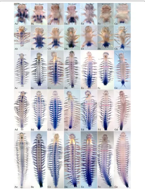
Nvi-Hoxgene expression patterns
To bring descriptive data for expression patterns into a system, we divided ten Hoxgenes into groups based on their expression behavior during larval development [4]. As in larval development [20], Nvi-Post1 is not detect-able in broad ectodermal domains of juvenile worms.
Nvi-Post1expression was observed in the chaetal sacs of developing chaetae (data not shown). Apparently, its ex-pression is not related to the AP patterning, and we will not discuss it here. In general, most Nvi-Hox genes are expressed in parapodia; this calls for a separate study and is not covered in this article.
Nvi-Hox1,Nvi-Hox4,Nvi-Hox5,Nvi-Lox5,Nvi-Post2
The first group includesNvi-Hox1, Nvi-Hox4, Nvi-Hox5,
Nvi-Lox5,and Nvi-Post2. These genes have colinear ex-pression patterns during larval development.
Nvi-Hox1
At the end of the larval development at the late nectochaete stage, expression domain of this gene has a sharp boundary in the first larval segment (Figure 3Aa).
Nvi-Hox1is detected in the ganglia of the ventral nerve cord (VNC) and parapodial ectoderm of all three larval
segments with the highest level in the second segment and the lowest level in the third segment. In addition,
Nvi-Hox1expression marks the bases of peristomial and pygidial cirri. After metamorphosis, the expression ex-pands to include the fourth and the fifth segments (first and second postlarval segments) (Figure 3Ab). Expres-sion in pygidial cirri is significantly reduced. In juvenile worms with six to ten segments, two newNvi-Hox1 ex-pression domains appear: in the digestive tube at the foregut-midgut boundary (Additional file 2) and in the lateral part of the ganglia of the new segments. The ex-pression patterns in anterior and posterior ganglia are different. In anterior segments, expression is detected in most of the ganglia but in posterior segments,Nvi-Hox1
expression appears as two spots in the lateral part of each ganglion (Figure 3Ac-Ae, Additional file 2). Despite significant changes in the first segment associated with the reorganization of the peristome, the anterior border of Nvi-Hox1 expression in this segment is persistent (Figure 3Ab-Ae; Figure 3A,E).Nvi-Hox1expression dur-ing later postlarval stages forms a gradient with two maxima: in the first body segment (second larval seg-ment) and in the ganglia of posterior third of the body.
Nvi-Hox1 expression in the medial segments is weak. The most posterior and youngest segments and the GZ areNvi-Hox1-negative (Figure 3Ab-Ae).
Nvi-Hox4
During larval development,Nvi-Hox4expression is asso-ciated with development of the second larval segment. At the nectochaete stage, expression extends to include
the third segment, but is not detected in the pygidium (Figure 3Ba).
At the onset of postlarval growth, strong Nvi-Hox4
expression is detectable in the ganglia and ectoderm of each nascent segment (Figure 3Bb). In small worms (6 or 7 seg-ments),Nvi-Hox4expression takes on a posterior-anterior gradient (data not shown). The interior border ofNvi-Hox4
expression is retained in the second larval segment. After the first larval segment has merged with the peristome, the second larval segment becomes the first adult body seg-ment.Nvi-Hox4 expression persists in this area during all postlarval stages (Figure 3Bb-Be). In animals larger than ten
segments, the VNC expression gradient changes its shape. NowNvi-Hox4 is expressed intensively in several anterior ganglia and in the most posterior ones, but staining is much weaker in central segments. In the ectoderm Nvi-Hox4 expression is strong in the most posterior newly formed segments and decreases gradually toward the ante-rior end so it is no longer detectable in the middle part of the body. Both gradients (in the VNC and the ectoderm) ‘stretch’ but keep their shapes at all analyzed stages (Figure 3Bc-Be). The anterior border ofNvi-Hox4 expres-sion in VNC is stable and located in the first body segment (Figure 3Bb-Be, Figure 4B,F).
Figure 4Anterior boundaries ofNvi-Hox1(A, E),Nvi-Hox4(B, F),Nvi-Hox5(C, G), andNvi-Lox5(D, H) have been stable since the larval stages.Anterior ends of juvenile worms are in the top row, nectochaetes are in the bottom row. See text for details.
(See figure on previous page.)
Nvi-Hox5
During larval development, expression of this gene begins later thanNvi-Hox1orNvi-Hox4[4] and is limited to the neuroectoderm of the third segment (Figure 3Ca). Since the beginning of postlarval growth, theNvi-Hox5domain expands to include the ectoderm of this segment and the fourth segment surface and the developing neuromere (Figure 3Cb). In juvenile Alitta virens, Nvi-Hox5 is expressed in each postlarval segment that already has parapodia. Several youngest, most posterior segments are
Nvi-Hox5-negative. Moreover,Nvi-Hox5expression is not detectable in the pygidium and the GZ at all analyzed stages (Figure 3Cb-Ce). The expression pattern of this gene takes on a broad bow-shaped gradient. The anterior border of Nvi-Hox5expression is retained in the second body segment (third larval segment) (Figure 3Cc-Ce; Figure 4C,G). The anterior boundary of the expression gradient in the ectoderm is located two or three segments posterior to that of the VNC. The Nvi-Hox5 expression pattern does not change significantly during postlarval growth (Figure 3Cc-Ce).
Nvi-Lox5
This gene displays early expression in larva during the third segment development [4]. At the late larval stages, expression persists in the third segment and expands to include the posterior GZ (Figure 3Da). After metamor-phosis, the nascent postlarval segment displays strong Nvi-Lox5 expression, which gradually decreases toward the anterior end. Staining in the third larval segment is slightly downregulated but does not disappear (Figure 3Db). In worms larger than ten segments, Nvi-Lox5 forms the expression gradient in VNC similar toNvi-Hox4.Nvi-Lox5
expression has two peaks: one in several anterior ganglia and one in posterior segments. The transcription level of
Nvi-Lox5 is weak in the central part of Alitta’s body
(Figure 3Dc-De). Expression in the second body segment is weak but persists at all analyzed postlarval stages (Figure 3Db-De; Figure 4 E,H).
Nvi-Post2
At the nectochaete stage, Nvi-Post2 expression is re-stricted to the pygidium and anal cirri (Figure 3Ga). At the onset of postlarval segmentation, expression of this gene expands to the GZ and new postlarval segments (Figure 3Gb). As the segment moves toward the anterior end, Nvi-Post2 expression weakens and gradually disap-pears (Figure 3Gb-Ge). Nvi-Post2 displays a short posterior-to-anterior expression gradient in the VNC, seg-mental ectoderm, mesoderm, and distal gut. The number ofNvi-Post2-positive segments increases with the growth of the worm, but their percentage of the total number remains virtually the same (Figure 3Gb-Ge).
Nvi-Hox7,Nvi-Lox4,Nvi-Lox2
Expression of these genes starts at the later stages of lar-val development. Their expression zones overlap in pro-spective posterior GZ [4]. They do not take part in larval morphogenesis.
Nvi-Hox7
In the late nectochaete and four-segment juvenile worm,
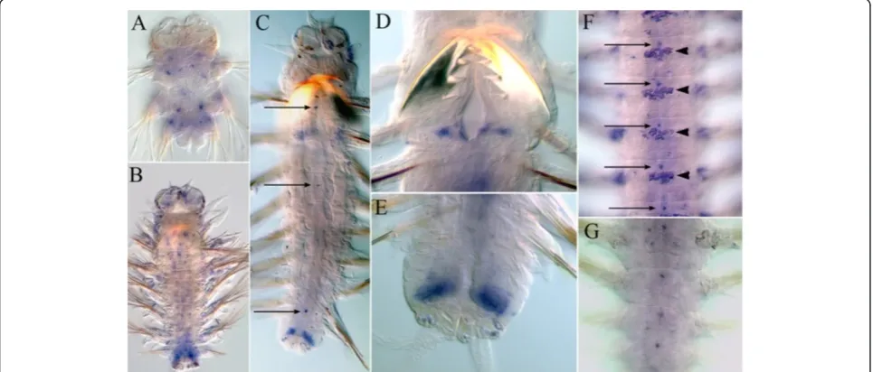
Nvi-Hox7 expression is restricted to the posterior GZ (Figure 3Ea). Expression is initiated in postlarval segments after the formation of the fourth segment (Figure 3Eb). In older worms, theNvi-Hox7 expression pattern takes on a posterior-to-anterior gradient in the VNC, segment ecto-derm, and parapodia (Figure 3Ec-Ee). The expression gradient is proportional to the length of the growing worm body. (Figure 3 Ec-Ee). The anterior expression boundary is in the third segment (Figure 3Eb) in four-segment worms, and moves backwards as the worm continues to grow. In animals with 15to 30 segments, it is located in the fifth to seventh segment (Figure 3Ec-Ee), but since the staining weakens towards the anterior end, we cannot de-termine the exact position of the anterior boundary.
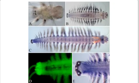
Nvi-Lox4
At all stages, detectedNvi-Lox4expression is very weak, which may be because of the small size of the RNA probe (302 b). Nvi-Lox4 is first detected at the late nectochaete stage in a small number of cells in the an-terior part of the pygidium (Figure 5A). In juvenile worms, the expression level increases with age. The Nvi-Lox4 transcript is spread diffusely in VNC and does not display a sharp metameric pattern (Figure 5B,C). Expres-sion is not detected in the ectoderm within the GZ and in the pygidium (Figure 5D). Low-level expression is detected in the mesoderm of the nascent segments (Figure 5E). Nvi-Lox4 displays a posterior-to-anterior expression gradient (Figure 5C,D). The anterior border of Nvi-Lox4 expression is loose and apparently not stable, likeNvi-Hox7.
Nvi-Lox2
This expression is very weak in the nectochaete (Figure 3Fa), but intensifies significantly with the onset of postlarval segmentation (Figure 3Fb-Fe). The Nvi-Lox2 transcript is detected in the posterior GZ, neural ganglia, and ectoderm of posterior postlarval segments and in the pygidium (Figure 3Fb).Nvi-Lox2also displays a posterior-to-anterior expression gradient, like that for
backwards as the worm continues to grow. In animals with 15 to 30 segments, it is located in the sixth to eighth segment (Figure 3Fc-Fe), but since the staining weakens towards the anterior end, we cannot determine the exact position of the anterior boundary.
Nvi-Hox2,Nvi-Hox3
These genes are expressed in an intensive and dynamic manner during early larval development [4], but at the nectochaete stage their expression domains narrow considerably.
Nvi-Hox2
In the nectochaete, Nvi-Hox2 is detected in small patches of cells in all larval segments (Figure 6A). A new expression domain appears in the mesodermal part of the GZ after metamorphosis (Figure 6B,C,E). Weak dif-fuse expression ofNvi-Hox2 is detected in nascent seg-ments in small juvenile worms (Figure 6B). Older worms do not have this expression domain. At the late juvenile stages, Nvi-Hox2 is expressed in the mesoderm of the
GZ and in small patches of 3 to 5 cells at the midline within each segment (Figure 6C,F,G). In worms of 8 to 10 segments and larger worms, Nvi-Hox2 is also detected in the pharynx (Figure 6E).
Nvi-Hox3
Since the early nectochaete stage, Nvi-Hox3 expression is present in the GZ. At all analyzed juvenile stages, strong, ring-like expression is detected in ectodermal cells of the GZ (Figure7A-E).
Antisense transcripts
We found antisense transcripts for some Nvi-Hoxgenes (Nvi-Hox1, Nvi-Hox4, Nvi-Hox5, Nvi-Hox7). Expression of Nvi-antiHox5 and Nvi-antiHox7 was analyzed in de-tail using WMISH.
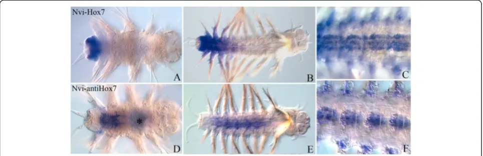
Nvi-antiHox5
After the beginning of postlarval segmentation, Nvi-antiHox5is detectable in the GZ and the posterior seg-ments. Sense and antisense transcripts have adjacent
and even slightly overlapping expression domains. The
Nvi-antiHox5expression domain is more posterior than senseHox5(Figure 8A,D). In a juvenile worm,antiHox5
is detected in the GZ and the most posterior segments, where sense Nvi-Hox5 is absent (Figure 8B,C,E,F). Dur-ing postlarval development, Nvi-antiHox5 expression does not have a stable anterior border. Its expression do-main constantly shifts toward the posterior end.
Nvi-antiHox7
After the beginning of postlarval growth, this transcript is detected in VNC of the third and fourth segments an-terior in relation to the sense transcript (Figure 9A,D). The anterior border of Nvi-antiHox7 expression is al-ways closer to the head than the sense transcript. Unlike the Nvi-Hox7 sense transcript, Nvi-antiHox7 is absent from nascent segments and the GZ (Figure 9B,E). In
Figure 7Nvi-Hox3expression pattern in nectochaete (A) and at different stages of postlarval development (B-D).The anterior end is to the right on all panels. (B) 4-segment worm. (C) 10-segment worm. (D) 25-segment worm. (E) Pygidium at higher magnification. See text for details.
older worms, sense and antisense Nvi-Hox7 domains seem to be broadly overlapping, but particular expres-sion patterns in the same ganglia seem to be different (Figure 9C,F). These RNAs are probably expressed in different neurons.
Discussion
Summary ofHoxgene expression
According to our results, mostNvi-Hoxgenes (Nvi-Hox1,
Nvi-Hox4,Nvi-Hox5,Nvi-Lox5,Nvi-Hox7,Nvi-Lox4, Nvi-Lox2, and Nvi-Post2) form expression gradients in the central nervous system and ectoderm ofA. virens. These gradients overlap significantly, but their span and shape are unique for each gene (Figures 3,5,10).
We do not have any data on physical linkage ofAlitta Hox genes. However, among Lophotrochozoa, the clus-ter organization of Hox genes was shown for Capitella sp. I[5]. We presume that the genomic order of theHox
genes ofA. virensis similar to that ofCapitella sp. I.
The spatial colinearity is clearly present for Nvi-Hox1,
Nvi-Hox2,Nvi-Hox4,Nvi-Hox5,andNvi-Lox5genes, which have well-defined anterior boundaries (Figures 4). For Nvi-Hox1,Nvi-Hox4,Nvi-Hox5,andNvi-Lox5, these boundaries were established during larval development (Figure 4). An-terior boundaries of otherHoxgenes (Nvi-Hox7,Nvi-Lox4,
Nvi-Lox2, and Nvi-Post2) are not so clear, but the spatial organization of their expression domains also shows some colinearity. TheNvi-Hox7expression gradient spreads fur-ther towards the anterior end of the body than the gradi-ents of Nvi-Lox2 and Nvi-Post2. Nvi-Post2 displays the shortest posterior-to-anterior gradient (Figure 3Ec-Ee,Fc-Fe,Gc-Ge). According to our results, theNvi-Lox4anterior boundary lies posterior to the anterior boundary of Nvi-Lox2expression. This seems to violate the spatial colinear-ity principle. However, the detected expression of Nvi-Lox4is very weak; therefore, we cannot ascertain the true localization of its anterior boundary. The only gene that does not display any colinear expression is Nvi-Hox3,
Figure 9Expression ofNvi-Hox7(top row, panels A, B and C) andNvi-antiHox7(bottom row, panels D, E and F).The anterior end is to the right on all panels. All views are ventral. Black and white asterisks mark the background. (A, D) Expression in 4-segment worm. (B, E) Expression in juvenile worms of about 10 segments. (C, F). Several central segments of the juvenile worms of about 20 segments at higher magnification are shown. Expression patterns forNvi-Hox7(C) andNvi-antiHox7in VNC are different. See text for details.
which is expressed in the GZ of the worm from the nectochaete stage.
Hoxgene expression in annelids
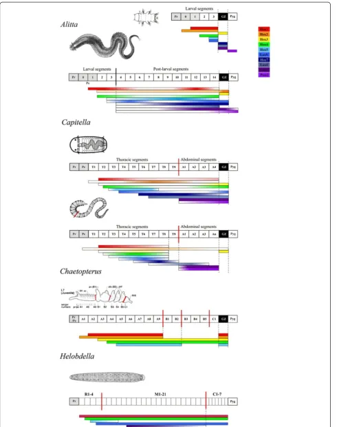
In most bilateral animals,Hoxgenes regionalize the AP axis of the body. A discrete distribution of Hox proteins divides the early embryo into separate domains by differential regu-lation of target genes. This leads to establishment of expres-sion domains correlated with morphological boundaries of the body regions [21-23].Hoxgene expression in previously studied annelids is consistent with this general principle [5]. At the moment there are only a few studies concerning expression of Hox genes in annelids. Among these are studies on the expression of some Hox genes in larval development of Chaetopterus sp. [7] and Platynereis dumerilii [4], development of the leech Helobdella sp.[24], and larval and postlarval development of the polychaeteCapitella sp.I[5].
Among all studied polychaetes, Chaetopterus has the most morphologically complex larva. It consists of three tagmata (A, B, and C), and there are morphological dif-ferences not only between segments in different tagmata, but also within a single tagma (В). At the late larval stages, posterior boundaries of СН-Нох1 and СН-Нох2
expression coincide with the boundary between tagmata A and B, and posterior boundary ofСН-Нох5expression coincides with the border between morphologically di-verse segments within tagma B (Figure 10) [7].
The Capitella sp. I body consists of a thoracic region, which includes nine larval segments, and an abdominal region, which includes four additional larval and all of the postlarval segments. There is no great morphological difference between thoracic and abdominal segments. During development, expression boundaries of some
Hox genes are stabilized in the region between thoracic and abdominal tagmata (Figure 10) [5].
Even leeches, being highly specialized annelids, retain the axial specification of the nervous system by means of the Hox cluster. The leech Helobdella sp. consists of 32 segments: 4 anterior R1-R4, 21 central М1-М21, and 7 caudal С1-С7. The body nervous system of the leech is
patterned by Hox genes according to the principle of spatial colinearity. Anterior expression boundaries of He-Hox7 (Hox1), He-Lox6(Hox4), He-Lox20(Hox5),and He-Lox5 genes correspond to nervous system structures in four sequential segments of the rostral region, while the posterior boundary ofHe-Lox2expression correlates with the anterior border of the caudal ganglion (Figure 10) [24].
A. virensandP. dumeriliihave larvae with morphologic-ally similar segments. The anterior expression boundaries of Hox genes coincide with the segments’ borders (Nvi-Hox1, Nvi-Hox2, Nvi-Hox4, Nvi-Hox5, Nvi-Lox5). The posterior boundaries are located between the seg-mented area and the pygidium. TheNvi-Post2gene marks the pygidium territory (Figure 10) [4].
During most of its postlarval life,A. virenscontinues to form morphologically identical segments, which are not divided into tagmata. In this case, mostHoxgene expres-sion domains do not possess the stable posterior boundar-ies that lie in the nascent segments or in the GZ. The expression domains ofA. virens Hoxgenes cover most of the body and overlap significantly. Comparison of theHox
gene expression in annelids with different body plans con-firms that the presence of posterior expression boundaries correlates with the presence of different body tagmata, as previously shown for other bilaterian animals, for example, Arthropoda [22].
Larval and postlarval developmental programs
As mentioned, there are significant differences in forma-tion of larval and postlarval segments of the polychaetes. These differences indicate the primary heteronomy of polychaete segments. Our data on Hox gene expres-sion during ontogenesis of A. virens reveals the differ-ences in molecular mechanisms of patterning of larval and postlarval segments.
First, a certain subset of Hox genes is expressed in each larval segment. Meanwhile, the postlarval segments express all Hox genes but not at the same time during postlarval growth. Each nascent segment expresses all
Hox genes except Nvi-Hox1, Nvi-Hox3, and Nvi-Hox5. As new segments continue to form, the older segments (See figure on previous page.)
Figure 10Comparison ofHoxgene expression patterns across annelids.Generalized diagram comparingHoxgene expression along the main body axis ofAlitta virenswith theHoxexpression inCapitella,Chaetopterus, andHelobdella. Body axes are shown as boxed diagrams next to the species names and include segments and tagmata for each species. Black vertical lines mark the boundary between larval and postlarval segments forAlitta virens, red vertical lines mark tagmata borders for other species. ForAlittaandCapitella, expression is shown at late larval stages and for juvenile animals. Solid color bars indicate uniform expression level, while gradient color bars indicate gradient expression. Larval Nvi-Hoxgene expression patterns have stable colinear expression boundaries like other polychaetesHoxgenes. During postlarval development, mostNvi-Hoxgenes have gradient expression patterns. During worm growth, anterior boundaries ofNvi-Hox7,Nvi-Lox4,Nvi-Lox2, andNvi-Post2 shift from the fourth (first postlarval) segment toward the posterior end, while the posterior boundaries ofNvi-Hox1andNvi-Hox5move toward the anterior end.CapI-Hoxgenes have stable expression boundaries corresponding to the morphological tagmata boundaries, but expression of manyHoxgenes is not uniform and forms a gradient. Abbreviations: GZ, growth zone; Pe, peristome; Pr, prostomium. Taxon-specific
move towards the head and begin to express Nvi-Hox1
and Nvi-Hox5, while the expression of other genes is gradually downregulated or upregulated according to the shape of gene expression gradients (Figure 11). Interest-ingly, neural expression of ParaHox cluster genes in A. virensis governed by the same principle; each ParaHox
gene is expressed at different periodsin all postlarval (but not the larval) neuromeres [25].
Second, three Hox genes, Nvi-Hox7, Nvi-Lox4 and
Nvi-Lox2, are not expressed during larval segment for-mation [4]. They are switched on in each postlarval seg-ment on its formation in the GZ (Nvi-Hox7, Nvi-Lox2) or at the beginning of growth (Nvi-Lox4).
Taken together, the existing morphological data and the differential character of Hox gene expression in lar-val and postlarlar-val development of A. virens support the idea of separate morphogenetic developmental programs in the segmented larva and adult worm.
Comparing Hox gene expression in different poly-chaetes (Chaetopterus, Capitella, Platynereis, Alitta), one can notice the fundamental similarity of Hox gene expression in their larval development. In all four cases (Figure 10),Hoxgenes are activated early in wide spatial domains of the larval body. The Hox gene expression corresponds to the location of primordial structures or-ganized along the main body axis, and the expression anterior boundaries are colinear. These features link the program of polychaete larval body organization to the programs utilizing the Hox genes during the embryonic
development of animals in other evolutionary clades, such as Deuterostomia and Ecdysozoa.
Apart from A. virens,Hoxgene expression in only one other postlarval polychaete has been studied to date,
Capitella sp. I[5]. Unfortunately, it is difficult to compare these two worms. All the segments of A. virens formed from the subterminal GZ are considered to be postlarval. In contrast, several segments ofCapitellathat are referred to as larval ones are formed sequentially from the poster-ior GZ [26]. The Hox gene expression inCapitella after metamorphosis is considerably different from that in
Alitta. First, in postlarval segments onlyCapI-Lox4, CapI-Lox2, andCapI-Post2genes are active. Second,Hoxgene expression in a juvenile worm is maintained only within the neural system ganglia. Third, expression boundaries in the juvenile worm are stable and correspond to the mor-phological tagma boundaries.
Nevertheless, a comparison of Hox ortholog expres-sion in late Capitellalarva, when it has already formed several segments from the posterior GZ, and juvenileA. virens reveals fundamental similarities in many gene patterns. Hox1, Hox4, and Lox5 genes have anterior as well as posterior expression domains in both species (Figure 11). In both cases, the expression has a gradient shape (Figure 3) [5]. We can speculate that this stage in
Capitella development is a separate phase that can be compared to the postlarval development ofA.virens.
There are also significant differences of Hoxgene ex-pression between larval and juvenile stages of Capitella
development. In juvenile stages of Capitella, almost all expression domains of Hox genes become limited to the VNC and discrete posterior expression boundaries appear [5].
Our data and results for Capitella suggest that there are actual differences between larval and postlarval de-velopmental programs. The question arises: which mode of segmentation, larval or postlarval, is more ancestral? Several authors [12,27] believe that larval segmentation (that is, the simultaneous metamerization of the somatic plate that creates larval segments) is a basic type, while sequential formation of postlarval segments from the GZ is an evolutionary novel segmentation type. Ander-son, on the other hand, believes that postlarval segmen-tation is a primitive type [14,27].
During A. virens nectochaete development, only the ‘ancient’ Hox genes are expressed. Indeed, the minimal
Hox gene complement of Urbilateria, the last common ancestor of bilateral animals, consisted of at least seven
Hox genes. It contained five anterior genes (PG1-5), at least one gene from a central group (PG6/8), and at least one posterior gene (PG9+) [19]. This is exactly the set of Hox paralogs that is expressed during development ofA. virens larva [4]. At the onset of postlarval growth, the genes of the central group, namelyHox7, Lox4, and
Lox2, are activated. This may support the notion that postlarval segmentation is an evolutionary novelty. How-ever, there is a certain contradiction with data for larval
Hox gene expression in Capitella. All CapI-Hox genes are expressed during larval segment formation, including
CapI-Hox7,CapI-Lox4, andCapI-Lox2.
The existing evidence is not sufficient to draw final conclusions about larval and postlarval developmental programs in the polychaetes. This emphasizes the need forHoxgene expression studies in other polychaete fam-ilies, since this will probably elucidate the evolution of postlarval morphogenesis in this class.
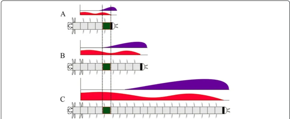
PossibleHoxgene function inA. virenspostlarval development
The nereid A. virens produces morphologically similar segments for almost its whole life. According to our data, most of A. virens Hox genes are expressed in the segment ectoderm and in the neural system of each postlarval segment. Hox gene transcripts are distributed as complex gene-specific gradients. As the worm grows, the position of each segment along the body AP axis changes, and this is accompanied by a change in Hox
gene expression profile (Figure 11). We assume that the system ofHoxgene transcriptional gradients is necessary not for specification of segment morphology, but to cre-ate and maintain the positional information for each seg-ment. Within the constantly growing polychaete body, this regulatory system allows one to assign each
metamere a unique‘Hox code’, which gradually changes as the segment moves with respect to the terminal struc-tures, that is, the head and pygidium (Figure 11). In this case, the ‘Hox code’ serves for positional, rather than morphological, specification.
Establishing and maintaining of positional information may be necessary for regeneration. Nereididae can re-generate posterior body parts. After a part of the body is lost, it is necessary to change the scale of the positional coordinates quickly in accordance with the new bound-aries of the body. Since genes in the Hox cluster are often coordinated by common regulatory elements and a common feedback system, one can expect that the ex-pression pattern of the whole complex would be easily reorganized during positional failure.
Planarian worms (Turbellaria; Platyhelminthes) also have gradientHoxgene expression. In planarianDugesia japonica,Plox4-Dj(PG5;Hox5),Plox5-Dj(PG6-8; Lox5), and Dj-Abd-Ba(PG9+, Post2) transcripts are distributed as gradients along the AP axis of the adult worm [28,29]. The key aspect is that in this caseHoxgene ex-pression does not correspond to any morphological structures, has no clear boundaries and is proportional to the length of the worm body. It was also experimen-tally shown that Dj-Abd-Ba gene quickly restores ex-pression pattern in head and tail regenerates, long before the new head and tail are formed, and this pat-tern is consistent with the new planarian body propor-tions [29].
Persistent 5'HoxC gene expression was surprisingly dis-covered in the spinal cord of an adult newtPleurodeles waltl
[30]. RT-PCR data showed thatPwHoxc13,PwHoxc12, and
PwHoxc10 expression is upregulated during tail regen-eration. The authors of this study suggest thatHoxgenes in an adult newt act as carriers for the positional memory necessary to achieve effective regeneration [30].
It is noteworthy that in newts and polychaetes the de-finitiveHoxgene expression is associated with the neural system. Expression patterns of variousHoxgenes in theA. virensVNC ganglia are somewhat different (Figure 3).Hox
Hoxgenes cooptions inA. virens
SomeAlitta Hoxgenes are likely to take part in patterning of particular structures rather than working in an orches-trated way together with other genes of the cluster. For ex-ample,Nvi-Hox1andNvi-Hox2genes are expressed in the pharynx and at the esophagus-midgut boundary. Interest-ingly, their orthologs in other studied polychaetes, such as
Capitella and Chaetopterus, are also expressed in the pharynx [5,7]. In addition,Hoxorthologs in deuterostomes are involved in establishing the posterior boundary of pharyngeal entoderm [35,36]. This cooption is likely to date back to the early stages of evolution, probably to the origins of all Eubilateria.
Hox1 orthologs work in peristomial and pygidial cirri in A. virens and P. dumerilii. Since Hox1 expres-sion is found in the notopodia and neuropodia of these polychaetes, one can assume that it is expressed in cirri as in serially homologous structures and thus is not purely a cooption. In general, most Nvi-Hox
genes are expressed in parapodia, and this calls for a separate study. It is noteworthy, however, that this ex-pression changes in accordance with general gradient pattern in the neural system and segment ectoderm of the worm.
The aforementioned Nvi-Post1 gene was identified in parapodial chaetal sacs not only inAlittaandPlatynereis, but even in Capitella [5,20], which is located far from Nereididae in the phylogenetic tree [37,38]. This suggests that this gene fell out of the common regionalization pro-gram early on.
In contrast to other Lophotrochozoa, Hox genes in
Alittahave a rather limited range of cooptions. For ex-ample, Nvi-Hox7 functions only in the neural system and in the segment ectoderm of postlarval segments, while its Capitella ortholog CapI-Antp has additional expression domains in the brain and pharynx [5]. In P. dumerilii, the Lox2 gene works in the coelomic epithe-lium; this sets it apart from other Pdu-Hox genes and the Nvi-Lox2 gene, which exhibits strong expression in ectodermal tissues [31].
In general, studiedHoxgene cooptions in different spe-cies within the Lophotrochozoa clade support an idea that these genes are easily involved in new morphogenetic pro-grams. They often play a role in the morphogenesis of structures, specific for large taxa within this animal group. For example, they pattern the shell gland in gastropods and the brachial crown in cephalopods [3,39].
Nvi-Hoxgenes antisense transcripts
Our results represent the first discovery ofHoxgene anti-sense transcripts in an animal from the Lophotrochozoa clade. These are long antisense RNA, complementary to the sense transcripts.
Hoxcluster regulation by noncoding RNA was reported for mammals and arthropods [40,41]. Over 200 noncoding RNAs of different sizes and directions are transcribed from four human Hox clusters [40]. Our finding sug-gests that this is a universal regulatory mechanism in bi-lateral animals.
The Nvi-antiHox5 transcript is expressed in a pattern almost complementary to the sense transcript (Figure 8). This resembles anti-Ubx expression in centipedes [41,42].Anti-Ubxis functional in embryogenesis and hibits a complex expression pattern that is mutually ex-clusive with the sense transcript. There have been no functional studies so far, but this early and specific na-ture of expression suggests a possible regulatory role of
anti-Ubx.
The Nvi-antiHox7 anterior expression boundary lies ahead of Nvi-Hox7 (Figure 9). Both transcripts are detected in VNC ganglia and in the segment ectoderm, but their expression patterns are different within the same ganglia (Figure 9C,F). The mutually exclusive terri-tories of sense and antisense transcript distribution sug-gest that one may be controlled by another.
So far, we have identified antisense transcripts for most A. virens Hox genes. Some of them are cloned (data are being prepared for publication). It should be noted that Hox genes long antisense RNAs in poly-chaetes are located in the cytoplasm. We do not know their function or biogenesis, but existing examples allow us to suppose that antisense RNAs take part in epigen-etic tuning of the Hox cluster. They can participate in transcriptional and translational repression of their tar-gets, protein-encoding RNAs. Nvi-antiHox5 and Nvi-antiHox7 expression patterns suggest that they may be controlling the anterior (Nvi-Hox7) or posterior ( Nvi-Hox5) expression boundaries of their sense counterparts. The worm is constantly growing, and its Hox genes change levels of expression in a coordinated manner in accordance with the body proportions. In this situation, turning the genes on and off quickly at a translational level may be very advantageous.
Conclusions
The data on expression patterns ofA. virensandCapitella sp.I Hoxgenes support the idea of different morphogenetic programs underlying larval and postlarval development of the polychaetes. The larval program seems to be more conservative among the polychaetes. The principles of
Hox gene expression during larval segmentation are similar to those during the body plan formation of other bilaterians. The postlarval programs display more differ-ences between species and seem to be more evolution-ary flexible.
maintaining the positional values along the AP axis of multi-segmented, constantly growing polychaete body. The role of A. virens Hoxcluster in positional specifica-tion can be studied in regeneraspecifica-tion experiments.
The system of long antisense RNAs is a possible clue to the question of ‘sliding’Hox gene expression boundaries in growing worms. This has not yet been described in existing model systems (planarian, mouse, drosophila), and we aim to investigate this phenomenon thoroughly.
Additional files
Additional file 1:Negative control with no probe.All these worms were processed in the same WMISH. (A,B,C) The worms with different numbers of segments.(D,E,F)The posterior ends of 25 to 35 segment worms in higher magnification. The anterior end is to the right on all panels. All views are ventral. (A) The small worms do not display any background. (B-E) Weak background is detected in the gut (arrowheads), epithelial glands (arrows), parapodia, pygidial glands (red arrows) and at the ventral surface of the heads of 15 to 35 segment animals. (F) Some worms display strong background in the parapodia and pygidial glands. We have never seen any background in the nervous system or mesoderm. WMISH, whole-mountin-situhybridization.
Additional file 2:Some details ofNvi-Hox1expression. (A)Nvi-Hox1 expression in the postlarval worm (overview). Images on panels(B)and (C)indicate the different expression patterns ofNvi-Hox1in the anterior (C) and posterior (B) ganglia of the VNC.(D)Nvi-Hox1expression in the peristomial cirri.(E)Nvi-Hox1expression in the pharynx and foregut-midgut boundary. VNC, ventral nerve cord.
Abbreviations
AP:Anterior-posterior; asRNA: Antisense RNA; bp: Base pair; DTT: Dithiothreitol; GZ: Growth zone; RT-PCR: Reverse transcriptase polymerase chain reaction; VNC: Ventral nerve cord; WMISH: Whole-mount in-situhybridization.
Competing interests
The authors declare that they have no competing interests.
Authors’contributions
NIB, ELN, and MAK performed the experiments and data analysis. AYN participated in material collection, maintenance of the worm culture and editing the manuscript. ELN, NIB, and MAK conceived the study and drafted the manuscript. All authors have read and approved the final manuscript.
Acknowledgements
We are grateful to Tanya Andreeva, who laid the foundation for this research; to Michael Akam and Charles Cook, who helped to clone Nvi-Hox -genes fragments and who supported us. We thank the staff of the Kartesh White Sea Biological Station (Zoological Institute, Russian Academy of Sciences) for help in collecting and maintaining theA. virens.
We thank Olga B. Lavrova and Victor Starunov for their help in maintaining theAlitta virensculture in the Laboratory of Experimental Embryology. We are grateful to the Chromas center for providing the opportunity to use the Leica microscope. The research was supported by RFBR grants: 06-04-49654-a 06-04-49654-and 09-04-01322-06-04-49654-a.
Received: 29 November 2012 Accepted: 29 January 2013 Published: 1 May 2013
References
1. Carroll SB:Homeotic genes and the evolution of arthropods and chordates.Nature1995,376:479–485.
2. Wray GA:Transcriptional regulation and the evolution of development.
Int J Dev Biol2003,47:675–684.
3. Lee PN, Callaerts P, de Couet HG, Martindale MQ:CephalopodHox
genes and the origin of morphological novelties.Nature2003,
424:1061–1065.
4. Kulakova M, Bakalenko N, Novikova E, Cook CE, Eliseeva E, Steinmetz PRH, Kostyuchenko RP, Dondua A, Arendt D, Akam M, Andreeva T:Hoxgene expression in larval development of the polychaetesNereis virensand
Platynereis dumerilii(Annelida, Lophotrochozoa).Dev Genes Evol2007,
217:39–54.
5. Fröbius AC, Matus DQ, Seaver EC:Genomic organization and expression demonstrate spatial and temporalHoxgene colinearity in the LophotrochozoanCapitella sp. I.PLoS One2008,3(12):e4004. doi:10.1371/ journal.pone.0004004.
6. Brusca RC, Brusca GJ:Invertebrates. Massachusetts: Sinauer; 2002. 7. Irvine SQ, Martindale MQ:Expression patterns of anteriorHoxGenes in
the polychaete Chaetopterus: correlation with morphological boundaries.DevBiol2000,217:333–351.
8. Tessmar-Raible K, Arendt D:Emerging systems: between vertebrates and arthropods, the Lophotrochozoa.CurrOpin Genet Dev2003,13:331–340. 9. Wilson EB:The cell lineage ofNereis.J Morph1892,6:361–480. 10. Ivanoff PP:Die Entwicklung der Larvalsegmentebei den Anneliden.Z
Morph Ökol Tiere1928,10:62–161.
11. Beklemishev WN:Principles of Comparative Anatomy in Invertebrates. Moscow: Nauka; 1969.
12. Fischer AHL, Henrich T, Arendt D:The normal development ofPlatynereis dumerilii(Nereididae, Annelida).Frontiers in Zoology2010,7:31. 13. Anderson TD:Embryology and Phylogeny in Annelids and Arthropods. Oxford:
Pergamon Press; 1973.
14. Winchell CJ, Valencia JE, Jacobs DK:Confocal analysis of nervous system architecture in direct-developing juveniles ofNeanthes arenaceodentata
(Annelida, Nereididae).Frontiers in Zoology2010,7:17.
15. Gibson GD, Gibson JF:Heterochrony and the evolution of poecilogony: generating larval diversity.Evol2004,58(12):2704–2717.
16. Kesäniemi JE, Geuverink E, Knott KE:Polymorphism in developmental mode and its effect on population genetic structure of a spionid polychaete,Pygospio elegans.Integr Comp Biol2010,52(1):181–196. 17. Dondua AK:Effect of actinomycin D and sibiromycin on the embryonic and
larval development ofNereis virens(Sars.).Ontogenez1975,6(5):475–484. 18. Andreeva TF, Kuk C, Korchagina NM, Akam M, Dondya AK:Cloning and
analysis of structural organization ofHoxgenes in the PolychaeteNereis virens.Ontogenez2001,32(3):225–233.
19. de Rosa R, Grenier JK, Andreeva T, Cook CE, Adoutte A, Akam M, Carroll SB, Balavoine G:Hoxgenes in brachiopods and priapulids and protostome evolution.Nature1999,399:772–776.
20. Kulakova MA, Kostyuchenko RP, Andreeva TF, Dondua AK:The abdominal-B -like gene expression during larval development ofNereis virens
(Polychaeta).Mech Dev2002,115(1–2):177–179.
21. Averof M, Akam M:HOM/Hoxgenes ofArtemia: implications for the origin of insect and crustacean body plans.Curr Biol1993,3:73–78.
22. Hughes CL, Kaufman TC:Hoxgenes and the evolution of the arthropod body plan.Evol Dev2002,4(6):459–499.
23. Burke AC, Nowicki JL:Hoxgenes and axial specification in vertebrates.
Amer Zool2001,41:687–697.
24. Kourakis MJ, Master VA, Lokhorst DK, Nardelli-Haefliger D, Wedeen CJ, Martindale MQ, Shankland M:Conserved anterior boundaries ofHoxgene expression in the central nervous system of the leechHelobdella.Dev Biol1997,190:284–300.
25. Kulakova MA, Cook CE, Andreeva TF:ParaHoxgene expression in larval and postlarval development of the polychaeteNereis virens(Annelida, Lophotrochozoa).BMC Dev Biol2008,8:61.
26. Seaver EC, Thamm K, Hill SD:Growth patterns during segmentation in the two polychaete annelids Capitella sp. I and Hydroides elegans: comparisons at distinct life history stages.Evol Dev2005,7(4):312–326. 27. Giangrande A, Gambi MC:Metamerism and life‐style within polychaetes:
morpho-functional aspects and evolutionary implications.Ital J of Zool 1998,65(1):39–50.
28. Orii H, Kato K, Umesono Y, Sakurau T, Agata K, Watanabe K:The planarian
HOM/HOXHomeobox genes (Plox) expressed along the anteroposterior axis.Dev Biol1999,210:456–468.
29. Nogi T, Watanabe K:Position-specific and non-colinear expression of the planarian posterior (Abdominal-B-like) gene.Dev Growth Differ2001,
30. Nicolas S, Papillon D, Perez Y, Caubit X, Le Parco Y:The spatial restrictions of 5'HoxC genes expression are maintained in adult newt spinal cord.
Biol Cell2003,95:589–594.
31. Pfeifer K, Dorresteijn AWC, Fröbius AC:Activation ofHoxgenes during caudal regeneration of the polychaete annelidPlatynereis dumerilii.Dev Genes Evol2012,222:165–179.
32. Ikuta T, Yoshida N, Satoh N:Ciona intestinalisHoxgene cluster: its dispersed structure and residual colinear expression in development.
PNAS2004,101(42):15118–15123.
33. Tara A, Christof N, Robb K:Hoxgenes and segmentation.Annu Rev Cell DevBiol2009,25:431–456.
34. Hinman VF, O’Brien EK, Richards GS, Degnan BM:Expression of anterior
Hoxgenes during larval development of the gastropodHaliotis asinina.
Evol Dev2003,5:508–521.
35. Schubert M, Yu JK, Holland ND, Escriva H, Laudet V, Holland LZ:Retinoic acid signaling acts viaHox1to establish the posterior limit of the pharynx in the chordate amphioxus.Dev2005,132:61–73. 36. Aronowicz J, Lowe CJ:Hoxgene expression in the hemichordate
Saccoglossus kowalevskiiand the evolution of deuterostome nervous systems.Integr Comp Biol2006,46(6):890–901.
37. Struck TH, Schult N, Kusen T, Hickman E, Bleidorn C, McHugh D, Halanych KM:Annelid phylogeny and the status of Sipuncula and Echiura.BMC Evol Biol2007,7:57.
38. Zrzavý J,Říha P, Piálek L, Janouškovec J:Phylogeny of Annelida (Lophotrochozoa): total-evidence analysis of morphology and six genes.
BMC Evol Biol2009,9:189.
39. Samadi L, Steiner G:Expression ofHoxgenes during the larval development of the snail,Gibbulavaria (L.)-further evidence of non-colinearity in molluscs.Dev Genes Evol2010,220(5):161–172. 40. Rinn JL, Kertesz M, Wang JK, Sharon L, Squazzo SL, Xu X, Brugmann SA,
Goodnough H, Helms JA, Farnham PJ, Segal E, Chang HY:Functional demarcation of active and silent chromatin domains in humanHOXloci by non-coding RNAs.Cell2007,129(7):1311–1323.
41. Brena C, Chipman AD, Minelli A, Akam M:Expression of trunkHoxgenes in the centipedeStrigamia maritima: sense and anti-sense transcripts.
Evol Dev2006,8(3):252–265.
42. Janssen R, Budd GE:Gene expression suggests conserved aspects ofHox
gene regulation in arthropods and provides additional support for monophyletic Myriapoda.Evo Devo2010,1:4.
doi:10.1186/2041-9139-4-13
Cite this article as:Bakalenkoet al.:Hoxgene expression during postlarval development of the polychaeteAlitta virens.EvoDevo2013
4:13.
Submit your next manuscript to BioMed Central and take full advantage of:
• Convenient online submission
• Thorough peer review
• No space constraints or color figure charges
• Immediate publication on acceptance
• Inclusion in PubMed, CAS, Scopus and Google Scholar
• Research which is freely available for redistribution