Tarekegn
et al. World Journal of Pharmaceutical and Life Sciences
HEPATIC APOPTOSIS: BASIC MOLECULAR CONCEPTS AND MECHANISMS
Tarekegn Gebreyesus Abisso1*, Yao Mawulikplimi Adzavon1*, Pengxiang Zhao1*, Xujuan Zhang1, Xin Zhang1, Mengyu Liu1, Limin Wang1 and Xuemei Ma1
1
College of Life Science and Bio-engineering, Beijing University of Technology, Beijing 100124, P. R. China.
Article Received on 13/02/2019 Article Revised on 05/03/2019 Article Accepted on 06/03/2019
1. INTRODUCTION
Liver is an organ with numerous roles, it has essential functions in biosynthesis, metabolism, secretion, excretion and detoxification.[1] Liver has maximum metabolic activity and hence its energy needs is substantial.[2] Liver is the biochemical hub utilizing 20% of oxygen in the whole body and is vital in the oxidation of fat, sugar, protein, vitamins and salt.[3] Because of its exceptional role and anatomical position, liver is susceptible to exogenous xenobiotics and toxins, comprising pollutants, medicines and alcohol, as well as to infection by viruses, and consequently, is highly vulnerable to tissue damage.[68] Elevated liver cell death and diminished renewal are undeniably the characteristics of many liver diseases.[1]
Apoptosis or programmed cell death (PCD) is a highly organized and genetically controlled type of cell death, which allows the elimination of damaged or surplus cells
in order to maintain tissue homeostasis and regulation of physiological functions.[4-6] Essentially any modification in this equilibrium, either in the direction of unnecessary multiplying or extreme apoptosis, results in the progression of disease situations.[100] Widespread varieties of both physiological and pathological conditions can activate apoptosis. Some of these conditions include: DNA fragmentation, intracellular damage, extracellular signals, toxins, heat, free radicals, nitric oxide, oxidative stress, and radiation.[7] Cells that die during normal development and tissue homeostasis have distinguishing morphological hallmarks, comprising cytoplasmic decline, chromatin shortening and marginalization and plasma membrane blebbing, which are accompanied by biochemical features such as DNA fragmentation, membrane alterations (that is, exposure of phosphatidylserine on the outside of the plasma membrane), and degradation of specific cellular proteins, as a result of the huge activation of a large number of intracellular proteases and endonucleases.[8,9]
World Journal of Pharmaceutical and Life Sciences
WJPLS
www.wjpls.org
SJIF Impact Factor: 5.088
*Corresponding Author: Tarekegn Gebreyesus Abisso
College of Life Science and Bio-engineering, Beijing University of Technology, Beijing 100124, P. R. China.
ABSTRACT
Elimination of unwanted, injured or diseased cells by apoptosis is the requirement for the maintenance of homeostasis and regulation of physiological functions in multicellular organisms. Because of its exceptional role and anatomical position, liver is susceptible to exogenous xenobiotics and toxins, and highly vulnerable to tissue apoptosis. Apoptosis, which is accompanied by biochemical features such as DNA fragmentation, membrane alterations, and degradation of liver cells can be initiated by extrinsic or intrinsic pathways by means of death signals from the cell exterior. The extrinsic pathway is activated by the binding of a group of transmembrane receptors (death receptors) to their related ligands. Some of the death receptors in liver comprise Fas or CD95, Tumor TNF-R1, TRAIL-R2 also called death receptor 4 and 5 (DR4 and DR5). The intrinsic pathway usually activates apoptosis through members Bcl-2 family, which control mitochondrial outer membrane permeabilization, cytochrome C release, and consequently caspase initiation. Caspases are a family of aspartate-specific cysteinyl proteases that are triggered during and assist the execution of apoptosis in liver cells. A group of caspases that are triggered first in the process (upstream) are called initiation caspases, while others that bring the important structural hits of apoptosis are called executioner caspases. Caspases, that is, caspase -2, -8, -9, and -10, belong to the group of initiator caspases, while caspases, that is, caspase- 3, -6, and -7, belong to the group of executioner enzymes. Any malfunctioning in the course of liver apoptosis might result in various kinds of liver disorders from auto-immune diseases to the high risk hepatocellular carcinoma (liver cancer). In this review, we addressed some common molecular concepts and mechanisms of hepatic apoptosis and circumstances that are linked to hepatic apoptosis.
It is vital to know that apoptosis is not all the time a deleterious process. It is essential during embryonic development to ensure proper organogenesis as well as for the health of adult organisms, and serves to eliminate damaged or malfunctioning cells from the body. Thus, apoptosis plays a vital part in ensuring the appropriate functioning of several organs.[10] Surplus of PCD could be detrimental and results in several deteriorating degenerative pathologies, while absence of PCD can also contribute to the progression of proliferative disease conditions like cancer.[88] In apoptosis, the dying cell is split into membrane bound vesicles comprising fairly intact organelles and chromatin remains named ‘apoptotic bodies’ which are easily engulfed by nearby cells and professional phagocytes, such as liver dwelling macrophages or Kupffer cells.[10] For these details, understanding of molecular mechanisms of the liver cells death could have apparent clinical significance. In this review article, we highlighted some common molecular events of hepatic apoptosis and circumstances that are linked to hepatic apoptosis.
2. Approaches of Hepatic Cell Death
As its name indicates, hepatic apoptosis means cell death in liver. The hepatocyte apoptosis designates cell death in hepatocytes (one type of liver cells). Liver is an organ comprising of numerous phenotypically different cell types, for instance, hepatocytes, cholangiocytes, hepatic stellate cells (HSCs), oval cells, sinusoidal endothelial cells, and so on.[11] Hepatocytes are dominant forms of liver cells where clinically documented apoptosis mainly happens. Principally hepatocytes make up 70–80% of the liver mass.[12,21] The two main forms of cell death in the liver are apoptosis and necrosis.[13] Chen et al.[13] indicated that apoptosis is identified to occur in liver in hepatocytes and is a significant part of the firmly controlled homeostatic mechanisms regulating liver role. One of the hallmarks of liver is that it has an incredible regenerative ability in reply to cell loss through infectious, physical, or hepatotoxic damage.[83]
In opposing to the controlled cellular death program called apoptosis, necrosis or currently retitled oncotic necrosis is a more disordered approach of cell death. It results from metabolic disruption with energy reduction and loss of adenosine triphosphate (ATP).[84] Rounding and inflammation of mitochondria, Cellular edema, dilations of the endoplasmic reticulum (ER), lysosomal disruption and development of plasma membrane protrusions called blebs are the results of loss of ATP.[15] Hence, necrotic cell death is morphologically characterized by an expansion in cell size (oncosis), swelling of organelles, plasma membrane rupture and resulting loss of intracellular contents.[16] Necroptosis (caspase- free cell death) has been used to designate this substitute cell death program.[66]
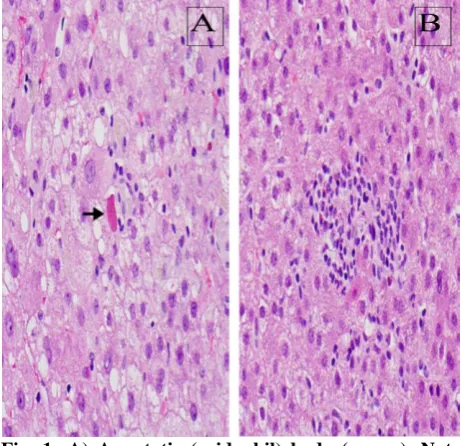
Fig. 1: A) Apoptotic (acidophil) body (arrow). Note the concentration and shady stain of the cytoplasm and lack of nucleus. B) Spotty necrosis. Numerous neighboring hepatocytes are lacking and substituted by inflammatory cells. Modified from.[46]
Table 1: Summary of morphological, molecular and biochemical features distinguishing hepatic apoptosis and necrosis.
Feature Apoptosis Necrosis Ref.
Cell size Reduced (shrinkage) Enlarged (swelling) [65]
Nucleus
Condensation and margination of
chromatin, often to characteristic crescent-shape. Very often breakup into sharply defined spherical chromatin masses that are finally scattered throughout the cytosol
Rarely compression (pyknosis) at early steps; later generally breakup
(karyorrhexis) and dissolution (karyolysis). On light microscopic level, early pyknotic stages may occasionally resemble apoptosis
[66]
Cell membrane Intact, changed structure specially orientation of lipids
breakdown and introduction of inflammation
[67]
DNA
fragmentation
Typical high molecular weight DNA fragmentation into 50kbp - 300kbp fragments.
Sometimes accidental DNA degradation. Often little DNA
degradation before lysis of the plasma membrane
[68]
Cellular contents Intact, may be released in apoptotic bodies Enzymatic digestion may leak out of cell [69] Nearby
inflammation No Frequent
Physiologic or pathologic role
Often physiologic, means of removing unwanted cells, may be pathologic after some form of cell injury particularly DNA damage
Always pathologic, ends with an irreversible cell injury
[70]
Phagocytosis Quick removal by professional phagocytes and by neighboring cells
Clearance of necrotic tissue by infiltrating phagocytes after breakdown of cells
[71]
Caspase initiation Caspase dependent No caspase participation [66]
Tissue reaction
Frequently unremarkable; mostly reconstitution and integrum; often
apoptosis necessary for tissue organization
Often inflammatory response, leukocyte infiltration, production of immune
mediators; often formation of scar tissue or disturbance of tissue organization
[72]
3. Mechanisms of Hepatic Apoptosis
Apoptosis denotes a programmed type of cell death that is essential to keep tissue homeostasis by balancing cell propagation and eradicating injured, diseased, or stressed cells.[5] This process is chiefly significant in the liver as an organ that is obviously exposed to toxins, drugs, and viruses.[17] Cell proliferation and tissue homeostasis that are ruled by various physiological growth-control mechanisms are related to apoptosis. Apoptosis mechanisms are extremely intricate and sophisticated, including an energy dependent cascade of molecular events.[18] The process of apoptosis in liver is controlled by diversity of cell signaling pathways and involved in regulation of cell fate, death or survival.[19]
Apoptosis might hence be regarded, in biochemical positions, as a caspase-mediated form of cell death.[20] To date, apoptotic signaling within the hepatocytes is transduced primarily through two distinct molecular pathways, even though it can be initiated by numerous stimuli: (a) extrinsic or death receptor related and (b) intrinsic or mitochondrial.[20,91] The final fate of both the extrinsic and intrinsic pathways is activation of a wide variety of intracellular proteases (especially a group of proteolytic enzymes called caspases) and endonucleases that eventually destroy the cellular ingredients.[6]
3.1. The Extrinsic pathway
The extrinsic pathway refers to a signaling pathway activated by the binding of a precise group of transmembrane receptors (death receptors) to their
related ligands. Certainly, apoptosis in the liver is principally facilitated by death receptors in disease conditions.[21] These receptors are expressed in several tissues and cells, as well as hepatic tissue. Apoptosis mediated by these receptors has the most important role in a diversity of biological practices, such as tissue injury, immunologic and lymphocytic homeostasis, and defense against pathogenic microorganisms.[22]
Some of the death receptors comprise first apoptotic signal (Fas) or CD95, tumor necrosis factor receptor - α-receptor 1 (TNF-R1) and tumor necrosis factor related apoptosis inducing ligand receptors 1 and 2 (TRAIL-R1 and TRAIL-R2) also called death receptor 4 and 5 (DR4 and DR5).[23] Their associated ligands (FasL/CD95L, TNF-α, and TRAIL) are chiefly expressed by cells of the immune system and play an essential role in the eradication of virally infected, distorted, or injured hepatocytes. Extrinsic pathway needs the enrolment of the Fas associated protein with death domain (FADD) that binds to the intracellular area of the death receptor.[25] The Ligand/receptor binding encourages the recruitment of numerous adapter proteins and proenzymes (procaspase-8 and -10) at the intracellular domain of the receptor to form a complex usually referred to as death inducing signaling complex (DISC). The signal produced at the DISC by triggered caspases results in cell death which, depending on the cell type, may or may not need the participation of mitochondria for its implementation.[24,25]
Table 2: Summary of the features of some death receptors.
Deathreceptor Alternativename Deathdomain Expressioninliver Ref.
Fas APO-1/CD-95 Yes Yes [61,82]
TNF-R1 APO-2 Yes Yes [62,63]
TNF-R2 - No Yes [64]
DR-3 TRAMP/ Apo-3 Yes No [75]
TRAIL- R1 DR-4 Yes ? [74]
TRAIL-R2 DR-5 Yes Yes [75]
TRAIL-R3 DcR1 No Yes [76]
TRAIL-R4 DcR2 Reduced Yes [77]
Fas and TNF-R1 expression have been evaluated in purified liver cell preparations by both Northern blot and immunoblot investigation. Expression of DR-3 and the TRAIL receptors have been assessed exclusively by
death receptor 4; DR-5 = death receptor 5; TRAIL-R1 = tumor necrosis factor related apoptosis inducing ligand receptors 1; TRAIL-R2 = tumor necrosis factor related apoptosis inducing ligand receptors 2; RTAIL-3 = tumor necrosis factor related apoptosis inducing ligand receptors 3; TRAIL-4 = tumor necrosis factor related apoptosis inducing ligand receptors 4; TRAMP = TNF receptor related apoptosis mediated protein; DcR1 = decoy receptor 1; DcR2 = decoy receptor 2). Modified from.[61]
3.2. The Intrinsic pathway
This pathway is influenced by an enormous range of non-receptor factors which may positively or negatively affect the initiation of apoptosis. These factors comprise but are not restricted to viral infections, toxins, physical stimuli, hormones, free radicals and reactive oxygen.[26] This pathway usually activates apoptosis through members of the B-cell lymphoma 2 (Bcl-2) family, which control mitochondrial outer membrane permeabilization, cytochrome C release, and consequently caspase initiation.[90] The main component of this pathway includes the release of mitochondrial contents due to increased alterations in mitochondrial outer membrane permeabilization (MOMP) to release a proapoptogenic factors, including cytochrome C, second mitochondria-derived activator of caspases (SMAC)/direct inhibitor apoptosis protein (IAP), and apoptosis-inducing factor (AIF), from the intermembrane space into the cytoplasm.[27-30] In the presence of adenosine triphosphate (ATP), the cytochrome C is combined with apoptosis protease-activating factor (Apaf-1) that is combined with caspase recruitment domain and caspase-9 precursor to trigger caspase-9, leading to activation of caspase-3 and caspase-7. Bcl-2 family, which plays a significant role in the mitochondrial pathway contains anti-apoptotic proteins including Bcl-2, Bcl- XL and Mcl-1, and pro-apoptotic proteins such as Bax, Bad and Bek, where Bcl-2 and Bax are more important proteins.[31]
This signaling cascade, termed as the mitochondrial (intrinsic) pathway of apoptosis, is triggered by the activation of pro-apoptotic B cell lymphoma Homology (BH3)-only (i.e., Bid, Bim, Bad, PUMA, Noxa) and multi-domain (Bax and Bak) members of the Bcl-2 family, which are accountable for the initiation of MOMP, and antagonized by the anti-apoptotic members of the same family (Bcl-2, Bcl-xL, Mcl-1).[7] The mitochondrial BAX and BAK move from the cytosol to the outer membrane to form permeability change pores, activating cascade of signaling actions such as the release of apoptotic factors and mitochondrial rupture.[89] Whereas, anti-apoptotic Bcl-2 and Bcl-xL inhibit BAX and BAK initiation by binding and impounding pro-apoptotic proteins, and preserve mitochondrial integrity.[32] Released cytochrome c links with Apaf-1 to form the apoptosome, a large multimeric complex which recruits procaspase 9 and facilitates its auto activation. Caspase 9 then cleaves and stimulates caspase 3 and 7,
which in turn, continue to degrade several cellular substrates, resulting in the morphological changes associated to apoptosis.[33] At the same time, endogenous cellular IAPs, usually inhibiting accidentally activated caspases, are neutralized by SMAC/DIABLO, which is released from the mitochondria together with cytochrome C. The extrinsic and intrinsic pathways are not mutually exclusive, as some cells, comprising hepatocytes and cholangiocytes, have been displayed to necessitate mitochondrial participation to increase the apoptotic sign from death receptors.[34,35]
Fig. 2: An overall diagrammatic illustration of the pathways of apoptosis. Modified from.[62]
4. Caspases and their Inhibitors
caspases, while short pro-domain caspases, that is, caspase- 3, -6, and -7, belong to the group of executioner enzymes.[38,39]
Majority of caspases involve in apoptosis through proteolytic stimulation of other caspases and by degrading cellular proteins, hence contributing to the progression of cell death. Certain caspases produce mature proinflammatory cytokines and, thus, regulate immune responses.[40] However, it becomes gradually obvious that caspases and apoptosis-regulatory molecules apply significant roles beyond that of cell death, comprising the regulation of T cell production and cell-cycle progress.[41] Besides the Bcl-2 family, caspase inhibitors, such as IAP family members, may protect against apoptotic cell death by interrupting the caspase cascade. IAPs, XIAP, and c-IAP-1 have been revealed to
bind procaspase 9 and stop its initiation, and can also directly bind and hinder active caspases.[42] Overexpression of the human homologue of cIAP2 did protect hepatocytes against apoptosis in vitro. IAP family members specifically inhibit caspases -3 and -9. Their action can be stopped by Second mitochondrial-derived activator of caspase/ Direct IAP-binding protein (Smac/DIABLO) and Omi/HtrA2.[43] As a result, the mild equilibrium between the comparative cytosolic concentrations of active caspases-3, -8, and -9 and Smac/DIABLO, OMI/HtrA2 compared with IAP family members determine cell fate. Heat shock proteins (HSPs) have likewise apoptosis regulatory functions. HSP27 binds to cytochrome c, and HSP70 and HSP90 bind to Apaf-1, resulting in the inhibition of apoptosome formation.[44]
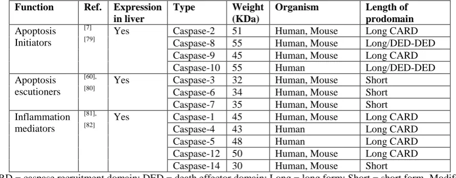
Table 3: Commonly known essential caspases participating in apoptosis.
Function Ref. Expression in liver
Type Weight
(KDa)
Organism Length of
prodomain Apoptosis
Initiators
[7] [79]
Yes Caspase-2 51 Human, Mouse Long CARD
Caspase-8 55 Human, Mouse Long/DED-DED
Caspase-9 45 Human, Mouse Long CARD
Caspase-10 55 Human Long/DED-DED
Apoptosis escutioners
[60], [80]
Yes Caspase-3 32 Human, Mouse Short
Caspase-6 34 Human, Mouse Short
Caspase-7 35 Human, Mouse Short
Inflammation mediators
[81], [82]
Yes Caspase-1 45 Human, Mouse Long CARD
Caspase-4 43 Human Long CARD
Caspase-5 48 Human Long CARD
Caspase-12 50 Human, Mouse Long CARD
Caspase-14 30 Human, Mouse Short
CARD = caspase recruitment domain; DED = death effector domain; Long = long form; Short = short form. Modified from.[60]
5. Apoptosis in some Liver Diseases
Liver cell apoptosis plays an essential part in the regulation of normal liver role, which is facilitated by numerous signal transduction pathways,[45] However, apoptosis serves as the cytological origin for the development of various liver diseases. Thus, diminishing non-physiological apoptosis in hepatocytes has vital clinical implication for keeping hepatic structure and function. Some researchers indicated that apoptosis may occur in response to viral infection, extreme alcohol
consumption, and contact to any sort of
hepatocarcinogen, or due to genetic mutations.[46] Elevated levels of cell death related receptors are expressed in liver dwelling cells, for example hepatocytes, activated stellate cells, Kupffer cells, and cholangiocytes express Fas. Prominently expressed Fas receptors aid to sustain liver homeostasis and also to remove virally infected cells of liver by the immunocytes.[20]
5.1. Apoptosis and Hepatocellular Carcinoma Hepatocellular carcinoma (HCC) is regarded as one of the well-known malignancies globally.[101] It is mainly
malignancy of the liver and the third top cause of cancer death worldwide.[47] Non-alcoholic steatohepatitis (NASH), Chronic viral hepatitis related liver cirrhosis, (Hepatitis B virus (HBV) and hepatitis C virus (HCV), hereditary diseases like hemochromatosis, ethanol consumption, contact to hepatotoxins (aflatoxin), obesity and diabetes denote the chief risk factors for HCC progression.[48,93-95] As in all other body parts, some extent of hepatocyte apoptosis is typical of a healthy liver.[94] Undeniably, currently, it has become obvious that development and evolution of HCC are connected in specific with both defective apoptosis and increased cell propagation.[102] Specially, tumor cells frequently display modifications in genes regulating the apoptotic mechanism.[49] Nevertheless, the exact molecular mechanisms of apoptosis regulation participated in hepatocarcinogenesis are still not well understood.
malignant cells, hyperplasia and tumor progression. Therefore, decreased apoptosis or its resistance plays an essential part in HCC. There are numerous ways a malignant cell can obtain decrease in apoptosis. Usually, the mechanisms by which avoidance of apoptosis happens could be roughly categorized into: 1) disordered balance of pro-apoptotic and anti-apoptotic proteins, 2) weakened death receptor signalling and 3) diminished caspase role.[51]
Inhibition of apoptosis in HCC needs IAPs, which hinder caspase initiation. Survivin, a class of the IAP family, can play a significant part in development of HCC by encouraging cell proliferation, and is clearly associated with extraordinary risk of disease reappearance and poor diagnosis in HCC.[47] For patients with HCC after hepatectomy, the appearance of surviving might consequently help as a predictive factor. Transfection of liver tumor cells (HepG2) with antisense oligonucleotide (ASO) against surviving results in substantial cells growth inhibition and lessening appearance of survivin. Additionally, systemic treatment with ASO meaningfully prevents tumour growth in an orthotopic transplant model of HCC in nude mice, signifying that ASO could possibly be an encouraging gene therapy approach to treatment of HCC.[52] To date, there are no recognized prognostic factors or standardized treatments for HCC relapse. The understanding of the causes of the disease and its pathogenesis is important for existence after HCC reappearance.[53]
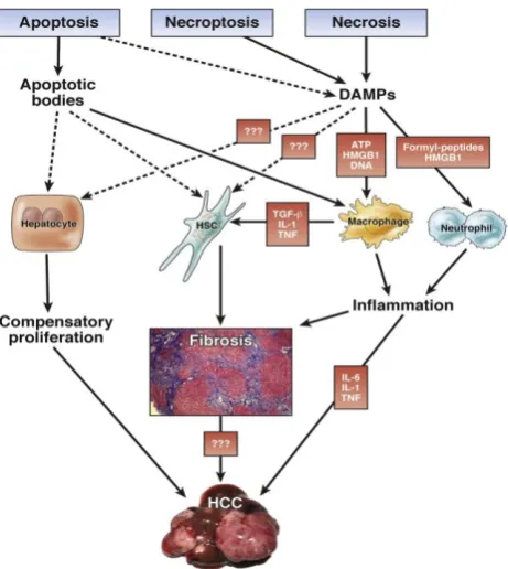
Fig. 3: Impact of specific cell death modes and subsequent cell death responses on development of liver disease. In acute and chronic liver diseases, apoptosis, necroptosis, and necrosis may promote hepatocyte proliferation, HSC activation, and inflammatory cell recruitment and activation. Although these responses are
beneficial in the short term, they result in maladaptive responses that result in development of fibrosis and HCC in the long term. Precise mechanisms by which apoptotic bodies and DAMPs promote development of chronic liver disease remain to be determined. (HSC = hepatic stellate cell; HCC = hepatocellular carcinoma; DAMPs = Damage-associated molecular patterns; TNF = tumor necrosis factor; IL = interleukin; TGF = transforming growth factor). Modified from.[27]
5.2. Acute liver failure
Acute liver failure (ALF) is a serious clinical disorder with high mortality degrees in which a formerly normal liver fails within short period of time like, few days or weeks.[54] ALF is a clinical condition defined by the abrupt start of severe liver damage and is characterized by encephalopathy and coagulopathy in individuals with previously normal liver function. Infections, toxins, or metabolic and genetic disease are the diverse causes of ALF regardless of their etiology.[55] ALF results from quick and widespread hepatic apoptosis and necrosis. The chief causal agents of the hepatic injury that activates the beginning of ALF comprise hepatotropic viral infections and the use of medications such as acetaminophen. To date, liver transplantation is effective option of treatment for ALF. Nevertheless, having to limited donor accessibility and the difficulty in choice of graft compatibility, liver transplantation is not feasible in majority of cases, hence, the pathogenesis of ALF needs to be further explored.[56]
Imbalance in immune system serves a serious role in the progression of ALF. Specifically, pro‑inflammatory and anti‑inflammatory cytokine imbalance in the liver and circulation elicits extreme immune response and brings adverse consequences.[56] Death receptor facilitated hepatic apoptosis is regarded as one of the most common pathologic mechanisms of the initiation of ALF from various causes, including fulminant hepatitis, viral hepatitis, autoimmune hepatitis, and endotoxic shock.[57] Being one of the death receptors, Fas/CD95 is plentifully expressed in hepatocytes and plays a key role in normal liver homeostasis by which virally infected or unhealthy hepatocytes are ruined via Fas/CD95 ligation. The expression of Fas is highly up-regulated in the livers of patients with fulminant hepatic failure and acute hepatitis.[58]
5.2. Apoptosis and Liver fibrosis
cells (HSCs) into myofibroblasts.[87] The activated HSCs quickly produce huge quantities of pro-fibrogenic cytokines, such as transforming growth factor-beta (TGF-β),[96] connective tissue growth factor (CTGF), and platelet-derived growth factor-beta (PDGF-β),[97] they then produce extracellular matrix (ECM) comprising collagen kinds I, III, and IV in hepatic tissues.[98,99]
TGF-𝛽1 from both paracrine and autocrine origins has been revealed to be an important facilitator of liver fibrogenesis.[88] Preventing HSCs activation is the key purpose in the therapy of alcohol induced apoptotic hepatic damage.[99]
6. CONCLUDING REMARKS
Apoptosis or programmed cell death is genetically controlled mode of cell death, which is essential for all multicellular organisms to balance tissues homeostasis and regulate cell propagation, also eliminate injured or unwanted cells. Apoptosis is usually accompanied by numerous distinctive metabolical and morphological modifications. Liver apoptosis is an exceptionally intricate process, with energy-dependent undertaking of events that takes place by two kinds of pathways: extrinsic and intrinsic that includes the stimulation of a number of cysteine proteases termed as caspases. The knowledge of the molecular pathways principal to stimulation of apoptosis and cell existence has increased substantially over the past few tens of years. Apoptosis mediates an enzymatic cascade system which plays vital roles in disease progression process. Significant advance has been made in our understanding of the mechanisms of apoptosis and the relative participation of apoptosis in some liver disease development.
7. REFERENCES
1. H. Malhi, G. J. Gores, J. J. Lemasters. Apoptosis and Necrosis in the Liver: A Tale of Two Deaths? Liver Biology And Pathobiology, 2006; 43(S1): 31-S44.
2. Z. F. Zaidi, M.B.B.S, D.Phil. Periportal Necrosis in Rat Liver Exposed to Sodium Nitrite-induced Hypoxia. Research Journal of Animal and Veterinary Sciences, 2010; 5(2): 111-116.
3. L.‑Y. Chen, B. Yang1, L. Zhou, F. Ren, Z.‑P. Duan, Y. ‑ J.Ma. Promotion of mitochondrial energy metabolism during hepatocyte apoptosis in a rat model of acute liver failure. Molecular Medicine Reports, 2015; 12(4): 5035-5041.
4. C. S. Mulvey, K. Zhang, W B. Liu, D. J. Waxman, I.J. Bigio. Wavelength-dependent backscattering measurements for quantitative monitoring of apoptosis, Part 1: early and late spectral changes are indicative of the presence of apoptosis in cell cultures. Journal of Biomedical Optics, 2011; 16(11): 1-10.
5. J. Skommer, T. Brittain, S. Raychaudhuri. Bcl-2 inhibits apoptosis by increasing the time-to-death and intrinsic cell-to-cell variations in the
mitochondrial pathway of cell death. Apoptosis, 2010; 15(10): 1223–1233.
6. M. E. Guicciardi, G. J. Gores. Apoptosis: a mechanism of acute and chronic liver injury. Recent Advances in Basic Science, Gut, 2005; 54(7): 1024–1033.
7. R.P. Rastogi, Richa, R.P. Sinha. Apoptosis: Molecular Mechanisms and Pathogenicity, EXCLI Journal, 2009
;
8: 155-181.8. Y. Fuchs, H. Steller. Live to die another way: modes of programmed cell death and the signals emanating from dying cells. Nat Rev Mol Cell Biol., 2015; 16 (6): 329–344.
9. J. M. Schattenberg, P. R. Galle, M. Schuchmann. Apoptosis in liver disease. Liver International, 2006; 26(8): 904–911.
10. S.Elmore. Apoptosis: A Review of Programmed Cell Death. Toxicol Pathol., 2007; 35(4): 495–516. 11. K.Wang, B. Lin. Pathophysiological significance of
Hepatic Apoptosis. Hindawi Publishing Corporation, ISRN Hepatology 2013, December, 2012; 1-14. 12. J. Gregory, M.D.Gores. Apoptosis and Hepatic
Necroinflammation. Gastroenterology & Hepatology, 2008; 4(6): 394-395.
13. K Wang. Molecular mechanisms of hepatic apoptosis. Cell Death and Disease, 2014; 5(1): 1-10.
14. W. Chen, K. T. Woodruff, K. E. Mayo. Activin A-Induced HepG2 Liver Cell Apoptosis: Involvement of Activin Receptors and Smad Proteins. Endocrinology, 2000; 141(3): 1263-1272.
15. H. Schulze-Bergkamen, M.Schuchmann, B.Fleischer, P.R. Galle. The role of apoptosis versus oncotic necrosis in liver injury:Facts or faith? Journal of Hepatology, 2006; 44(5): 984-993.
16. G. Kroemer, L. Galluzzi, P. Vandenabeele, J .Abrams, E.S. Alnemri, E.H.Baehrecke, M.V. Blagosklonny, W.S. El-Deiry, P. Golstein,D.R. Green, M. Hengartner, R.A. Knight, S.Kumar, S.A. Lipton, W. Malorni, G. Nunez, J .Tschopp, J .Yuan, M. Piacentini, B. Zhivotovsky, G. Melino. Classification of cell death. Cell Death Differ, 2009; 16(1): 3–11.
17. H. Bantel, K. Schulze-Osthoff. Mechanisms of cell death in acute liverfailure, frontiers in physiology Gastrointestinal Sciences, 2012; 3(79): 1-8.
18. G.O. Arıcan. Apoptosis signalling: A life or death decision. Advances in Molecular Biology, 2008; 2: 57-66.
19. Z. Hongmei, Extrinsic and Intrinsic Apoptosis Signal Pathway Review. Apoptosis and Medicine, June 2012; 21-34.
20. J.B. Chakraborty, F.Oakley, and M. J.Walsh. Mechanisms and Biomarkers of Apoptosis in Liver Disease and Fibrosis. Hindawi Publishing Corporation International Journal of Hepatology 2012, January, 2012; 1-10.
22. M. I. Schinoni, R. Parana, D. Cavalcante. Apoptosis and Progression of Hepatic Fibrosis in Hepatitis C Patients. BJID, April, 2006; 10: 117-121.
23. Z.Su, Z.Yang, Y. Xu, Y. Chen, Q. Yu. Apoptosis, autophagy, necroptosis, and cancer metastasis. Molecular Cancer, 2015; 14 (1): 1-14.
24. H.A. Khan, M. Z. Ahmad, J.A. Khan, M. I Arshad. Crosstalk of liver immune cells and cell death mechanisms in different murine models of liver injury and its clinical relevance. Hepatobiliary Pancreat Dis Int, 2017; 16(3): 245-256.
25. J. Lee, K. Jang, H. Kim, Y. Lim, S. Kim, H. Yoon, I. Chung, J. Roth , N. K Predisposition to apoptosis in keratin 8-null liver is related to inactivation of NF-kB and SAPKs but not decreased c-Flip. Biology Open, 2018; 2(7): 695–702.
26. Y. Kiraz, A. Adan, M. K. Yandim, Y. Baran. Major apoptotic mechanisms and genes involved in apoptosis. Tumor Biology, 2016; 37(7): 8471–8486. 27. T. Luedde, N. Kaplowitz, R. F. Schwabe. Cell Death
and Cell Death Responses in Liver Disease:
Mechanisms and Clinical Relevance.
Gastroenterology, 2014; 147(4): 765–783.
28. H. Jaeschke, J. J. Lemasters. Apoptosis Versus Oncotic Necrosis in Hepatic Ischemia/Reperfusion Injury. Gastroenterology, 2003; 125(4): 1246–1257. 29. J. Tower. Programmed cell death in aging. Ageing
Res Rev. 23, September, 2015; 90–100.
30. M.E. Guicciardi, G. J. Gores. Apoptosis as a Mechanism for Liver Disease Progression. Semin Liver Dis., 2010; 30(4): 402–410.
31. J. Shi, X. Jia, M. Li, N.Yang, Y. Li, X. Zhang, N. Gao, S. Dang. Guggulsterone induces apoptosis of human hepatocellular, carcinoma cells through intrinsic mitochondrial pathway. World J Gastroenterol, 2015; 21(47): 13277-13287.
32. C.H Chen, M.F.Chen, S. J. Huang, C.Y.Huang, H. K.Wang, W. C. Hsieh, C.H. Huang, L. F. Liu, L.Y. Shiu. Saikosaponin A Induces Apoptosis through Mitochondria-Dependent Pathway in Hepatic Stellate Cells. The American Journal of Chinese Medicine, 2017; 45(2): 351-368.
33. J.T Opferman. Apoptosis in the development of the immune system. Cell Death and Differentiation, 2008; 15(2): 234–242.
34. U.Wazir, M. W Orakzai, Z. S Khanzada, W. G Jiang, A. K Sharma, A. Kasem, K. Mokbel. The role of death-associated protein 3 inapoptosis, anoikis and human cancer. Cancer Cell International, 2015; 15(39): 1-11.
35. D. R. McIlwain, T. Berger, T. W. Mak. Caspase Functions in Cell Death and Disease. Cold spring Harbor perspectives in Biology, 2018; 5(4): 21-34. 36. M. J. White, S. M. Schoenwaelder, E. C. Josefsson,
K, E. Jarman, K. J. Henley, C. James, M.A. Debrincat, S.P. Jackson, D. S. Huang, B.T. Kile. Caspase-9 mediates the apoptotic death of megakaryocytes and platelets, but is dispensable for their generation and function. BLOOD, 2012; 119(18): 4283-4290.
37. M. Bilodeau. Liver cell death: Update on apoptosis. Can J Gastroenterol, 2003; 17(8): 501-506.
38. S. Orrenius, P. Nicotera, B. Zhivotovsky. Cell Death Mechanisms and their implications in toxicology. Toxicological Sciences, 2010; 119(1): 3–19.
39. D. R. McIlwain, T. Berger, T. W. Mak. Caspase Functions in Cell Death and Disease. Cold Spring Harbor Perspectives in Biology, 2013; 5(4): 1-29. 40. S. Kumar, B. J. van Raam, G. S. Salvesen, P.
Cieplak. Caspase Cleavage Sites in the Human Proteome: CaspDB, a Database of Predicted Substrates. PLoS ONE, 2014; 9(10): 1-7.
41. K. Sadowski-Debbing, J. F. Coy, W. Mier, H. Hug, M. M. Los. Caspases: Their Role in Apoptosis and Other Physiological Processes as Revealed by Knock-Out Studies. Archivum Immunologiae Therapiae Experimentalis, 2002; 50: 19–34. 42. S. Ghavami, M. Hashemi K. Kadkhoda, S.M.
Alavian, G.H Bay, M Los. Apoptosis in liver diseases – detection and therapeutic applications. Med Sci Monit, 2005; 11(11): 337-345.
43. J.-H. Yoon, G. J. Gores. Death receptor-mediated apoptosis and the liver. Journal of Hepatology, 2002; 37: 400–410.
44. R. Arya, M. Mallik, S. C. Lakhotia. Heat shock genes – integrating cell survival and death. J. Biosci, 2007; 32(3): 595–610.
45. B. Tang, Y. Zhang, R. Liang1, P. Yuan, J. Du, H. Wang, L. Wang. Activation of the δ-opioid receptor inhibits serum deprivation-induced apoptosis of human liver cells via the activation of PKC and the mitochondrial pathway. International Journal Of Molecular Medicine, 2011; 28: 1077-1085.
46. M. Krishna. Patterns of Necrosis in Liver Disease. Clinical Liver Disease, 2017; 10(2): 53-56.
47. M.-X. Liu1, L. Jin, S.-J. Sun, P. Liu, X. Feng, Z.-L. Cheng, W.-R. Liu, K.-L.Guan, Y.-H, Shi, H.-X. Yuan, Y. Xiong. Metabolic reprogramming by PCK1 promotes TCA cataplerosis, oxidative stress and apoptosis in liver cancer cells and suppresses hepatocellular carcinoma. Oncogene, 2018; 39(12): 1637-1653.
48. H. Sun, Y. Gaol, K. Lu, G. Zhao, X. Li, Z. Li, H. Chang. Overexpression of Klotho suppresses liver cancer progression and induces cell apoptosis by negatively regulating wnt/β-catenin signaling pathway. World Journal of Surgical Oncology, 2015; 13(1): 1-8.
49. Y. Zhang, L. Wang. Nuclear Receptor Small Heterodimer Partner in Apoptosis Signaling and Liver Cancer, Cancers, 2011; 3(1): 198-212.
51. R. S.Y .Wong. Apoptosis in cancer: from pathogenesis to treatment. Journal of Experimental & Clinical Cancer Research, 2011; 30(87): 1-14. 52. J. L. Mauriz, M.J. Tunon, J. G.-Gallego. Apoptotic
Signaling Pathways as a Target for the Treatment of Liver Diseases. Medicinal Chemistry, 2008; 8(14): 1485-1493.
53. D. Song, X. Zhao, S.Yu, H.Yu, H. Wei, Y. Wang. Effect of cytokine-induced apoptosis inhibitor 1 on liver cancer cell apoptosis. Int J Clin Exp Pathol, 2016; 9(2): 1776-1781.
54. L. Zender, S. Hutker, C. Liedtke, H. L. Tillmann, S. Zender, B. Mundt, M. Waltemathe, T. Gosling, P. Flemming, N. P. Malek, C. Trautwein, M. P. Manns, F. Kuhnel, S. Kubicka. Caspase 8 small interfering RNA prevents acute liver failure in mice. MEDICAL SCIENCES, 2003; 100(13): 7797–7802. 55. L. Zhang, F. Ren, X. Zhang, X. Wang, H. Shi, L. Zhou, S. Zheng, Y.Chen, D.Chen, L. Li, C. Zhao, Z. Duan. Peroxisome proliferator-activated receptor alpha acts as a mediator of endoplasmic reticulum stress-induced hepatocyte apoptosis in acute liver failure. Disease Models & Mechanisms, 2016; 9(7): 799-809.
56. X. Yang, Y. Chen, J. Zhang, T. Tang, Y. Kong, F. Ye, X. Zhang, X. Liu, S. Lin. Thymosin α1 treatment reduces hepatic inflammation and inhibits hepatocyte apoptosis in rats with acute liver failure. Experimental And Therapeutic Medicine, 2018; 15: 3231-3238.
57. A. E. Rutherford, L. S. Hynan, B. S. Borges, D. G. Forcione, J. T. Blackard, W. Lin, A. R. Gorman, O. S. Shaikh, A. Reuben, E. Harrison, K. Rajender Reddy, W. Le, R. T. Chung. Serum Apoptosis Markers in Acute Liver Failure: A Pilot Study. Clinical Gastroenterology And Hepatology, 2007; 5(12): 1477–1483.
58. W. Liu, Z.-T. Jing, S.-X. Wu, Y. He, Y.-T. Lin, W.-N. Chen, X.-J. Lin, X. Lin. A Novel AKT Activator, SC79, Prevents Acute Hepatic Failure Induced by Fas-Mediated Apoptosis of Hepatocytes. The American Journal of Pathology, 2018; 188(5): 1171-1182.
59. R. Issa, E. Williams, N. Trim, T. Kendall, M. J. P. Arthur, J. Reichen, R. C. Benyon, J. P. Iredale. Apoptosis of hepatic stellate cells: involvement in resolution of biliary fibrosis and regulation by soluble growth factors. Gut, 48, October, 2001; 548–557.
60. P. Behzadi, R. Ranjbar. Caspases and Apoptosis, Molecular Enzymology and Drug targets, 2015; 1(2): 1-5.
61. W. A. Faubion, G J. Gores. Death Receptors in Liver Biology and Pathobiology. Hepatology, 1999; 29(1): 1-4.
62. A. Dabbagh, S, Rajaei. The Role of Anesthetic Drugs in Liver Apoptosis. Hepatitis Monthly, 2013; 13(8): 23-32.
63. F.J. Cubero, A. Singh, E. Borkham-Kamphorst, Y.A .Nevzorova1, M. A. Masaoudi, U .Haas, M.V.
Boekschoten, N. Gassler, R. Weiskirchen, M. Muller, C. Liedtke, C .Trautwein. TNFR1 determines progression of chronic liver injury in the IKKc/Nemo genetic model. Cell Death and Differentiation, 2013; 20(11): 1580–1592.
64. N.Tarrats, A. Moles, A. Morales, C. Garcı´a-Ruiz, J.C. Ferna´ndez-Checa, M. Mari. Critical Role of Tumor Necrosis Factor Receptor 1, but not 2, in Hepatic Stellate Cell Proliferation, Extracellular Matrix Remodeling, and Liver Fibrogenesis. Hepatology, 2011; 54(1): 319–327.
65. B. Ham, N.Wang, Z. D’Costa, M.C.Fernandez, F. Bourdeau, P. Auguste, P. Brodt. TNF Receptor-2
Facilitates an Immunosuppressive
Microenvironment in the Liver to Promote the Colonization and Growth of Hepatic Metastases. Cancer Research, 2015; 75(24): 5235–5247. 66. Y.S. Cho, S.Y. Park, H.S. Shin, F.K.-M Chan.
Physiological consequences of programmed necrosis, an alternative form of cell demise. Molecules and Cells, 2010; 29(4): 327–332.
67. S.A. Elmore, D. Dixon,J.R. Hailey, T. Harada, R.A.
Herbert, R.R.Maronpot, D. M.Creasy.
Recommendations from the INHAND
Apoptosis/Necrosis Working Group. Toxicologic Pathology, 2016; 44(2): 173–188.
68. Q. Chen, X. Xia, S. Wu, A. Wu, D. Qi, W. Liu, J. Cao. Apoptosis, necrosis, and autophagy in mouse intestinal damage after 15-Gy whole body irradiation. Cell Biochemistry and Function, 2014; 32(8): 647–656.
69. R. Mizuta, S. Araki, M. Furukawa, Y. Furukawa, S. Ebara, D. Shiokawa, D. Kitamura. DNase γ Is the Effector Endonuclease for Internucleosomal DNA Fragmentation in Necrosis. PLoS ONE, 2013; 8(12): 1-8.
70. S. L. Fink, B.T. Cookson. Apoptosis, Pyroptosis, and Necrosis: Mechanistic Description of Dead and Dying Eukaryotic Cells. Infection And Immunity, 2005; 7(4): 1907–1916.
71. M.E. Guicciardi, H. Malhi, J.L.Mott, G.J. Gores. Apoptosis and Necrosis in the Liver. Comprehensive Physiology, 2013; 2(3): 977-1010.
72. J.M. Blander. The many ways tissue phagocytes respond to dying cells. Immunological Reviews, 2017; 277(1): 158–173.
73. Y.-S. Yoon, Y.-J. Lee, Y.-H Choi, Y.M. Park, J.L. Kang, J. L. Macrophages programmed by apoptotic cells inhibit epithelial-mesenchymal transition in lung alveolar epithelial cells via PGE2, PGD2, and HGF. Scientific Reports, 2016; 6(1): 1-18.
74. A. Eggert, M. Grotzer, T. Zuzak, N. Ikegaki, H. Zhao, G. Brodeur. Expression of Apo-3 and Apo-3L in primitive neuroectodermal tumours of the central and peripheral nervous system. European Journal of Cancer, 38 (1), September, 2002; 92–98.
prognosis, Int J Clin Exp Med, 2017; 10(9): 12995-13002.
76. Y. Oh, O.Park, M. Swierczewska, J. P. Hamilton, J.-S. Park, T.H. Kim, J.-S.-M.Lim, H. Eom, D. G. Jo, C.-E. Lee, R. Kechrid, P. Mastorakos, C. Zhang, S. K. Hahn, O.-C.Jeon, Y. Byun, K. Kim, J. Hanes, K. C. Lee, M. G. Pomper, B. Gao, S. Lee. Systemic PEGylated TRAIL Treatment Ameliorates Liver Cirrhosis in Rats by Eliminating Activated Hepatic Stellate Cells. HEPATOLOGY, 2016; 64(1): 209-223.
77. S. Brost, A. Zimmermann, R. Koschny, J. Sykora, W. Stremmel, P. Schirmacher, T. Ganten, T. M. Hepatocyte expression of TRAIL pathway regulators correlates with histopathological and clinical parameters in chronic HCV infection. Pathology - Research and Practice, 2014; 210(2): 83–91.
78. S.-J. Zheng, G. Tsabary, Y. H. Chen. Critical roles of TRAIL in hepatic cell death and hepatic inflammation. J Clin Invest, 2004; 113(1): 58-64. 79. R. Guo, B. Lin, J.F. Pan, E.C. Liong, A.M. Xu, M.
Youdim, T.L. Tipoe. Inhibition of caspase-9 aggravates acute liver injury through suppression of cytoprotective autophagy. Scientific Reports, 2016; 6(1): 1-13.
80. H.J. Baek, Y.M. Lee, T.H. Kim, J.-Y. Kim, E.J. Park, K. Iwabuchi, S.S.Kim. Caspase-3/7-mediated Cleavage of β2-spectrin is required for
Acetaminophen-induced Liver Damage.
International Journal of Biological Sciences, 2016; 12(2): 172–183.
81. L.J. Dixon, M. Berk, S. Thapaliya, B.G. Papouchado, A.E. Feldstein. Caspase-1-mediated regulation of fibrogenesis in diet-induced steatohepatitis. Laboratory Investigation, 2012; 92(5): 713–723. 82. X. Liu, J. Lieberman. A Mechanistic Understanding
of Pyroptosis: The Fiery Death Triggered by Invasive Infection. Advances in Immunology, 2017; 137: 81–117.
83. C. T. Tan, Q.-L. Zhou, Y.-C. Su, K. Sabapathy, C.-D. Yu, V. C. Yu. MOAP-1 Mediates Fas-Induced Apoptosis in Liver by, facilitating tBid Recruitment to Mitochondria. Cell Reports, 2016; 16(1): 174–185. 84. S. Agrawal, D. Gupta. A Study of Regenerative
ability of Liver after repetitive heat stress induced Liver injury. International Journal of Medical Science and Public Health, 2014; 3(1): 19-23. 85. V. Nikoletopoulou, M. Markaki, K. Palikaras, N.
Tavernarakis. Crosstalk between apoptosis, necrosis and autophagy. Biochimica et Biophysica Acta (BBA) - Molecular Cell Research, 2013; 1833(12): 3448–3459.
86. J. B. Chakraborty, F. Oakley, M.J. Walsh. Mechanisms and Biomarkers of Apoptosis in Liver Disease and Fibrosis. International Journal of Hepatology, 2012; 1–10.
87. J.X. Jiang, X. Chen, N. Serizawa, C. Szyndralewiez, P.Page, K. Schröder, N.J. Török. Liver fibrosis and hepatocyte apoptosis are attenuated by GKT137831,
a novel NOX4/NOX1 inhibitor in vivo. Free Radical Biology and Medicine, 2012; 53(2): 289–296. 88. Q. Wang, H. Du, M. Li, Y. Li, S. Liu, P. Gao, J.
Cheng. MAPK Signal Transduction Pathway Regulation: A Novel Mechanism of Rat HSC-T6 Cell Apoptosis Induced by Fuzhenghuayu Tablet. Evidence-Based Complementary and Alternative Medicine, 2013; 1–13.
89. M. C. Abraham, S. Shaham. Death without caspases, caspases without death. TRENDS in Cell Biology, 2004; 14(4): 185-193.
90. J. K. Brunelle, B. Zhang. Apoptosis assays for quantifying the bioactivity of anticancer drug products. Drug Resistance Updates, 2010; 13(6): 172–179.
91. L. Cao, X.-B. Quan, W.-J. Zeng, X.-O. Yang, M.-J. Wang. Mechanism of Hepatocyte Apoptosis. Journal of Cell Death, 2016; 9: 19–29.
92. G. Albertoni1, C. P. Arnoni, F. R. M. Latini1, S. S. Andrade, P. R. B. Araújo, F. K. Rodrigues, P. B. Rozenchan, M. C. M.-Correa, O. H. M. Leite, N. Schor, M. J. C. B. Girão, J. A. Barreto. Altered of apoptotic markers of both extrinsic and intrinsic pathways induced by hepatitis C virus infection in peripheral blood mononuclear cells. Virology Journal, 2012; 9(314): 1-8.
93. I. Fabregat. Dysregulation of apoptosis in hepatocellular carcinoma cells. World J Gastroenterol, 2009; 15(5): 513-520.
94. R. Cardin, M. Piciocchi, M. Bortolami, A. Kotsafti, L. Barzon, E. Lavezzo, A. Sinigaglia, K. I. Rodriguez-Castro, M. Rugge, F. Farinati. Oxidative damage in the progression of chronic liver disease to hepatocellular carcinoma: An intricate pathway. World J Gastroenterol, 2014; 20(12): 3078-3086. 95. R. R.-Tagle, C. A. Escobar, V. Romero, I.
Montorfano, R. Armisén, V. Borgna, E. Jeldes, L. Pizarro, F. Simon, C. Echeverria. Chalcone-Induced Apoptosis through Caspase-Dependent Intrinsic Pathways in Human Hepatocellular Carcinoma Cells. Int. J. Mol. Sci., 2016; 17(2): 1-18.
96. J. -C. Nault, G. Amaddeo, J. Z.-Rossi, J. When activated oncogene meets immunity: A fight to prevent liver tumor initiation. Hepatology, 2012; 56(1): 387–389.
97. K. Q. Andrade, F. A. Moura, J. M. dos Santos, O. R. P. de Araújo, J. C. de Farias Santos, lM. O. F. Goulart, Oxidative Stress and Inflammation in Hepatic Diseases: Therapeutic Possibilities of N-Acetylcysteine. Int. J. Mol. Sci., 2015; 16(12): 30269–30308.
98. H.-G. Kim, J.-M. Kim, J.-M. Han, J.-S. Lee, M.-K. Choi, D.-S. Lee, Y.-H. Park, C.-G. Son. Chunggan extract, a traditional herbal formula, ameliorated alcohol-induced hepatic injury in rat model. World J Gastroenterol, 2014; 20(42): 15703-15714.
100. H. C. Masuoka, M. E. Guicciardi, G.J. Gores. Caspase Inhibitors for the Treatment of Hepatitis C. Clin Liver Dis., 2009; 13(3): 467–475.
101. S. Xu, P. Shu, S. Zou, X. Shen, Y. Qu, Y. Zhang, K. Sun, J. Zhang. NFATc1 is a tumor suppressor in hepatocellular carcinoma and induces tumor cell apoptosis by activating the FasL-mediated extrinsic signaling pathway. Cancer Medicine, 2018; 7: 4701–4717.
![Fig. 2: An overall diagrammatic illustration of the pathways of apoptosis. Modified from.[62]](https://thumb-us.123doks.com/thumbv2/123dok_us/9786798.1964233/4.595.315.537.214.399/fig-overall-diagrammatic-illustration-pathways-apoptosis-modified.webp)