IRPMS | VOL-3 | No. 2 | APR-JUN | 2017 90
COMPARATIVE STUDY OF IMAGING EVALUATION IN
SUSPECTED BOWEL OBSTRUCTION
1D. Sunil Kumar,2Kushal Gehlot, 3Narendra Kardam, 4Alsaba Khan, 5Nidhi Aggarwal
1,4,5Resident Doctor, 2Associate Professor, 3Professor & Head,
Department of Radiodiagnosis, R.N.T. Medical College, Udaipur, Rajasthan, India.
Corresponding author: Dr. D. Sunil, Resident Doctor, Department of Radiodiagnosis, R.N.T. Medical College, Udaipur-313001, Rajasthan, India. Mob.: +91-9043740654 Email: [email protected]
INTRODUCTION
Acute abdominal pain represents one of the most common, and most difficult practical problems that the general surgeon has to face. Intestinal obstruction is responsible for approximately 20% of surgical
admissions for acute abdominal conditions. Bowel obstruction is a common clinical condition that occurs secondary to mechanical or functional obstruction of the bowel, preventing normal transit of its contents. Simple obstruction implies that the lumen is partially or completely ABSTRACT
Introduction: Intestinal obstruction is responsible for approximately 20% of surgical admissions for acute abdominal conditions. The early diagnosis of obstruction is critical. The aim of our study is to calculate and compare sensitivity, specificity and accuracy of plain films, USG and CT scan in suspected cases of bowel obstruction, and determining the level and cause of obstruction.
Methodology: In this prospective study, 67 patients between 8-85 years of age with clinical suspicion of bowel obstruction were included, who underwent plain radiography, USG and CT scan Examination in M. B. Hospital, Udaipur.
Result:61 of 67 patients had obstruction, out of whom 46 (75%) had small bowel and 15 (25%) had a large bowel obstruction. Adhesion (28%) and neoplasm (66.7%) were the commonest cause of obstruction in small bowel and large bowel, respectively. Our study showed that CT has higher sensitivity, specificity and accuracy than USG and plain radiographs in detecting cause and the level of obstruction.
Conclusion: Our study showed that CT is the modality of choice in evaluating suspected bowel obstruction. However, in a developing country like India, where CT is less available and costly than USG, significant contribution can be made by USG in patients with suspected bowel obstruction.
IRPMS | VOL-3 | No. 2 | APR-JUN | 2017 91 occluded, but the blood flow is preserved.
Strangulation or strangulated obstruction means that blood flow is compromised, leading to bowel wall edema, intestinal edema and eventually necrosis to perforation.
The early diagnosis of bowel obstruction is critical, and imaging plays an important role in diagnosis and management of these patients by preventing complications, particularly perforation and ischemia. Modern diagnostic imaging is charged with the multifaceted task of verifying the presence of obstruction and providing relevant information on the site, severity, possible causes and potential complications of the obstruction. Plain abdominal radiography continues to be the initial examination in patients of suspected bowel obstruction due to its wide availability and relatively low cost. However, the diagnostic accuracy of plain radiographs alone is low, varying from 55% to 80%; in up to 20% of patients, there may not be any plain radiographic evidence of intestinal obstruction.1
USG remains the primary imaging modality in the Emergency Department for evaluation of acute abdomen. In the Western countries, sonography is not commonly the first choice for the initial work-up of patients with bowel obstruction. This is because the presence of abundant gas in the intestinal tract prevents satisfactory examination of the abdomen. CT scans now is considered to be one of the most valued tools in the diagnostic work-up of trauma patients and patients with non traumatic emergency conditions. In the past several years, several studies have described the value of CT in confirming the diagnosis and revealing the cause of
small-bowel obstruction; in these studies.2,3 In our
prospective study, we aimed to calculate and compare sensitivity, specificity and accuracy of plain films, USG and CT scan in the diagnosis of bowel obstruction, and determining the level and cause of obstruction.
MATERIALS AND METHODS
This prospective randomized hospital based observational study was carried out during the period from January 2016 to December 2016. In this study, 67 patients between 8-85 years of age, irrespective of sex, with clinical suspicion of bowel obstruction were included, who underwent plain radiography, USG and CT scan examination in M.B. Hospital, Udaipur, Rajasthan. The plain radiograph examination included supine and erect radiographs of the abdomen. They were evaluated for the presence of dilated bowel loops, the relative amount of air and fluid, and their distribution. Intestinal obstruction was diagnosed when plain films showed more than 2 air fluid levels in dilated bowel loops (>2.5 cm 13 diameter in the case of small bowel and >6.0 cm in large bowel). The level of obstruction was also assessed by noting the location of the dilated loops and the presence of valvulae, haustra and fecal matter in the dilated loops.
IRPMS | VOL-3 | No. 2 | APR-JUN | 2017 92 bowel) with or without peristaltic activity
and a zone of transition beyond which the bowel was normal. The study was considered inconclusive when proper evaluation was not possible because of gas-filled bowel loops. The level of obstruction was identified by assessing specific mucosal structures like valvulae
denoting ileal loop) and haustrations (denoting colon). The transition zone was carefully evaluated to ascertain the cause of obstruction.
CT Scan performed in 64-slice MDCT (Siemens Somatom) Scanner. Axial as well as reformatted coronal, sagittal and oblique scans were studied. Intravenous contrast (Iodine content 300mg/ml) was given at a dose of 1.5 2mL/kg body weight. Oral contrast was given in selected cases. Delayed scans from 6 - 24 hours were taken in selected patients. On CT, the diagnosis of obstruction was made based on the presence of dilated bowel loops (>2.5 cm in the small bowel and >6.0 cm in large bowel) and change in the calibre of bowel loops from distended segments proximal to the point of obstruction to a collapsed segment distal to the obstruction with or without a definite transition zone. The transition zone was assessed for the presence or absence of thickening, bowel loop kinking, tumour, intussusception, inflammation, abscess, hernia etc.
The plain films and CT images were interpreted independently by two radiologists. USG was performed and interpreted by an experienced radiologist. All three radiologists were blinded to the results of other imaging techniques. The final diagnosis was obtained by surgery, or
by clinical follow-up in those patients who were managed conservatively. Sensitivity, specificity and accuracy of plain radiograph, USG and CT in diagnosis of bowel obstruction was calculated and compared. The percentage of cases in which the level and cause of obstruction could be determined was also compared.
RESULTS
Our study consisted of 67 patients, with maximum number of cases belonged to 4th and 7th decade. Out of 67 Cases, 46.3% cases were female and 53. 7% cases were male. 61 of 67 patients had intestinal obstruction, out of whom 46 (75%) had small bowel and 15 (25%) had a large bowel obstruction. The findings were compared to the final diagnosis obtained by surgery (n=52), and by clinical follow-up in those patients managed conservatively (n=9). The specific aetiologies of small bowel included adhesions in 13 patients, Tuberculosis in 8 patients, Hernia in 6 patients, stricture in 4 patients (2 cases of inflammatory stricture and 2 cases of radiation stricture), abdominal cocoon in 3 patients, neoplasm in 3 patients, volvulus in 3 patients (2 ileal volvulus, 1 midgut volvulus), appendicular abscess in 2 patients, gall stone ileus in 2 patients, bezoar in 1 patient and congenital band in 1 patient.
IRPMS | VOL-3 | No. 2 | APR-JUN | 2017 93
Distribution
of
Level
of
Obstruction
Distribution
of
Causes
of
Obstruction in Small Bowel
Distribution
of
Causes
of
Obstruction in Large Bowel
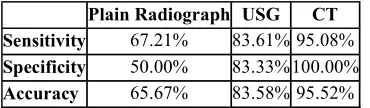
In 6 patients no mechanical obstruction was found; 2 had pancreatitis; 2 had paralytic ileus (1 due to scleroderma other due to peritonitis); 1 had mesenteric ischemia and 1 had cholecystitis. Plain radiography was able to diagnose obstruction in 41 (67%) of the 61 patients. Of the 6 patients with no obstruction, plain radiograph correctly identified 2 patients and 3 other patients were wrongly interpreted as showing obstruction. The sensitivity, specificity and accuracy of plain radiography in diagnosing bowel obstruction were 67.21%, 50.00% and 65.67% respectively.
USG showed the presence of obstruction in 51 (83.6%) out of 61 patients. USG assessment was inadequate in 2 patients because of predominantly gas-filled bowel loops. In 5 other patients with low grade intermittent partial obstruction, bowel loops were not dilated at the time of the USG examination. 3 patients were misdiagnosed as having ileus because of sparse peristaltic activity. Out of 6 patients without obstruction 5 patients were correctly identified as not having obstruction, 1 patient with mesenteric ischemia was misdiagnosed as having obstruction. The Sensitivity, Specificity and Accuracy of USG in diagnosing bowel obstruction were 83.61%, 83.33% and 83.58% respectively.
IRPMS | VOL-3 | No. 2 | APR-JUN | 2017 94 Table 1: Sensitivity and Specificity of
Imaging Modalities
Plain Radiograph USG CT Sensitivity 67.21% 83.61% 95.08%
Specificity 50.00% 83.33% 100.00%
Accuracy 65.67% 83.58% 95.52%
Level of obstruction was correctly predicted on plain films in 29 of these 41 patients. In the other 12 patients, the level was wrongly identified; in 1 patient with malrotation with midgut volvulus was misdiagnosed as distal bowel obstruction due to the abnormal location of bowel loops, 2 cases of large bowel obstruction was misinterpreted as distal small bowel obstruction because of fluid filled, gas less colon; 9 cases of distal small bowel obstruction were misinterpreted as proximal small bowel obstructions.
Table 2: Efficacy of Imaging Modalities in Detecting the Level of Obstruction
Level Radiograph USG CT Scan Diagnosis Final
Colon 10 12 15 15
Ileum 16 32 34 37
Jejunum 3 6 9 9
Total 29 50 58 61
USG correctly identified the level of obstruction in 50 patients. Level of obstruction was wrongly interpreted in 1 patient: A case of small bowel obstruction was interpreted as large bowel obstruction because the hugely dilated small bowel loops were mistaken as the large bowel. Level of obstruction was correctly identified by CT in all the 58 patients in whom CT diagnosed the presence of obstruction. The cause of obstruction could be correctly identified by plain radiography in 3 patients only; plain films showed the characteristic coffee-bean sign in 2 patients with sigmoid volvulus and dilated small bowel loops with air fluid levels and a radio
opacity in RIF in a patient with gall stone ileus. USG could correctly identify the cause of obstruction in 22 patients. In 2 patients, in whom the findings were false negative for obstruction USG was able to diagnose the cause as Tuberculosis.
CT correctly identified the etiology of obstruction in 50 of these 58 patients. In the 3 patients in whom CT was false-negative for obstruction (because of absence of bowel dilatation at the time of imaging), the underlying disease process was correctly diagnosed as tuberculosis in the 2 patients and abdominal cocoon in 1 patient. Hence, the overall efficacy of CT in the diagnosis of etiology of obstruction was 85% (53/61).
Table 3: Efficacy of Imaging Modalities in Detecting Cause of Obstruction
Cause Radiograph USG Scan CT Diagnosis Final
Adhesion - - 10 13
Neoplasm - 9 13 13
Tuberculosis - 8 8 8
Hernia - 3 6 6
Stricture - 1 4 6
Volvulus 2 1 6 6
Cocoon - - 1 3
Appendicular
abscess - 2 2 2
Gall stone
ileus 1 1 1 2
Bezoar - - 1 1
Congenital
Band - - - 1
Total 3 24 52 61
DISCUSSION
IRPMS | VOL-3 | No. 2 | APR-JUN | 2017 95 cases. The problems encountered while
predicting the site and underlying etiology of obstruction resulting either from fluid-filled bowel loops, or hugely dilated small bowel with effacement of folds mimicking large bowel, due to abnormal positioning of small bowel loops or due to lack of specific features are well described in literature.4
We faced these problems in 12 patients. KO et al reported the efficacy of plain film in the diagnosis of the cause of obstruction to be 2%.5 Suri et al4 reported the efficacy of
plain radiography in the diagnosis of the cause of obstruction to be 7% (2/30). In the present study also, the cause of obstruction could be correctly identified in 3 (4%) patients only.
USG have been commonly used in evaluating cases of suspected bowel obstruction in developing countries like India and it is easily available. Danse et al6
50 found USG to be 96% sensitive in diagnosing obstruction; the level and cause were correctly predicted at 86% and 42% of the cases, respectively. In another study in India by Suri et al4, USG had a sensitivity
of 83% specificity of 100 % specificity in diagnosing obstruction. The level and cause were correctly identified in 70% and 23% of patients respectively. Our study showed a sensitivity of 83% and 83% specificity. The level and cause were correctly identified in 81% and 39% of patients respectively.
A major limitation of USG is the presence of gas-filled bowel loops which prevent adequate assessment. This was seen in only 2 of our patients, which may be attributed to the special techniques used to bypass gaseous bowel and experience of the radiologist. In 5 patients, the bowel loops were not dilated during USG examination.
3 were due to low grade obstruction and 2 due to decompression by nasogastric tube prior to examination. Similar to previous studies, our results also show that USG is useful in diagnosing conditions like Tuberculosis, neoplasm, hernia, Midgut volvulus, appendicular abscesses and gall stone ileus; however, it is of limited value in depicting obstructions secondary to adhesions.USG also gave some information about extra intestinal pathology like lymphadenopathy, necrotic changes within nodes and free fluid in the peritoneal cavity.
Reasons for misinterpretation of the site of obstruction on USG were hugely dilated small bowel loops mimicking large bowel loops and gas-filled bowel loops not permitting evaluation on USG. However, USG can still significantly contribute in developing countries like India because it is cheap, portable, does not use ionising radiation and expertise in USG is also high. In our study USG was able to identify all cases of tuberculosis, which is significant in a country like India where TB is endemic. Sonography was also found to be better than plain radiographs at establishing the diagnosis of bowel obstruction and differentiating between paralytic ileus and mechanical obstruction. Hence, sonography where expertise and equipment is available can be an initial investigation in patients presenting with bowel obstruction and that plain radiography can only be used as a complementary tool.
CT scanning over time has proven to be better at confirming the diagnosis of bowel obstruction than most of the other radiological modalities. Megibow et al7 in
IRPMS | VOL-3 | No. 2 | APR-JUN | 2017 96 obstruction were 94%, 96% and 95%
respectively. Miyazaki et al8 showed that
the sensitivity of CT for bowel obstruction was 83%. Our study showed a sensitivity 95%, specificity 100% and accuracy 95%, which was similar to the previous studies. The level of obstruction was also correctly identified in all cases of obstruction by CT. When the bowel obstruction was classified as high grade and low grade, the reported sensitivities by Maglinte et al9 for high
grade and low grade obstructions were 81% and 48% respectively. This shows that CT is less sensitive for low grade obstruction. This is important because in our study 3
time of the study had a low grade obstruction. This again shows that CT is less sensitive for low grade obstruction. In our study, 2 other patients with low grade obstruction had mildly dilated focal bowel loops at the initial study. Delayed scans confirmed the presence of obstruction in these patients by showing persistent focal mildly dilated loops and partial hold up of contrast. This showed that delayed scans with oral contrast may help in detecting low grade obstruction.
CT also ruled out obstruction in all cases with no obstruction (specificity 100%). It also detected the underlying pathology in all six cases which was valuable regarding patient management. The ability of CT in revealing the cause of obstruction has been shown in previous studies to be varied from 75% to 95%. Shakil et al10 in 2011 showed
the efficacy of a 64 slice CT scan to identify the cause of obstruction to be 73%. In our study the CT was able to identify the cause of obstruction in 85% of patients. CT identified neoplasm, tuberculosis, hernia, volvulus, bezoar causing obstruction with 100% accuracy. In addition to these CT was
also valuable in providing information regarding extraluminal pathology like lymphadenopathy, necrotic changes, mesenteric edema, free fluid, tumour extent and distant metastasis. Thus, our study showed that CT should be the modality of choice in evaluating suspected bowel obstruction and should be used when USG and Plain Radiograph are inconclusive.
CONCLUSION
Intestinal obstruction is responsible for approximately 20% of the surgical admissions for acute abdominal conditions. Early detection of bowel obstruction will allow the early initiation of proper treatment, thus preventing complications and achieving a better outcome. Our study showed that CT has higher sensitivity, specificity and accuracy than USG and plain radiography in detecting the cause and level of obstruction. It is also the most efficient tool in detecting the alternate diagnosis in patients with no obstruction and in addition gives information regarding extraluminal pathology.
IRPMS | VOL-3 | No. 2 | APR-JUN | 2017 97 Conflicts of Interest: None.
Source of Funding: Nil.
REFERENCES
1. Maglinte DD, Balthazar EJ, Kelvin FM, Megibow AJ. The role of radiology in the diagnosis of small bowel obstruction. AJR Am J Roentgenol 1997; 168 (5): 1171 1180.
2. Fukuya 1. Hawes DR. Lu CC, Chang PJ. Barloon TJ. CT diagnosis of small bowel obstruction: efficacy in 60 patients. AJR 1992: 158: 765-772. 3. Gazelle GS, Goldberg MA, Wittenberg
J, et al. Efficacy of CT in distinguishing small bowel obstruction from other causes of small bowel dilatation. AJR 1994; 162: 43-47.
4. Suri S, Gupta S, Sudhakar PJ, et al. Comparative evaluation of plain films, ultrasound and CT in the diagnosis of intestinal obstruction. Acta Radiol 1999; 40: 422 8.
5. Ko YT, Lim JH, Lee DH. Small bowel obstruction. Sonographic evaluation. Radiology 1993;188: 649.
6. Danse EM, Van Beers BE, Goncette L, Dardenne AN, Detry R, Pringot J Value of echography in the diagnosis of acute intestinal occlusion J Radiol. 1996 Dec; 77 (12): 1223-7.
7. Megibow AJ, Balthazar EJ. Cho KC, et al. Bowel obstruction: evaluation with CT. Radiology 1991: 180: 3, 13-3 18. 8. Miyazaki O Efficacy of abdominal
plain film and CT in bowel obstruction Nihon Igaku Hoshasen Gakkai Zasshi. 1995 Mar; 55 (4): 233-9.
9. Maglinte DD, Reyes BL, Harmon BH, et al. Reliability and role of plain film radiography and CT in the diagnosis of small-bowel obstruction. AJR Am J Roentgenol 1996; 167: 1451-1455.