Tolerability of BRAF/MEK inhibitor
combinations: adverse event evaluation
and management
Lucie Heinzerling,1 Thomas K Eigentler,2 Michael Fluck,3 Jessica C Hassel,4
Daniela Heller-Schenck,5 Jan Leipe,6 Matthias Pauschinger,7 Arndt Vogel,8
Lisa Zimmer,9,10 Ralf Gutzmer 11
To cite: Heinzerling L, Eigentler TK, Fluck M, et al. Tolerability of BRAF/MEK inhibitor combinations: adverse event evaluation and management. ESMO Open 2019;4:e000491. doi:10.1136/ esmoopen-2019-000491
►Prepublication history and additional material for this paper are available online. To view these files, please visit the journal online (http:// dx. doi. org/ 10. 1136/ esmoopen- 2019- 000491).
Received 15 January 2019 Revised 6 March 2019 Accepted 31 March 2019
For numbered affiliations see end of article.
Correspondence to Ralf Gutzmer; Gutzmer. Ralf@ mh- hannover. de
© Author (s) (or their employer(s)) 2019. Re-use permitted under CC BY-NC. No commercial re-use. Published by BMJ on behalf of the European Society for Medical Oncology.
AbstrAct
The inhibition of the mitogen-activated protein kinases signalling pathway through combined use of BRAF and MEK inhibitors (BRAFi+MEKi) represents an established therapeutic option in patients with BRAF-mutated, advanced melanoma. These efficient therapies are well tolerated with mostly moderate and reversible side effects and a discontinuation rate due to adverse events of 11.5%–15.7%. Median duration of therapy ranges between 8.8 and 11.7 months. Based on data from confirmatory trials, safety profiles of three BRAFi+MEKi combinations were reviewed, that is, dabrafenib plus trametinib, vemurafenib plus cobimetinib and encorafenib plus binimetinib. Many adverse events are class effects, such as cutaneous, gastrointestinal, ocular, cardiac and musculoskeletal events; some adverse events are substance associated. Fever (dabrafenib) and photosensitivity (vemurafenib) are the most common and clinically prominent examples. Other adverse events are less frequent and the association to one substance is less strong such as anaemia, facial paresis (encorafenib), neutropenia (dabrafenib), skin rash, QTc prolongation and increased liver function tests (vemurafenib). This narrative review provides recommendations for monitoring, adverse event evaluation and management focusing on the clinically relevant side effects of the three regimens.
IntroduCtIon
Combination therapy with BRAF plus MEK inhibitors (BRAFi+MEKi) offers an effective therapy in BRAF-mutated metastatic1–6 and, for adjuvant therapy, in BRAF-mutated stage III melanoma.7
Investigator-assessed response rates in BRAF-mutated, metastatic melanoma were 64%–67% for dabrafenib+trametinib (D+T),2 3 68% for vemurafenib+cobimetinib
(V+C)5 and 75% for encorafenib+binimetinib
(E+B)6; landmark survival analyses showed
2-year overall survival rates of 53% for D+T,8
48% for V+C9 and 58% for E+B.10 However,
direct comparison of these findings cannot be made since the trials show differences in patient populations; for example, for V+C, a significantly higher number of patients with
elevated lactate dehydrogenase levels was included.11
Side effects occurring in all three BRAFi+MEKi combinations were assessed during the confirmatory clinical trials.1–6
Some events can be attributed to BRAFi adverse reactions12–15 and some to MEKi
adverse reactions.16–19 Class effects of MEKi
include the induction of a papulopustular exanthema in almost all treated patients, neuroretinal detachment, muscular prob-lems, hypertension and ventricular ejection fraction decrease. Certain side effects are drug associated and may also occur with the respective combination; examples comprise phototoxicity,14 15 20 21 fever12 22 or transient
facial paresis.23
With extensive adverse event (AE) data sets for E+B becoming publicly available now,6
the safety profiles of D+T, V+C and E+B are reviewed here with recommendations for monitoring, evaluation and management of the most common or most critical AE.
Methods and MaterIals
The tolerability of the three combination regimens D+T, V+C and E+B is described here by following safety parameters: (1) AE frequencies (all grades and grade 3–4); (2) selected AEs of special interest; (3) serious AE rates; (4) deaths that occurred within 90 days after end of treatment and that are considered ‘related’ or ‘possibly related’ to study treatment and (5) dose reduction, dose interruption and dose discontinuation rates in relation to AEs of any causality.
Event rates for the three combinations were extracted from reports of the pivotal, confir-mative phase III trials COMBI-V, COMBI-D, coBRIM and COLUMBUS (Part I); rates were determined by the common terminology criteria for AEs of the US National Cancer Institute, V.4.0. A recently published indirect
on September 12, 2020 by guest. Protected by copyright.
treatment comparison of D+T versus V+C in previously untreated patients with melanoma provided method-ical guidance and updated safety data for the COMBI-V trial24; the comparison focused exclusively on trials with
vemurafenib (dose regimen 960 mg twice daily) used as a control arm for null-hypothesis testing.
All incidence variables for tolerability were collected from publicly available data sets. Recent data sets were used in this narrative review to compile AE frequency, considering the cut-off dates for safety analyses, length of AE observation and proportion of patients still under therapy (see online supplementary file 1 for additional information). Follow-up times for AE data sets analysed in our paper, calculated as period from day of last-patient-in-cluded until safety-data cut-off-date, were 19.8 months (D+T),24 25 18.5 months (V+C)9 26 and 16.5 months (E+B).6
This retrospective, non-experimental analysis of anony-mised, pooled patient data did not require institutional review board approval. Standard recommendations to enhance the quality of evidence-based judgements were followed as previously described.27 28
Pharmacological drug profiles
The various BRAFi and MEKi differ in kinase-binding properties, structural and pharmacodynamic characteris-tics. These parameters influence the inhibitory potency towards the V600-mutated BRAF kinase and determine off-target effects.29
For the predominant BRAFV600E-mutant kinase, the half
maximal inhibitory concentration (IC50)—a measure of the potency of a drug to inhibit a specific biological or biochemical function—is 0.65 nM for dabrafenib, 10 nM for vemurafenib and 0.35 nM for encorafenib. All three BRAFi were also active against other BRAF mutations as well as against wild-type BRAF. Encorafenib inhibited most cell lines at an IC50 of <40 nmol/L; slightly higher concen-trations of dabrafenib (<100 nmol/L), but significantly higher concentrations of vemurafenib (<1000 nmol/L) were required in the same assay to inhibit proliferation of most cell lines with BRAF mutations.23
The dissociation half-life (t½diss), a measure describing target inhibition and its durability, is relevant to deter-mine drug-dosing intervals. In vitro investigations on drug–receptor interactions indicated that t½diss for encorafenib is with >30 hour considerably longer than for dabrafenib (2 hour) or vemurafenib (0.5 hour), resulting in longer lasting pharmacodynamic target inhibition of encorafenib.23
For the MEKi trametinib, IC50 values for in vitro inhibi-tion of MEK1 and MEK2 range between 0.7 and 0.9 nM.30
The IC50 of cobimetinib for MEK1 is with 0.95 nM much lower than for MEK2 (199 nM).30 In target inhibitory
assays, binimetinib was a potent inhibitor of MEK1 and MEK2 with an enzyme IC50 of 12–46 nM.30
Bioavailability, a measure how much of the adminis-tered drug reaches systemic circulation, was 95% and 85% for BRAFi dabrafenib and encorafenib, respectively.23 31–33
For vemurafenib with its low solubility and permeability,
it is unknown.20 For MEKi trametinib, cobimetinib and
binimetinib, bioavailability was 72%,34 35 46%18 and
50%,36 respectively.
Further pharmacokinetic properties of the BRAFi and MEKi, as determined in their early clinical trials,18 20 23 34–36
are outlined in figure 1. Due to their differing bioavail-ability and pharmacokinetics, drug doses and administra-tion schedules vary: 150 mg dabrafenib are administered twice daily 1 hour before or 2 hour after the morning and evening meal (ie, 75 mg capsules 2-0-2), vemurafenib twice daily (960 mg per dose; 240 mg tablets 4-0-4), and encorafenib once daily in the morning or evening inde-pendent of food intake (450 mg; 75 mg capsules 6-0-0 or 0-0-6).
Administration of trametinib (2 mg; 2 mg tablets 1-0-0) as well as of cobimetinib (60 mg; 20 mg tablets 3-0-0) is once daily, whereas binimetinib is taken twice daily (45 mg per dose; 15 mg tablets 3-0-3). In combination, D+T and E+B are administered continuously. V+C is admin-istered in combination for 21 days plus 7 days rest for cobimetinib.4
toxicity profiles of BraFi and MeKi monotherapies
For dabrafenib, treatment-related fever has been iden-tified in 20% of patients in its first-in-human phase I trial22; no dose–response correlation was noted for fever
of grade 2 or higher in the monotherapy trials, but phar-macokinetic analyses conducted in the context of the BRAFi+MEKi combination studies showed a possible asso-ciation between fever and exposure to the hydroxy-dab-rafenib metabolite and, to a lesser extent, to dabhydroxy-dab-rafenib.3
Common toxicities included cutaneous side effects, arthralgia, fatigue and headache.13
Most treatment-related side effects of vemurafenib appeared to be proportional to dose and exposure of the drug20; the photosensitivity induced by vemurafenib
is considered to be a property of the chemical structure of the drug, not related to its BRAF-inhibiting activity.22
Common side effects in early monotherapy trials were arthralgia, nausea, fatigue, rash, cutaneous squamous-cell carcinoma, pruritus and palmar–plantar dysesthesia.20
Most frequent drug-related AEs occurring with encorafenib monotherapy included myalgia, nausea, palmoplantar erythrodysesthesia, arthralgia, alopecia and hyperkeratosis. Transient Bell’s palsy, the most common disorder affecting a single nerve and associated with facial nerve weakness/paralysis, was reported in 8% of patients treated with encorafenib, whereas it has rarely been reported in association with other BRAFi.33
Trametinib showed an early onset of dose-limiting toxic effects, as observed in trametinib’s first-in-human trial including 206 patients34 with two cases of rash, one
case of diarrhoea and three cases of central serous reti-nopathy. As two of the three ocular toxicity events arose either 1 day after a loading dose or within days of admin-istration of the highest once-daily dose, this treatment-re-lated effect was thought to be potentially retreatment-re-lated to the Cmax of trametinib.34 Similarly, a reported papulopustular
on September 12, 2020 by guest. Protected by copyright.
Figure 1 Structural and population pharmacokinetic properties (single drug) of BRAF indicator dabrafenib, vemurafenib and encorafenib and of MEK indicator trametinib, cobimetinib and binimetinib. AR, accumulation ratio; AUC 0–24, area under the curve for 0–24 h (AUC 0–8 : 0-8 h); BCS, biopharmaceuticsclassification system; C max, maximum concentration; C 24, concentration after 24 h; d, days; h, hours; MTD,maximum tolerated dose; od, once daily; RP2D, recommended phase 2 dose; t 1/2 eff, effective half-life,calculated as –0.693*tau/(ln[1–{1/AR}]); td, twice daily; t max, time taken to reach maximum concentration,reported as median. In brackets: (%CV b), between-subject coefficient of variation; (nc), not calculated; (nr),not reported; […], range. All reported values are means, if not indicated otherwise (i.e. median).
exanthema adverse reaction emerging after cessation of combined D+T treatment was proposed to be associated with trametinib’s long half-life37 (figure 1).
Cobimetinib’s most frequent AEs attributed in its phase I monotherapy trial were diarrhoea, rash, fatigue, oedema, nausea and vomiting.18
Dose-limiting toxicities of binimetinib in early mono-therapy trials were single events of mono-therapy-resistant papulopustular rash and of central serous-like retinop-athy, respectively. Other frequent treatment-related AEs included gastrointestinal (GI) (diarrhoea, nausea) and skin disorder as well as peripheral oedema, increased creatine phosphokinase (CPK) and retinal disorders.38
toxicity profile of BraFi+MeKi combinations
Overall AE and serious AE frequencies for the three combi-nations D+T, V+C and E+B and the respective median drug exposure times are displayed in table 1. Although compar-ison across trials has its limitations, that is, with respect to different methodological AE evaluation across trials, AE
rates in the respective vemurafenib comparator arms are nearly identical.6 9 24 AEs of any grade occurred in almost all
patients treated with the combination therapy. In patients treated with a BRAFi+MEKi, grade 3–4 AEs occurred in 46%–56% for D+T (COMBI-D, COMBI-V), 69% for V+C (coBRIM) and 58% for E+B (COLUMBUS, Part I).
Median duration of therapy ranged between 10.0 and 11.0 months for D+T, 8.8 months for V+C and 11.7 months for E+B, with median dose intensities ranging between 94% and 100% for all substances.
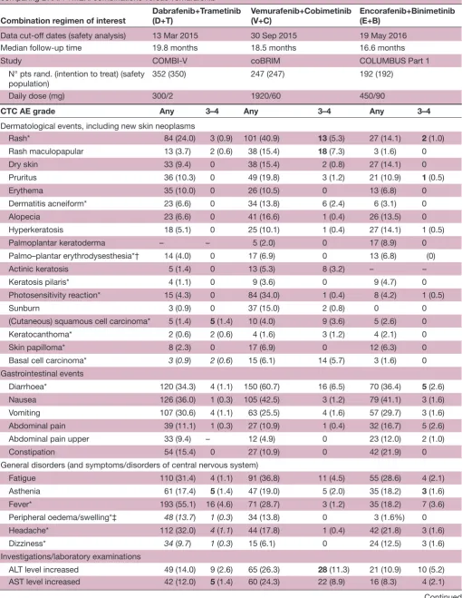
The detailed AE frequencies of D+T (COMBI-V), V+C (coBRIM) and E+B (COLUMBUS Part I) for relevant organ systems are listed in table 2. To facilitate the anal-ysis and interpretation of the data, AE frequencies for the combination regimen versus frequencies of the respective vemurafenib monotherapy arms of each trial6 9 24 were
compared: the respective shifts, expressed as an increase or decrease in BRAFi+MEKi AE rates compared with vemurafenib, are displayed in figure 2.
on September 12, 2020 by guest. Protected by copyright.
Table 1
Overall fr
equencies of adverse events in the safety populations of the pivotal clinical trials and median drug exposur
e times and rates
Combination r
egimen of inter
est
Dabrafenib+T
rametinib
(D+T)
Vemurafenib+Cobimetinib (V+C) Encorafenib+Binimetinib (E+B)
Date of (safety) analysis
13 Mar 2015
24 25
12 Jan 2015
2
19 Sep 2014
24
19 May 2016
6
Study
COMBI-V
COMBI-D
coBRIM
COLUMBUS Part 1
Arms D+T V D+T D+placebo V+C V+placebo E+B V E
N° pts (ITT) rand (N° safety population)
352 (350) 352 (349) 211 (209) 212 (211) 247 (247) 248 (246) 192 (192) 191 (186) 194 (192)
Daily dose (mg)
300/2 1920 300/2 300 1920/60 1920 450/90 1920 300 Events (fr equencies)
AE (all grades, any causality) (%)
345 (98.6) 345 (98.9) 203 (97.1) 205 (97.2) 244 (98.8) 240 (97.6) 188 (97.9) 185 (99.5) 191 (99.5)
AE (°3–4) (%)
195 (55.7) 228 (65.3) 95 (45.5) 106 (50.2) 171 (69.2) 143 (58.1) 111 (57.8) 118 (63.4) 127 (66.1)
AE leading to death (‘fatal AE’; (°5)) (%)
4 (1.1) 4 (1.2) 5 (2.4) 1 (0.5) 5 (2.0) 3 (1.2) 3 (1.7) 2 (1.1) 2 (1.0)
AE leading to discontinuation (%)
55 (15.7) 48 (13.8) 24 (11.5) 14 (6.6) 37 (15.0) 20 (8.1) 24 (12.5) 31 (16.7) 27 (14.1)
AE leading to dose r
eduction (%) 192 (54.9) 197 (56.4) 59 (28.2) 29 (13.7) 110 (44.5) 87 (35.4) 22 (11.5) 42 (22.6) 52 (27.1)
AE leading to dose interruption (%)
118 (56.5)
78 (37.0)
88 (45.8)
98 (52.7)
122 (63.5)
SAE (all grades, any causality) (%)
151 (43.1) 136 (39.0) 88 (42.1) 78 (37.0) 85 (34.4) 64 (26.0) 66 (34.4) 69 (37.1) 65 (33.9) Dose exposur
e rates/times (months)
Median duration of therapy (range)
10.0 (0–21) 6.0 (0–18) 11.0 (0–30) 8.0 (0–32) 8.8 (0–18) 5.7 (0–17) 11.7 (0–27) 6.2 (0–28) 7.2 (0–26)
Median dose intensity (in %)
97.3%+100% 96.1% 98.2%+100% 99.9%+ 100% 94.4%+96.6% 96.7%+ 99.0% 100%+99.6% 94.5% 86.2%
AE, adverse events; B, binimetinib; C, cobimetinib; D, dabrafenib; E, encorafenib; ITT
, intention-to-tr
eat; SAE, serious adverse event; T
, trametinib; V
, vemurafenib; pts rand., patients
randomized.
on September 12, 2020 by guest. Protected by copyright.
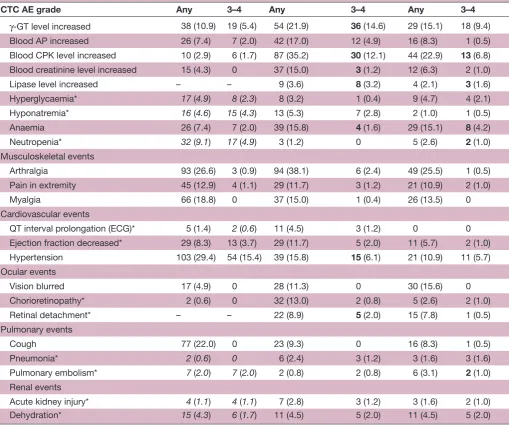
Table 2 Frequencies of AE of combination therapy arms, as observed in the safety population of pivotal clinical trials comparing BRAFi +MEKi combinations versus vemurafenib6 9 24
Combination regimen of interest
Dabrafenib+Trametinib (D+T)
Vemurafenib+Cobimetinib (V+C)
Encorafenib+Binimetinib (E+B)
Data cut-off dates (safety analysis) 13 Mar 2015 30 Sep 2015 19 May 2016 Median follow-up time 19.8 months 18.5 months 16.6 months Study COMBI-V coBRIM COLUMBUS Part 1 N° pts rand. (intention to treat) (safety
population) 352 (350) 247 (247) 192 (192) Daily dose (mg) 300/2 1920/60 450/90
CTC AE grade Any 3–4 Any 3–4 Any 3–4
Dermatological events, including new skin neoplasms
Rash* 84 (24.0) 3 (0.9) 101 (40.9) 13 (5.3) 27 (14.1) 2 (1.0) Rash maculopapular 13 (3.7) 2 (0.6) 38 (15.4) 18 (7.3) 3 (1.6) 0 Dry skin 33 (9.4) 0 38 (15.4) 2 (0.8) 27 (14.1) 0 Pruritus 36 (10.3) 0 49 (19.8) 3 (1.2) 21 (10.9) 1 (0.5) Erythema 35 (10.0) 0 26 (10.5) 0 13 (6.8) 0 Dermatitis acneiform* 23 (6.6) 0 34 (13.8) 6 (2.4) 6 (3.1) 0 Alopecia 23 (6.6) 0 41 (16.6) 1 (0.4) 26 (13.5) 0 Hyperkeratosis 18 (5.1) 0 25 (10.1) 1 (0.4) 27 (14.1) 1 (0.5) Palmoplantar keratoderma – – 5 (2.0) 0 17 (8.9) 0 Palmo–plantar erythrodysesthesia*† 14 (4.0) 0 17 (6.9) 0 13 (6.8) (0) Actinic keratosis 5 (1.4) 0 13 (5.3) 8 (3.2) – –
Keratosis pilaris* 4 (1.1) 0 9 (3.6) 0 9 (4.7) 0 Photosensitivity reaction* 15 (4.3) 0 84 (34.0) 1 (0.4) 8 (4.2) 1 (0.5) Sunburn 3 (0.9) 0 37 (15.0) 2 (0.8) 0 0 (Cutaneous) squamous cell carcinoma* 5 (1.4) 5 (1.4) 10 (4.0) 9 (3.6) 5 (2.6) 0 Keratocanthoma* 2 (0.6) 2 (0.6) 4 (1.6) 3 (1.2) 4 (2.1) 0 Skin papilloma* 8 (2.3) 0 17 (6.9) 0 12 (6.3) 0 Basal cell carcinoma* 3 (0.9) 2 (0.6) 15 (6.1) 14 (5.7) 3 (1.6) 0 Gastrointestinal events
Diarrhoea* 120 (34.3) 4 (1.1) 150 (60.7) 16 (6.5) 70 (36.4) 5 (2.6) Nausea 126 (36.0) 1 (0.3) 105 (42.5) 3 (1.2) 79 (41.1) 3 (1.6) Vomiting 107 (30.6) 4 (1.1) 63 (25.5) 4 (1.6) 57 (29.7) 3 (1.6) Abdominal pain 39 (11.1) 1 (0.3) 27 (10.9) 1 (0.4) 32 (16.7) 5 (2.6) Abdominal pain upper 33 (9.4) – 12 (4.9) 0 23 (12.0) 2 (1.0) Constipation 54 (15.4) 0 27 (10.9) 0 42 (21.9) 0 General disorders (and symptoms/disorders of central nervous system)
Fatigue 110 (31.4) 4 (1.1) 91 (36.8) 11 (4.5) 55 (28.6) 4 (2.1) Asthenia 61 (17.4) 5 (1.4) 47 (19.0) 5 (2.0) 35 (18.2) 3 (1.6) Fever* 193 (55.1) 16 (4.6) 71 (28.7) 3 (1.2) 35 (18.2) 7 (3.6) Peripheral oedema/swelling*‡ 48 (13.7) 1 (0.3) 34 (13.8) 0 3 (1.6%) 0 Headache* 112 (32.0) 4 (1.1) 44 (17.8) 1 (0.4) 42 (21.8) 3 (1.6) Dizziness* 34 (9.7) 1 (0.3) 15 (6.1) 0 24 (12.5) 3 (1.6) Investigations/laboratory examinations
ALT level increased 49 (14.0) 9 (2.6) 65 (26.3) 28 (11.3) 21 (10.9) 10 (5.2) AST level increased 42 (12.0) 5 (1.4) 60 (24.3) 22 (8.9) 16 (8.3) 4 (2.1)
Continued
on September 12, 2020 by guest. Protected by copyright.
CTC AE grade Any 3–4 Any 3–4 Any 3–4
γ-GT level increased 38 (10.9) 19 (5.4) 54 (21.9) 36 (14.6) 29 (15.1) 18 (9.4) Blood AP increased 26 (7.4) 7 (2.0) 42 (17.0) 12 (4.9) 16 (8.3) 1 (0.5) Blood CPK level increased 10 (2.9) 6 (1.7) 87 (35.2) 30 (12.1) 44 (22.9) 13 (6.8) Blood creatinine level increased 15 (4.3) 0 37 (15.0) 3 (1.2) 12 (6.3) 2 (1.0) Lipase level increased – – 9 (3.6) 8 (3.2) 4 (2.1) 3 (1.6) Hyperglycaemia* 17 (4.9) 8 (2.3) 8 (3.2) 1 (0.4) 9 (4.7) 4 (2.1) Hyponatremia* 16 (4.6) 15 (4.3) 13 (5.3) 7 (2.8) 2 (1.0) 1 (0.5) Anaemia 26 (7.4) 7 (2.0) 39 (15.8) 4 (1.6) 29 (15.1) 8 (4.2) Neutropenia* 32 (9.1) 17 (4.9) 3 (1.2) 0 5 (2.6) 2 (1.0) Musculoskeletal events
Arthralgia 93 (26.6) 3 (0.9) 94 (38.1) 6 (2.4) 49 (25.5) 1 (0.5) Pain in extremity 45 (12.9) 4 (1.1) 29 (11.7) 3 (1.2) 21 (10.9) 2 (1.0) Myalgia 66 (18.8) 0 37 (15.0) 1 (0.4) 26 (13.5) 0 Cardiovascular events
QT interval prolongation (ECG)* 5 (1.4) 2 (0.6) 11 (4.5) 3 (1.2) 0 0 Ejection fraction decreased* 29 (8.3) 13 (3.7) 29 (11.7) 5 (2.0) 11 (5.7) 2 (1.0) Hypertension 103 (29.4) 54 (15.4) 39 (15.8) 15 (6.1) 21 (10.9) 11 (5.7) Ocular events
Vision blurred 17 (4.9) 0 28 (11.3) 0 30 (15.6) 0 Chorioretinopathy* 2 (0.6) 0 32 (13.0) 2 (0.8) 5 (2.6) 2 (1.0) Retinal detachment* – – 22 (8.9) 5 (2.0) 15 (7.8) 1 (0.5) Pulmonary events
Cough 77 (22.0) 0 23 (9.3) 0 16 (8.3) 1 (0.5) Pneumonia* 2 (0.6) 0 6 (2.4) 3 (1.2) 3 (1.6) 3 (1.6) Pulmonary embolism* 7 (2.0) 7 (2.0) 2 (0.8) 2 (0.8) 6 (3.1) 2 (1.0) Renal events
Acute kidney injury* 4 (1.1) 4 (1.1) 7 (2.8) 3 (1.2) 3 (1.6) 2 (1.0) Dehydration* 15 (4.3) 6 (1.7) 11 (4.5) 5 (2.0) 11 (4.5) 5 (2.0)
Listed are AE with event frequencies (of any grade) ≥10%, and/or with event frequencies (≥2% for grade ≥3 AE) and frequency independent, clinically relevant ‘AE of specific interest’.
Values in bold indicates CTC grade 3 and grade 4 toxicity reported; values in italics indicates different data cut-off dates for safety analysis (D+T only).
*D+T: values and frequencies reported at initial safety analysis (14 April 2014) only,2 extended listings are published in the European Public
Assessment Report (EMA/589140/2015, dated 2 Septmber 2015).
†D+T: term ‘hand–foot syndrome’ was reported, including the terms palmo–plantar erythrodysesthesia, planto–palmar hyperkeratosis and palmoplantar keratoderma.
‡E+B: frequencies for peripheral swelling were reported.
–, not reported; AE, adverse event; ALT, alanine aminotransferase; AP, alkaline phosphatase; AST, aspartate aminotransferase; CPK, creatine phosphokinase; CTC, common toxicity criteria; GT, γ-gamma-glutamyl transferase; pts rand., patients randomised.
Table 2 Continued
Remarkably, most cutaneous side effects, particularly secondary neoplasms (cutaneous squamous cell carci-noma and its specific variant keratoacanthoma and skin papilloma), decreased considerably compared with BRAF monotherapy (figure 2); this effect is explained in the literature by the suppression of the paradoxical activa-tion of the mitogen-activated protein kinases (MAPK) pathway in BRAF wild-type cells in various tissues through MEKi coadministration.39 On the other side, an increase
of GI side effects, particularly vomiting, is noted due to
the addition of the MEKi. Increases in cardiac and ocular side effects are related to MEKi coadministration as well.
therapy related, organ class-specific aes Dermatological events, secondary skin neoplasms
The most frequent cutaneous side effects are rash, itching, dry skin, hair loss, photosensitivity reaction, keratinocytic proliferation and panniculitis.
Different conditions such as maculopapular exan-thema, papulopustular exanthema or even eczema
on September 12, 2020 by guest. Protected by copyright.
Figure 2 Differences in adverse event frequencies: combination therapies versus vemurafenib monotherapy. CPK, creatine phosphokinase; cSCC, cutaneous squamous cell carcinoma; PPH, palmoplantar hyperkeratosis.
are often summed up by ‘rash’; however, a differentia-tion is desirable to apply the most adequate treatment approach. Rash was most often reported for V+C with 41% and was less common for D+T (24%) and for E+B (14%) (table 2). Although ‘rash’ is often a low-grade AE, severe and even life-threatening side effects of the skin have been reported, including erythema exsudativum multiforme, Stevens-Johnson syndrome, toxic epidermal necrolysis, drug rash with eosinophilia and systemic symp-toms, drug-induced hypersensitivity syndrome and acute generalised exanthematous pustulosis.40
Dry skin (xerosis cutis) and itching (pruritus) are side effects that occur regularly among all three combinations with a frequency of 10%–20% (table 2). Pruritus is often the result of xerosis cutis.
In 34% of patients treated with V+C, ultraviolet A (UVA)-mediated photosensitivity reactions with erythema, blistering and painful burning were reported, whereas this occurred only in 4% of patients treated with D+T or E+B. Radiosensitivity was observed in patients treated concom-itantly with radiotherapy and BRAFi/MEKi, mainly with vemurafenib.41 Effectiveness does not seem to be reduced
by interruption of BRAF therapy during radiotherapy, as shown for vemurafenib.42
BRAFi+MEKi therapy associated ‘alopecia’ generally means diffuse hair loss. Interestingly, curly hairs often found under BRAFi monotherapy are not observed under BRAFi+MEKi therapy.
Keratinocytic proliferations including keratosis pilaris, actinic keratoses, cutaneous squamous cell carcinoma (cSCC), keratoacanthoma and skin papilloma were observed in up to 7% of patients treated with BRAFi+MEKi (table 2). Keratosis pilaris with disseminated small hyper-keratotic follicular papules on the face or proximal extremities was reported in 4%–7% of patients (table 2). With regard to hand–foot syndrome, there is a spectrum of clinical variants: palmoplantar erythrodysesthesia (PPE) with inflammatory and painful lesions not restricted to pressure points and palmoplantar hyperkeratosis (PPH, also called palmoplantar keratoderma) with hyperkera-totic and painful lesions at pressure points. They are also called type I and type II hand–foot syndrome.43 Within
the clinical studies, this was not clearly differentiated (table 2). While encorafenib seems to induce both PPE and PPH more often than vemurafenib or dabrafenib, the BRAFi+MEKi combination therapy is well tolerated: mainly PPH occurs.44 Frequency of PPH is lowered with
D+T and E+B, compared with vemurafenib (figure 2). D+T, V+C or E+B induce benign acanthotic skin papil-lomas, keratoacanthomas and well-differentiated cSCC in 2%–7%, 1%–2% and 1%–4% of patients, respectively (table 2); these rates are lower than the AE rates induced by BRAFi monotherapy (figure 2). New primary mela-nomas were observed in less than 1% of all patients treated with BRAFi+MEKi. Moreover, panniculitis with painful erythematous subcutaneous nodules predominantly located on the extremities and buttocks—which can occur with or without fever, arthralgia or joint swelling— have been described under combined BRAFi+MEKi with unknown frequency.45
Cutaneous side effects are usually well treatable and should not immediately lead to dose reduction or discon-tinuation of therapy. Exanthema, xerosis cutis and pruritus can be successfully treated by regular application of moisturisers containing urea or glycerine or topical application of class II–III glucocorticoids. Severe cases of exanthema require systemic steroids, dose interruptions or permanent discontinuation.
For ultraviolet (315–380 nm: UVA) mediated photosen-sitivity, strict avoidance of UVA and sun protection with UVA filter-containing sun screen and protective clothing (including hat, sunglasses) is crucial even behind windows since UVA can penetrate the window glass. Sunburn can be treated with topical steroids and possibly by non-ste-roidal anti-inflammatory drugs (NSAIDs).
Keratosis pilaris and PPH can be treated with creams containing urea or salicylic acid and in cases of inflam-mation with topical steroids. Patients with PPH should avoid pressure and friction. Panniculitis can be treated symptomatically with non-steroidal anti-inflammatory
on September 12, 2020 by guest. Protected by copyright.
drugs (eg, etoricoxib), topical steroids and compres-sion; some severe cases require systemic steroids and temporary dose interruption. Keratoacanthomas, cSCC and new primary melanomas should be surgically removed.
GI events
GI toxicities are commonly seen during therapy with BRAFi+MEKi and include diarrhoea and nausea and vomiting and can be accompanied by abdominal pain and GI bleeding. Frequencies of GI AE are, with shifts, for example, for diarrhoea up to 27%, higher for the combi-nation of BRAFi+MEKi compared with either mono-therapy (figure 2). The underlying pathophysiological mechanisms are not completely understood. The MAPK pathway is activated via EGFR in GI normal mucosa and there is evidence that this pathway is a negative regulator of chloride secretion. Inhibitors of the EGFR pathway could therefore increase chloride secretion and thereby induce secretory diarrhoea.46
Alternative aetiologies for GI toxicities such as progres-sive disease or infection (eg, Clostridium difficile infection or other bacterial/viral pathogens) need to be ruled out; the time of onset is of diagnostic relevance to assess whether the AE is treatment related. A cytomegalovirus DNA PCR performed in blood can diagnose cytomega-lovirus infection or reactivation. For persistent grade 2 diarrhoea, colonoscopy with colonic biopsies can be considered. All grade diarrhoea occurred much more frequently in patients treated with V+C (61%), compared with D+T or E+B (34% and 36%); nausea (36%–41%) and vomiting (26%–31%), however, occurred at similar frequencies (table 2).
Management includes rehydration since vomiting and diarrhoea can lead to dehydration, hypotension and in severe cases to kidney failure. Grades 1 and 2 diar-rhoeas may be managed with antidiarrhoeal medications including loperamide and octreotide, oral hydration and electrolyte supplements. In grade >3 cases, BRAFi/MEKi therapy should be withheld in addition to symptomatic treatment. Common GI adverse reactions usually resolve within a few days after cessation of treatment. Systemic treatment and prophylaxis of nausea and vomiting should follow established guidelines.
Hepatic AE manifest as asymptomatic increase of liver function tests, mainly aspartate aminotransferase (AST), alanine aminotransferase (ALT) and gamma-glutamyl transferase, rarely bilirubin. They are very frequent (table 2). The very common frequency of AST and ALT events may be related to the addition of the MEKi (figure 2). Regular laboratory controls are required with treatment interruption in case of elevations of grade 3 or higher. In some cases with grade 2 elevation, specifically with simultaneous elevated bilirubin levels, treatment may be paused earlier. Other causes of acute liver injury such as infection should be ruled out.
General disorders and haematological events
Fever, fatigue and peripheral oedema are very frequent with BRAFi+MEKi therapy. Fever is one of the main symptoms under D+T treatment, occurring in more than half of the patients (table 2) and is more frequent than under BRAFi monotherapy with vemurafenib (21%) or dabrafenib (28%).3 24 However, the grade >3 fever rate
remains below 5%. E+B and V+C can also induce fever, but less frequent (ie, in 18%–29% of patients). Fatigue can accompany this AE or may occur independently, with similar frequencies throughout the three combinations (28%–37%).
Fever occurs early after treatment start—mainly within the first 4 weeks.26 47 A dose interruption of both drugs is
recommended if the fever exceeds 38.5°C. The aetiology of BRAFi+MEKi induced fever is still unclear, and an infectious cause should be excluded. No baseline clinical characteristics predict fever, and it is not associated with treatment outcome.47 The fever can be treated with
anti-pyretics such as ibuprofen and paracetamol and, if not effective, with low-dose corticosteroids. To avoid hypovo-laemia with hypotension and possible acute renal failure, sufficient fluid substitution is important. As soon as the fever ceased for at least 24 hours, BRAFi+MEKi therapy can be restarted.48 Patients experience a median of two
events of fever, with 21% of patients having >4 events.47 A
dose reduction is recommended if fever recurs. However, clinical experience shows that short ‘drug holidays’ (about 2–7 days) are much more effective and the full dose can be maintained.48 In recurrent cases,
intermit-tent treatment might be an option to avoid additional fever events.
Peripheral oedema is a typical side effect of the MEKi (table 2). Mostly extremities are affected, but facial oedema especially of the eye lids is also common. In mild cases, symptomatic treatment with compression therapy and head elevation while sleeping is adequate after exclu-sion of other causes, for example, hypalbuminaemia or reduced kidney function.
Haematological events occur in the form of disorders in the different cell subsets of the peripheral blood, most frequently anaemia with around 15% for V+C and E+B and 7% for D+T. Severe anaemia occurs in 2.0, 1.6 and 4.2% for D+T, V+C and E+B, respectively. In contrast, D+T induces neutropenia more frequently, followed by E+B and rarely occurring in V+C (table 2). In general, neutropaenia occurs with a late onset (up to 2 months after treatment initiation) and resolves without dose modifications. All other reported differential blood count changes such as thrombocytopenia, lymphopenia and eosinophilia occur with AE frequencies below 5% and are rarely severe.
Musculoskeletal/rheumatic events
Musculoskeletal/rheumatic side effects primarily include arthralgia, myalgia and vasculitis.
Arthralgia—the main musculoskeletal event—is a very common AE associated with BRAF monotherapy. In
on September 12, 2020 by guest. Protected by copyright.
combination, incidences of arthralgia were lowered for D+T, V+C and E+B (figure 2): arthralgia of grade 1–2 remains, however, a very common AE (table 2). Grade
≥3 arthralgia events occur at frequencies of 0.5%–2%; drug withdrawal or adjustment may be considered. Arthritis also appears to occur more frequently in BRAF monotherapy compared with combination therapy9 and
can be managed with dose reduction and corticosteroid treatment.49
Myalgia is another very common AE, occurring with BRAFi+MEKi in 14%–19% (table 2); frequencies are similar for the different BRAFi (figure 2).
Vasculitis is mainly described in single patients treated with BRAFi as cutaneous side effects in the context of panniculitis,45 50–53 and as leukocytoclastic vasculitis,45 54
but also involving the kidney as glomerulonephritis55 56
and the eye as retinal vasculitis.57
For treatment of mild symptoms of arthralgia or myalgia, NSAIDs or low-dose corticosteroids can be applied. For more accentuated musculoskeletal/rheu-matic side effects that require intra-articular or high-dose steroids, rheumatologists should be consulted.
Severe AEs, for example, myositis or vasculitic organ-threatening manifestations require moderate to high-dose (1 mg/kg) corticosteroids; BRAFi/MEKi therapy discontinuation can be considered. If symptoms persist, corticosteroid-sparing therapies like leflunomide or methotrexate can be applied. Based on findings from in vitro and in vivo models, in which leflunomide prevents melanoma growth in combination with BRAFi+MEKi,58
leflunomide might be preferred.
Cardiovascular events
Cardiovascular side effects have been described for BRAFi and MEKi including QT-prolongation, cardiomyopathy with reduced pump function and hypertension. While QT prolongation is mainly an issue in treatment with BRAFi, decreased ejection fraction has been described for MEKi, however, with different frequencies. The MAPK pathway in cardiomyocytes is a protective signal-ling pathway and its inhibition interferes with intramy-ocytic repair mechanisms by inhibition of extracellular signal-regulated kinases 1/2.59 Immunotherapy-mediated
subclinical cardiotoxicity or damage induced by radi-otherapy may therefore increase the risk for significant left ventricular systolic dysfunction or even heart failure induced by concomitant or subsequent use of BRAFi and MEKi.60 However, despite fatal events considered to be
due to arrhythmias or sudden cardiac death (10/139 in an analysis of the FDA Adverse Event Reporting System database60 61), most of the cardiac side effects can be
adequately managed and are reversible.
QT prolongation was observed in up to 3%–7% of patients treated with vemurafenib and 2% treated with V+C5 62 and has been shown to be dose dependent.63
Grade ≥3 QT interval prolongation occurred in 1% of patients treated with vemurafenib monotherapy or with V+C.9 Such negative effects on QT prolongation were not
seen with dabrafenib or encorafenib, considered to be due to an additional fluorinated phenyl ring.6 64 65 The
MEKi trametinib caused no QT prolongation.66 Impor-tantly, other factors like electrolyte disturbances, long QT syndrome and concomitant medications can potentiate the AE.
Thus, electrolyte dysbalances, for example, due to diar-rhoea, should be corrected (including magnesium) and other QT-prolonging drugs (eg, pantoprazole, ciproflox-acin) omitted.67 ECG should be assessed before therapy
and monthly during the first 3 months, then every 12 weeks; doses should be withheld in case of QTc >500 ms, or more than 60 ms increase from baseline.
It is important to detect left ventricular dysfunction in treatment with BRAFi+MEKi, since it often results in discontinuation of therapy. The degree of left ventricular dysfunction can range from asymptomatic changes, best diagnosed by echocardiographic strain analysis, to severe cardiac failure. Although BRAFi+MEKi were not included in the European Society for Medical Oncology guidelines on cardiotoxicity, the guideline outlines the diagnostic procedures including cardiac MRI and multigated acqui-sition scans.68
Grade ≥3 decrease in ejection fraction (ie, <40% or decrease of >20% from baseline) was reported in 4%, 2% and 1% in D+T, V+C, and E+B, respectively (table 2). Myocardial dysfunction is modified by genetic factors and impaired myocardial function before initiating cancer treatment, arterial hypertension, >65 years of age, body mass index >30 kg/m2 and radiotherapy increase the risk.
The onset of left ventricular dysfunction after application of MEKi or BRAFi+MEKi therapy ranges from 2 weeks to 5 months and 1–13 months, respectively, and resolved in the majority of cases.61
Thus, risks and benefits of BRAFi+MEKi therapy should be carefully evaluated in patients with significant heart disease, and hypertension should be controlled before initiation of therapy. Ejection fraction, troponin and the N-terminal prohormone of brain natriuretic peptide (NT-proBNP) should be checked at therapy start. While troponin indicates myocardiac damage and thus represents an early marker, NT-proBNP is associated with myocardial insufficiency and a marker of chronic heart failure. Further controls can be adapted based on risk factors, symptoms and CPK findings. Manage-ment includes withholding treatManage-ment in case of an ejec-tion fracejec-tion reducejec-tion of >10%, with rechallenge at a lower dose and discontinuation in case of ejection frac-tion reducfrac-tion of >20%. Symptomatic treatment, for example, with beta-blockers, can be given as advised by the cardiologist.
Arterial hypertension can be caused by BRAFi; for BRAFi+MEKi combinations, the MEKi also contributes to this side effect. The incidence for combination ther-apies with D+T, V+C and E+B were 29%, 16%, and 11%, respectively (table 2). Antihypertensive treatment should be pursued according to the existing guidelines.
on September 12, 2020 by guest. Protected by copyright.
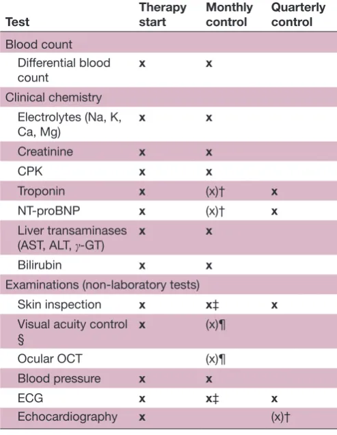
Table 3 Recommendations for routine* monitoring of BRAF/MEK combination therapy
Test Therapy start Monthly control Quarterly control
Blood count Differential blood
count x x Clinical chemistry
Electrolytes (Na, K, Ca, Mg)
x x
Creatinine x x
CPK x x
Troponin x (x)† x
NT-proBNP x (x)† x
Liver transaminases
(AST, ALT, γ-GT) x x Bilirubin x x
Examinations (non-laboratory tests)
Skin inspection x x‡ x
Visual acuity control §
x (x)¶
Ocular OCT (x)¶ Blood pressure x x
ECG x x‡ x
Echocardiography x (x)†
*In patients/situations without clinical particularities.
†In case of clinically abnormal signs (eg, heart, chest) or increasing CPK.
‡In months 1 and 2.
§Visual acuity (at therapy start) to be checked/noted (ie, in patient history).
¶in case of patient-reported visual disturbances.
γ-GT, gamma-glutamyl transferase;ALT, alanine transaminase; AST, aspartate transaminase; CPK, creatine phosphokinase; LDH, lactate dehydrogenase; NT-proBNP, N-terminal prohormone of brain natriuretic peptide; OCT, optical coherence tomography; x, test recommended; (x), test optional.
Ocular events
Fluid accumulation (oedema) in the retina resulting in a serous neuroretinal detachment (SND) is a regular AE during treatment with BRAFi+MEKi (table 2). Although this AE is sometimes called serous retinopathy, chori-oretinopathy, macular oedema or retinal pigment epithe-lial detachment, the term SND is preferable since the other terms refer to established diagnoses, with in part different clinical manifestations.69 SND associated with
BRAFi+MEKi is often asymptomatic, but may rarely cause transient visual disturbances, that is, blurred vision, reduced visual acuity, dyschromatopsia and photo-phobia.70 The diagnosis is made with optical coherence
tomography (OCT); the oedema is often bilateral and multifocal.69 71
After initiation of BRAF+MEK inhibition, SND occurs early within hours, days or weeks and is mostly caused by the MEKi.69 71 Frequency of diagnosis depends on time
and method of the ophthalmology examinations with an incidence of 13% or 8% of patients in the V+C and E+B trials, where regular OCT examinations were included (table 2). The pathogenetic mechanism is currently still unknown. SND is usually transient, but long-term expe-rience is limited and retinal atrophy without functional relevance has been described.72 Regular controls without
any treatment are usually sufficient; however, in severe cases, dose interruption or reduction of the MEKi might be necessary.
Ocular inflammation (uveitis, conjunctivitis) is rarely induced by BRAFi. Here, topical steroids are usually suffi-cient for treatment; in severe cases, dose interruption or reduction of the BRAFi might be necessary.73 In single
cases, retinal vein occlusions have been described both during BRAFi and BRAFi+MEKi therapy.73 In these cases,
study protocols required discontinuation of BRAFi+MEKi and ophthalmological treatment. However, the frequency of retinal vein occlusions is similar in patients not treated with BRAFi+MEKi74; thus, the relation and the
thera-peutic consequences are unclear.
Taken together, ophthalmologic AEs (in particular SND) are frequent but often asymptomatic and transient; during treatment, ophthalmologic checks including OCT should be performed depending on visual disturbances.
Pulmonary events
Lung toxicity under BRAFi+MEKi therapy is infrequent. Cough, pneumonitis, sometimes accompanied by fever and pulmonary embolism may occur commonly.75–77
Grade 3/4 events of interstitial lung disease or pneumo-nitis, subsumed rather under the term pneumonia in clin-ical studies, occurred in up to 2% of the cases (table 2). Pneumonitis is thought to be mainly caused by the MEKi.
In case of cough, shortness of breath or abnormal auscultation, chest CT should be performed. Lung infil-trates sometimes show a discrepancy to patient-reported symptoms. When an infectious origin has been ruled out via bronchoalveolar lavage, high dose of corticosteroids should be applied and tapered over time. While in severe
cases, the MEKi has to be permanently discontinued, reinduction of the BRAFi after recovery is possible.
Renal events
Combined BRAFi+MEKi therapy can cause renal impair-ment, mostly as increase of serum creatinine. Acute renal failure or electrolyte disorders have been reported for all combination therapy78 79 (table 2). A recent system-atic review of BRAFi-mediated nephrotoxicity including pathology reports of kidney biopsies indicated tubule-in-terstitial damage with an acute and chronic compo-nent.80–83
In cases of higher grade acute kidney injury, BRAFi +MEKi therapy should be interrupted and reintroduced after improvement of renal function at a lower dose. As therapy with D+T frequently causes fever that may lead to dehydration of the patient—in cases of insufficient
on September 12, 2020 by guest. Protected by copyright.
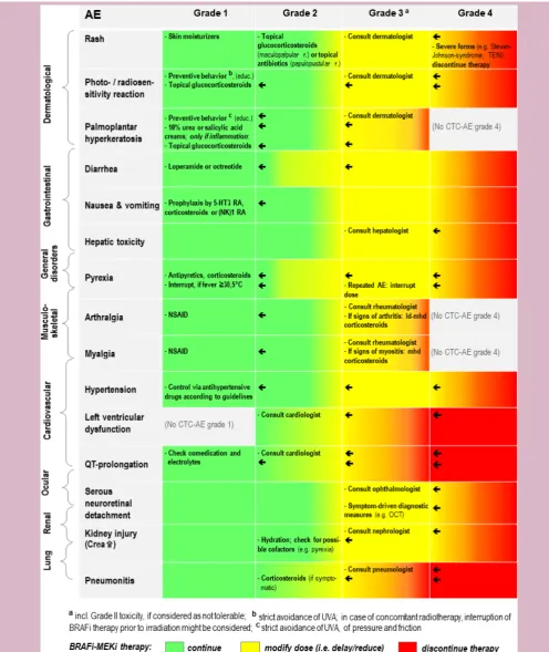
Figure 3 Recommendations for the management of clinically relevant and/or very frequent AE of BRAFi+MEKi therapy. Disclaimer: Official recommendations for the management of the three combinations might differ (please consider first the detailed specifications and recommendations as outlined in the summary of product characteristics (European Union) or in the respective, official prescribing information documents (USA and other countries outside European Union)). CTC-AE, common terminology criteria for adverse events; Crea, creatinine; ld, low-dose; mhd, middle-to-high-dose; NSAID, nonsteroidal anti-inflammatory drugs; OCT, optical coherence tomography; RA,receptor antagonist; TEN, toxic epidermal necrolysis; UVA, ultraviolet A (radiation).
on September 12, 2020 by guest. Protected by copyright.
oral fluid intake—pre-renal insufficiency may occur that ameliorates after rehydration of the patient.
dIsCussIon
BRAFi+MEKi combinations are highly effective in the therapy of metastatic melanoma84 85 and D+T has
addi-tionally been shown to prolong overall survival in the adjuvant setting.7 However, AEs occur in almost all (ie, ≥97%) of patients treated with D+T, V+C or E+B (table 1), with grade 3–4 AE rates of 46%–56%, 69% and 58%, respectively. Knowledge of side effect profiles, moni-toring and management strategies are important to tailor the therapy to patients, diagnose side effects early, keep discontinuation rates low and guide patients through occurrences of such effects. This review is based on the available safety data from phase III clinical studies: our group’s recommendations for the monitoring of side effects are provided in table 3, our recommendations for the management of clinically relevant and/or very frequent AEs of BRAFi +MEKi therapy are summarised in figure 3.
While some of the side effects are class effects, others are substance-specific. Differences in drug tolerability might partly be explained by their individual pharmaco-kinetic (figure 1) and pharmacodynamics characteristics. Class-effects of BRAFi include gastrointestinal side effects, increases in transaminases and arthralgia as well as cutaneous toxicities with formation of secondary neoplasms.20 22 23 For MEKi, class-effects encompass
gastrointestinal side effects, increases in transaminases, oedema, ocular, cardiovascular AEs and cutaneous toxicity with the occurrence of papulopustular rash.86–89
The frequency of papulopustular rash, described in MEKi monotherapy trials,18 34 36 is decreased in combination
regimens (figure 2). This is similar for the cutaneous side effects of BRAFi which are reduced by the suppression of the ‘paradoxical activation’ of the MAPK pathway, when adding a MEKi to the BRAFi. A systematic review indi-cates higher risk ratios for all grade diarrhoea, decreased ejection fraction and fever as well as a higher risk ratio for high grade diarrhoea when comparing BRAFi+MEKi versus BRAFi monotherapy.90
Substance specific, very common side effects include fever (for D+T) and photosensitivity (for V+C), attributed to the pharmacological properties of the BRAF inhibitors dabrafenib and vemurafenib. They were already identi-fied during initial clinical development in BRAFi mono-therapy trials.12–15 20 22 Differences in side effect profiles
can be used to adjust prescription to the individual patient, but also to switch between BRAFi+MEKi combi-nations when side effects occur.
Because frequencies of documented side effects also depend on the design of a given clinical study, its defi-nition of monitoring intervals and its methods to assess safety parameters, comparisons across studies have limited validity. Since there are no head-to-head trials comparing D+T, V+C and E+B, the indirect comparison
against vemurafenib is currently the best approxima-tion (figure 2). The only published indirect treatment comparison, comparing BRAFi+MEKi combinations so far, concluded that D+T shows a more favourable toxicity profile than V+C.24
Like the published indirect treatment comparison between D+T and V+C, which itself provides a less strong evidence than a randomised head-to-head trial could do, our review has certain limitations. Since the confirmatory trials COMBI-V (D+T), CoBRIM (V+C) and COLUMBUS Part I (E+B) ended at different time points and had follow-up intervals of variable length (see online supple-mentary file 1), there is some analysis bias in favour of E+B, authorised for use in the USA and the European Union since July and September 2018, respectively, only. In addition, some publication bias must be considered, as case reports and case series, describing, for example, rare and very rare side effects as discussed in our review, have been issued so far mainly for D+T and V+C: late or addi-tional cases reporting on a new BRAFi or a newly approved combination might maybe be less easily publishable too. By choosing—as the main source for the appraisal of AE and serious AE frequencies (tables 1 and 2)—three confirmatory trials comparing all against a common comparator, we attempted, however, to diminish analysis and publication bias.
In conclusion, BRAF plus MEK inhibitor therapy is an effective and safe therapy, if monitored adequately. There-fore, the treating physicians need to be familiar with side effect management to reduce morbidity and mortality as well as premature treatment discontinuation.
author affiliations
1Department of Dermatology, University of Erlangen, Erlangen, Germany 2Department of Dermatology, Center for Dermatooncology, University Medical
Center Tübingen, Tübingen, Germany
3Department of Internal Medicine, Fachklinik Hornheide, Münster, Germany 4Department of Dermatology, University Hospital Heidelberg, Heidelberg, Germany 5MVZ Augenärzte am Kröpcke, Hannover, Germany
6Division of Rheumatology and Clinical Immunology, Medizinische Klinik und
Poliklinik IV, University of Munich, Munich, Germany
7Department of Cardiology, Klinikum Nürnberg Süd, Paracelsus Medical University
Nürnberg, Nuremberg, Germany
8Gastroenterology, Hepatology and Endocrinology, Hannover Medical School,
Hannover, Germany
9Department of Dermatology, University Hospital, University Essen-Duisburg, Essen,
Germany
10German Cancer Consortium (DKTK), Heidelberg, Germany
11Department of Dermatology, Skin Cancer Center Hannover, Hannover Medical
School, Hannover, Germany
acknowledgements The authors vouch for the content of this publication and confirm that recommendations made by consensus are matching their viewpoints. The corresponding author prepared the initial draft of the manuscript with editorial and writing assistance, all authors and particularly the first author contributed to the subsequent drafts. All authors contributed to manuscript writing, reviewed the drafted manuscript and approved the final version as well as the submission. Editorial and medical writing assistance was provided by Dr Markus Hartmann from Ecc-Oncology, funded by Pierre Fabre Pharma GmbH. Pierre Fabre Pharma GmbH did not influence the content of the manuscript, nor did the authors receive financial compensation for authoring this manuscript.
Contributors All authors made essential contributions to the conception of the article and drafted one or several paragraphs and/or chapters, were revising the
on September 12, 2020 by guest. Protected by copyright.
content of the manuscript critically, agreed on final approval of the version and agreed to be accountable for all aspects of the work. The first author consolidated the draft manuscript in conjunction with the corresponding author who also co-ordinated the manuscript drafting and finalisation process.
Funding The authors have not declared a specific grant for this research from any funding agency in the public, commercial or not-for-profit sectors.
Competing interests LH: Personal fees and advisory role: Amgen, Bristol-Myers Squibb, MSD, Novartis, Roche, Pierre Fabre, Sanofi; Travel grants: Bristol-Myers Squibb, MSD; Grants: Novartis. TE: Consultancy fees from Amgen, Bristol-Myers Squibb, Celgene, Merck KGaA, MSD, Pierre Fabre, Novartis, Roche, Sanofi. MF: Consultant/advisory role: Bristol-Myers Squibb, Merck KGaA, MSD, Novartis, Roche, Sanofi. JH: Personal fees: Bristol-Myers Squibb, MSD, Novartis, Pfizer, Roche; Scientific Support/Grants: Bristol-Myers Squibb. DHS: None. JL: Speaker fees: AbbVie, Bristol-Myers Squibb, Celgene, Janssen-Cilag, MSD, Pfizer, Roche, Sanofi, USB; Consultant fees: AbbVie, AstraZeneca, Bristol-Myers Squibb, Celgene, Hospira, Janssen-Cilag, Leo Pharma, Lilly, Novartis, Roche, Sanofi. MP: None. AV: Consultancy and speaker fees: Amgen, Array, Merck KGaA, Novartis, Roche; Research grants: none. LZ: Consultant/advisory role: Bristol-Myers Squibb, MSD, Novartis, Roche, Sanofi; Honoraria: Bristol-Myers Squibb, Merck KGaA, MSD, Pierre Fabre, Novartis, Roche; Support for scientific meetings: Amgen, Bristol-Myers Squibb, MSD, Novartis, Pierre Fabre. RG: Honoraria: Almirall Hermal, Amgen, AstraZeneca, Bristol-Myers Squibb, Incyte, Leo Pharma, Merck Serono, MSD, Novartis, Pierre Fabre, Pfizer, Roche, Sanofi, SUN; Research funding: Johnson & Johnson, Novartis, Pfizer.
Patient consent for publication Not required.
Provenance and peer review Not commissioned; externally peer reviewed. open access This is an open access article distributed in accordance with the Creative Commons Attribution Non Commercial (CC BY-NC 4.0) license, which permits others to distribute, remix, adapt, build upon this work non-commercially, and license their derivative works on different terms, provided the original work is properly cited, any changes made are indicated, and the use is non-commercial. See: http:// creativecommons. org/ licenses/ by- nc/ 4. 0/.
RefeRences
1. Flaherty KT, Infante JR, Daud A, et al. Combined BRAF and MEK inhibition in melanoma with BRAF V600 mutations. N Engl J Med
Overseas Ed 2012;367:1694–703.
2. Robert C, Karaszewska B, Schachter J, et al. Improved overall survival in melanoma with combined dabrafenib and trametinib. N
Engl J Med 2015;372:30–9.
3. Long GV, Stroyakovskiy D, Gogas H, et al. Combined BRAF and MEK inhibition versus BRAF inhibition alone in melanoma. N Engl J Med
2014;371:1877–88.
4. Ribas A, Gonzalez R, Pavlick A, et al. Combination of vemurafenib and cobimetinib in patients with advanced BRAF(V600)-mutated melanoma: a phase 1b study. Lancet Oncol 2014;15:954–65. 5. Larkin J, Ascierto PA, Dréno B, et al. Combined vemurafenib and cobimetinib in BRAF-mutated melanoma. N Engl J Med
2014;371:1867–76.
6. Dummer R, Ascierto PA, Gogas HJ, et al. Encorafenib plus binimetinib versus vemurafenib or encorafenib in patients with BRAF-mutant melanoma (COLUMBUS): a multicentre, open-label, randomised phase 3 trial. Lancet Oncol 2018;19:603–15.
7. Long GV, Hauschild A, Santinami M, et al. Adjuvant dabrafenib plus trametinib in stage III BRAF-mutated melanoma. N Engl J Med
2017;377:1813–23.
8. Schadendorf D, Long GV, Stroiakovski D, et al. Three-year pooled analysis of factors associated with clinical outcomes across dabrafenib and trametinib combination therapy phase 3 randomised trials. Eur J Cancer 2017;82:45–55.
9. Ascierto PA, McArthur GA, Dréno B, et al. Cobimetinib combined with vemurafenib in advanced BRAF(V600)-mutant melanoma (coBRIM): updated efficacy results from a randomised, double-blind, phase 3 trial. Lancet Oncol 2016;17:1248–60.
10. Dummer R, Ascierto PA, Gogas H, et al. Overall survival in patients with BRAF-mutant melanoma receiving encorafenib plus binimetinib versus vemurafenib or encorafenib (COLUMBUS): a multicentre, open-label, randomised, phase 3 trial. Lancet Oncol
2018;19:1315–27.
11. Petrelli F, Ardito R, Merelli B, et al. Prognostic and predictive role of elevated lactate dehydrogenase in patients with melanoma treated
with immunotherapy and BRAF inhibitors: a systematic review and meta-analysis. Melanoma Res 2019;29:1–12.
12. Ascierto PA, Minor D, Ribas A, et al. Phase II trial (BREAK-2) of the BRAF inhibitor dabrafenib (GSK2118436) in patients with metastatic melanoma. J Clin Oncol 2013;31:3205–11.
13. Hauschild A, Grob J-J, Demidov LV, et al. Dabrafenib in BRAF-mutated metastatic melanoma: a multicentre, open-label, phase 3 randomised controlled trial. Lancet 2012;380:358–65.
14. Sosman JA, Kim KB, Schuchter L, et al. Survival in BRAF V600-mutant advanced melanoma treated with vemurafenib. N Engl J Med
2012;366:707–14.
15. Chapman PB, Hauschild A, Robert C, et al. Improved survival with vemurafenib in melanoma with BRAF V600E mutation. N Engl J Med
2011;364:2507–16.
16. Kim KB, Kefford R, Pavlick AC, et al. Phase II Study of the MEK1/ MEK2 Inhibitor Trametinib in Patients With Metastatic BRAF -Mutant Cutaneous Melanoma Previously Treated With or Without a BRAF
Inhibitor. JCO 2013;31:482–9.
17. Flaherty KT, Robert C, Hersey P, et al. Improved survival with MEK inhibition in BRAF-mutated melanoma. N Engl J Med
2012;367:107–14.
18. Rosen LS, LoRusso P, Ma WW, et al. A first-in-human phase I study to evaluate the MEK1/2 inhibitor, cobimetinib, administered daily in patients with advanced solid tumors. Invest New Drugs
2016;34:604–13.
19. Ascierto PA, Schadendorf D, Berking C, et al. MEK162 for patients with advanced melanoma harbouring NRAS or Val600 BRAF mutations: a non-randomised, open-label phase 2 study. Lancet
Oncol 2013;14:249–56.
20. Flaherty KT, Puzanov I, Kim KB, et al. Inhibition of mutated, activated BRAF in metastatic melanoma. N Engl J Med 2010;363:809–19. 21. Dummer R, Rinderknecht J, Goldinger SM. Ultraviolet A and
photosensitivity during vemurafenib therapy. N Engl J Med
2012;366:480–1.
22. Falchook GS, Long GV, Kurzrock R, et al. Dabrafenib in patients with melanoma, untreated brain metastases, and other solid tumours: a phase 1 dose-escalation trial. The Lancet
2012;379:1893–901.
23. Delord J-P, Robert C, Nyakas M, et al. Phase I Dose-Escalation and -Expansion Study of the BRAF Inhibitor Encorafenib (LGX818) in Metastatic BRAF-Mutant Melanoma. Clin Cancer Res
2017;23:5339–48.
24. Daud A, Gill J, Kamra S, et al. Indirect treatment comparison of Dabrafenib plus trametinib versus vemurafenib plus cobimetinib in previously untreated metastatic melanoma patients. J Hematol Oncol
2017;10.
25. Robert C, Karaszewska B, Schachter J, et al. 3301 two year estimate of overall survival in COMBI-v, a randomized, open-label, phase III study comparing the combination of Dabrafenib (D) and trametinib (T) with vemurafenib (Vem) as first-line therapy in patients (PTS) with unresectable or metastatic BRAF V600E/K mutation-positive cutaneous melanoma. Eur J Cancer 2015;51.
26. Dréno B, Ribas A, Larkin J, et al. Incidence, course, and
management of toxicities associated with cobimetinib in combination with vemurafenib in the coBRIM study. Ann Oncol 2017;28:1137–44. 27. Eigentler TK, Hassel JC, Berking C, et al. Diagnosis, monitoring and management of immune-related adverse drug reactions of anti-PD-1 antibody therapy. Cancer Treat Rev 2016;45:7–18.
28. Hassel JC, Heinzerling L, Aberle J, et al. Combined immune checkpoint blockade (anti-PD-1/anti-CTLA-4): evaluation and management of adverse drug reactions. Cancer Treat Rev
2017;57:36–49.
29. Karoulia Z, Gavathiotis E, Poulikakos PI. New perspectives for targeting Raf kinase in human cancer. Nat Rev Cancer
2017;17:676–91.
30. Cheng Y, Tian H. Current development status of MEK inhibitors.
Molecules 2017;22.
31. Falchook GS, Long GV, Kurzrock R, et al. Dose selection, pharmacokinetics, and pharmacodynamics of BRAF inhibitor dabrafenib (GSK2118436). Clin Cancer Res 2014;20:4449–58. 32. Ouellet D, Gibiansky E, Leonowens C, et al. Population
pharmacokinetics of Dabrafenib, a BRAF inhibitor: effect of dose, time, covariates, and relationship with its metabolites. J Clin
Pharmacol 2014;54:696–706.
33. Koelblinger P, Thuerigen O, Dummer R. Development of encorafenib for BRAF-mutated advanced melanoma. Curr Opin Oncol
2018;30:1–33.
34. Infante JR, Fecher LA, Falchook GS, et al. Safety, pharmacokinetic, pharmacodynamic, and efficacy data for the oral MEK inhibitor trametinib: a phase 1 dose-escalation trial. Lancet Oncol
2012;13:773–81.
on September 12, 2020 by guest. Protected by copyright.
35. Falchook GS, Lewis KD, Infante JR, et al. Activity of the oral MEK inhibitor trametinib in patients with advanced melanoma: a phase 1 dose-escalation trial. Lancet Oncol 2012;13:782–9.
36. Bendell JC, Javle M, Bekaii-Saab TS, et al. A phase 1 dose-escalation and expansion study of binimetinib (MEK162), a potent and selective oral MEK1/2 inhibitor. Br J Cancer 2017;116:575–83. 37. Carlos G, Anforth R, Clements A, et al. Cutaneous toxic effects of BRAF inhibitors alone and in combination with MEK inhibitors for metastatic melanoma. JAMA Dermatol 2015;151:1103–9. 38. Koelblinger P, Dornbierer J, Dummer R. A review of binimetinib
for the treatment of mutant cutaneous melanoma. Future Oncol
2017;13:1755–66.
39. Holderfield M, Nagel TE, Stuart DD. Mechanism and consequences of Raf kinase activation by small-molecule inhibitors. Br J Cancer
2014;111:640–5.
40. Chen C-B, Wu M-Y, Ng CY, et al. Severe cutaneous adverse reactions induced by targeted anticancer therapies and immunotherapies. Cancer Manag Res 2018;10:1259–73. 41. Hecht M, Zimmer L, Loquai C, et al. Radiosensitization by BRAF
inhibitor therapy-mechanism and frequency of toxicity in melanoma patients. Ann Oncol 2015;26:1238–44.
42. Hecht M, Meier F, Zimmer L, et al. Clinical outcome of concomitant vs interrupted BRAF inhibitor therapy during radiotherapy in melanoma patients. Br J Cancer 2018;118:785–92.
43. Degen A, Alter M, Schenck F, et al. The hand-foot-syndrome associated with medical tumor therapy - classification and management. J Dtsch Dermatol Ges 2010;8:652–61.
44. Graf NP, Koelblinger P, Galliker N, et al. The spectrum of cutaneous adverse events during encorafenib and binimetinib treatment in B-rapidly accelerated fibrosarcoma-mutated advanced melanoma. J
Eur Acad Dermatol Venereol 2019;33.
45. Mössner R, Zimmer L, Berking C, et al. Erythema nodosum-like lesions during BRAF inhibitor therapy: report on 16 new cases and review of the literature. J Eur Acad Dermatol Venereol
2015;29:1797–806.
46. Keely SJ, Barrett KE. P38 mitogen-activated protein kinase inhibits calcium-dependent chloride secretion in T84 colonic epithelial cells.
Am J Physiol Cell Physiol 2003;284:C339–C348.
47. Menzies AM, Ashworth MT, Swann S, et al. Characteristics of pyrexia in BRAFV600E/K metastatic melanoma patients treated with combined dabrafenib and trametinib in a phase I/II clinical trial. Ann
Oncol 2015;26:415–21.
48. Atkinson V, Long GV, Menzies AM, et al. Optimizing combination dabrafenib and trametinib therapy in BRAF mutation-positive advanced melanoma patients: guidelines from Australian melanoma medical oncologists. Asia Pac J Clin Oncol 2016;12 Suppl 7:5–12.
49. Babacan T, Türkbeyler IH, Balakan O, et al. A case of vemurafenib-induced polyarhritis in a patient with melanoma: how to manage it?
Int J Rheum Dis 2017;20:398–401.
50. Maldonado-Seral C, Berros-Fombella JP, Vivanco-Allende B, et al. Vemurafenib-associated neutrophilic panniculitis: an emergent adverse effect of variable severity. Dermatol Online J 2013;19. 51. Zimmer L, Livingstone E, Hillen U, et al. Panniculitis with arthralgia in
patients with melanoma treated with selective BRAF inhibitors and its management. Arch Dermatol 2012;148:357–61.
52. Ueno M, Namiki T, Inui K, et al. Neutrophilic panniculitis with vasculitis in a melanoma patient treated with vemurafenib: a case report and its management. Int J Dermatol 2017;56:e163–5. 53. Chaminade A, Conte H, Jouary T, et al. BRAF inhibitors-induced
panniculitis: a cutaneous side effect mimicking subcutaneous melanoma metastasis. J Eur Acad Dermatol Venereol 2015;29:392–3. 54. Novoa RA, Honda K, Koon HB, et al. Vasculitis and panniculitis
associated with vemurafenib. J Am Acad Dermatol 2012;67:e271–2. 55. Maanaoui M, Saint-Jacques C, Gnemmi V, et al. Glomerulonephritis
and granulomatous vasculitis in kidney as a complication of the use of BRAF and MEK inhibitors in the treatment of metastatic melanoma: a case report. Medicine 2017;96:e7196.
56. Mirouse A, Savey L, Domont F, et al. Systemic vasculitis associated with vemurafenib treatment: case report and literature review.
Medicine 2016;95:e4988.
57. Niro A, Recchimurzo N, Sborgia A, et al. Bilateral ischemic retinal vasculitis in metastatic cutaneous melanoma patient treated with dabrafenib and trametinib: a case report. Ocul Immunol Inflamm
2018;26:783–5.
58. Hanson K, Robinson SR, Al-Yousuf K, et al. The anti-rheumatic drug, leflunomide, synergizes with MEK inhibition to suppress melanoma growth. Oncotarget 2018;9:3815–29.
59. Lips DJ, Bueno OF, Wilkins BJ, et al. MEK1-ERK2 signaling pathway protects myocardium from ischemic injury in vivo. Circulation
2004;109:1938–41.
60. Banks M, Crowell K, Proctor A, et al. Cardiovascular effects of the MEK inhibitor, trametinib: a case report, literature review, and consideration of mechanism. Cardiovasc Toxicol 2017;17:487–93. 61. Shah RR, Morganroth J. Update on cardiovascular safety of tyrosine
kinase inhibitors: with a special focus on QT interval, left ventricular dysfunction and overall risk/benefit. Drug Saf 2015;38:693–710. 62. Flaherty L, Hamid O, Linette G, et al. A single-arm, open-label,
expanded access study of vemurafenib in patients with metastatic melanoma in the United States. Cancer J 2014;20:18–24. 63. Kim G, McKee AE, Ning Y-M, et al. FDA approval summary:
vemurafenib for treatment of unresectable or metastatic melanoma with the BRAFV600E mutation. Clin Cancer Res 2014;20:4994–5000. 64. Nebot N, Arkenau H-T, Infante JR, et al. Evaluation of the effect of
dabrafenib and metabolites on QTc interval in patients with BRAF
V600-mutant tumours. Br J Clin Pharmacol 2018;84:764–75. 65. Bronte E, Bronte G, Novo G, et al. What links BRAF to the heart
function? New insights from the cardiotoxicity of BRAF inhibitors in cancer treatment. Oncotarget 2015;6:35589–601.
66. Patnaik A, Tolcher A, Papadopoulos KP, et al. Phase 1 study to evaluate the effect of the MEK inhibitor trametinib on cardiac repolarization in patients with solid tumours. Cancer Chemother
Pharmacol 2016;78:491–500.
67. Haverkamp W, Mönnig G, Schulze-Bahr E, et al. Physician-induced torsade de pointes--therapeutic implications. Cardiovasc Drugs Ther
2002;16:101–9.
68. Curigliano G, Cardinale D, Suter T, et al. Cardiovascular toxicity induced by chemotherapy, targeted agents and radiotherapy: ESMO clinical practice guidelines. Ann Oncol 2012;23 Suppl 7:vii155–66. 69. Francis JH, Habib LA, Abramson DH, et al. Clinical and morphologic
characteristics of MEK inhibitor-associated retinopathy: differences from central serous chorioretinopathy. Ophthalmology
2017;124:1788–98.
70. de la Cruz-Merino L, Di Guardo L, Grob J-J, et al. Clinical features of serous retinopathy observed with cobimetinib in patients with BRAF-mutated melanoma treated in the randomized coBRIM study. J Transl Med 2017;15.
71. Urner-Bloch U, Urner M, Stieger P, et al. Transient MEK inhibitor-associated retinopathy in metastatic melanoma. Ann Oncol
2014;25:1437–41.
72. Urner-Bloch U, Urner M, Jaberg-Bentele N, et al. MEK inhibitor-associated retinopathy (MEKAR) in metastatic melanoma: Long-term ophthalmic effects. Eur J Cancer 2016;65:130–8.
73. Choe CH, McArthur GA, Caro I, et al. Ocular toxicity in BRAF mutant cutaneous melanoma patients treated with vemurafenib. Am J
Ophthalmol 2014;158:831–7.
74. Rogers S, McIntosh RL, Cheung N, et al. The prevalence of retinal vein occlusion: pooled data from population studies from the United States, Europe, Asia, and Australia. Ophthalmology 2010;117:313–9. 75. Welsh SJ, Corrie PG. Management of BRAF and MEK inhibitor
toxicities in patients with metastatic melanoma. Ther Adv Med Oncol
2015;7:122–36.
76. Daud A, Tsai K. Management of Treatment‐Related adverse events with agents targeting the MAPK pathway in patients with metastatic melanoma. The Oncologist 2017;22:823–33.
77. Schmitt L, Schumann T, Löser C, et al. Vemurafenib-induced pulmonary injury. Onkologie 2013;36:10–16.
78. Teuma C, Pelletier S, Amini-Adl M, et al. Adjunction of a MEK inhibitor to Vemurafenib in the treatment of metastatic melanoma results in a 60% reduction of acute kidney injury. Cancer Chemother
Pharmacol 2017;79:1043–9.
79. Launay-Vacher V, Zimner-Rapuch S, Poulalhon N, et al. Acute renal failure associated with the new BRAF inhibitor vemurafenib: a case series of 8 patients. Cancer 2014;120:2158–63.
80. Teuma C, Perier-Muzet M, Pelletier S, et al. New insights into renal toxicity of the B-Raf inhibitor, vemurafenib, in patients with metastatic melanoma. Cancer Chemother Pharmacol
2016;78:419–26.
81. Wanchoo R, Devoe C, Jhaveri KD. BRAF inhibitors - do we need to worry about kidney injury? Expert Opin Drug Saf 2016;15:1–3. 82. Jhaveri KD, Sakhiya V, Fishbane S. Nephrotoxicity of the BRAF
inhibitors vemurafenib and dabrafenib. JAMA Oncol 2015;1:1133–4. 83. Wanchoo R, Jhaveri KD, Deray G, et al. Renal effects of BRAF
inhibitors: a systematic review by the cancer and the kidney international network. Clin Kidney J 2016;9:245–51.
84. Mai R, Zhou S, Zhong W, et al. Therapeutic efficacy of combined BRAF and MEK inhibition in metastatic melanoma: a comprehensive network meta-analysis of randomized controlled trials. Oncotarget
2015;6:28502–12.
85. Pasquali S, Chiarion-Sileni V, Rossi CR, et al. Immune checkpoint inhibitors and targeted therapies for metastatic melanoma: a network meta-analysis. Cancer Treat Rev 2017;54:34–42.
on September 12, 2020 by guest. Protected by copyright.