S H O R T R E P O R T
Open Access
Identification of
TCR V
β
11-2-
D
β
1-
J
β
1-1 T
cell clone specific for WT1 peptides using
high-throughput
TCR
β
gene sequencing
Yikai Zhang
1,2†, Ling Xu
1,2†, Shaohua Chen
1, Xianfeng Zha
3, Wei Wei
4*and Yangqiu Li
1*Abstract
We previously identified aTCR Vβ21 T cell clone which was specific to CML patients, and demonstrated that TCR Vα13/β21 gene-modified CD3+T cells had specific cytotoxicity for HLA-A11+K562 cells. However, it remains unclear which antigen is specifically recognized by theTCR Vβ21 T cell clone. In this study, CD3+T cells from healthy donor peripheral blood were stimulated with the WT1 peptide or mixed BCR-ABL peptides in the presence or absence of IL-2 and IL-7. The distribution of theTCR Vβrepertoire was analyzed after different stimulations. We found that the mixed BCR-ABL peptides induced clonally expandedVβ7–9-Dβ2-Jβ2–7 T cells while the Wilms Tumor 1 peptide induced clonally expandedVβ11–2-Dβ1-Jβ1–1 T cells by high-throughputTCRβsequencing and GeneScan.
Interestingly, the sequence and CDR3 motif ofVβ11–2 T cell clone are similar to theTCR Vβ21 (a differentTCR Vregion naming system) T cell clone that we previously found in CML patients. Thus, our findings suggest that theTCR Vβ21 T cell clone found in CML patients might be a T cell clone that specifically recognizes WT1.
Keywords:Chronic myelogenous leukemia, Wilms tumor 1, BCR-ABL, T cell repertoire, T-cell receptor beta-chain sequencing
Background
Chronic myelogenous leukemia (CML) is a common hematological malignancy in adults and has the molecu-lar characteristic of BCR-ABL fusion proteins, which exhibit abnormal kinase activity [1]. Although many CML patients benefited from the development and ap-plication of tyrosine kinase inhibitors (TKIs) [2], a part of patients still suffer from primary and acquired resist-ance to TKIs [3, 4]. Another therapeutic approach for CML is hematopoietic stem cell transplantation (HSCT), including allogenic-HSCT and haploidentical-HSCT; however, their use is limited for older patients [5, 6]. There is evidence demonstrating that CML patients who have undergone recurrent allo-HSCT could be aided by donor lymphocyte infusion (DLI) [7–9]. These findings
suggest that adoptive T cell immunotherapy may be a potentially effective strategy for CML patients.
Mechanistically, infusing donor-derived cytotoxic T lymphocytes (CTLs) induces CTL-mediated leukemia cell death through the recognition of leukemia-associated an-tigens. However, DLI also causes graft-versus-host disease (GVHD), mainly because CTLs are multi-clonal T cells that also recognize allo-antigens expressed in host-normal tissues [10]. Therefore, infusing leukemic antigen-specific CTLs is a better strategy for overcoming GVHD for adop-tive T cell immunotherapy.
Wilms Tumor 1 (WT1) is a tumor suppressor gene involved in the etiology of Wilms’ tumor. It is overex-pressed mainly in myeloid leukemias, such as acute myeloid leukemia (AML) and CML, myelodysplastic syndrome (MDS), and several solid tumors [11–14]. There are evidences demonstrating that WT1 overex-pression is closely associated with CML progression, and the poor therapeutic effect of TKIs [15–19]. These re-sults highlight WT1 as a common therapeutic target for leukemia. For example, a clinic trail showed that WT1 peptide vaccination in WT1-expressing AML and MDS
© The Author(s). 2019Open AccessThis article is distributed under the terms of the Creative Commons Attribution 4.0 International License (http://creativecommons.org/licenses/by/4.0/), which permits unrestricted use, distribution, and reproduction in any medium, provided you give appropriate credit to the original author(s) and the source, provide a link to the Creative Commons license, and indicate if changes were made. The Creative Commons Public Domain Dedication waiver (http://creativecommons.org/publicdomain/zero/1.0/) applies to the data made available in this article, unless otherwise stated.
* Correspondence:[email protected];[email protected]
†Yikai Zhang and Ling Xu contributed equally to this work.
4Guangzhou Municipality Tianhe Nuoya Bio-engineering Co. Ltd, Guangzhou 510663, China
1Key Laboratory for Regenerative Medicine of Ministry of Education, Institute of Hematology, School of Medicine, Jinan University, 601 Huang Pu Da Dao Xi, 510632 Guangzhou, People’s Republic of China
patients without curative treatment option had clinical benefit including complete remission or stable diseases (SDs) with more than 50% blast reduction. The effect is accompanied by the emergence of a predominant TCR
Vβ+T cell clone both in blood and bone marrow [20–23]. However, the types of leukemia associated-antigens in AML patients are relatively complex, thus not allowing a clear definition of the types of antigens recognized by clonally expanded TCR Vβ T cells after injection with a WT1 vaccine.
We previously identified a TCR Vβ21 monoclone in blood from patients with CML and demonstrated that
TCR Vα13/β21 gene-modified T cells could induce cell death in HLA-A11+ K562 cells [24, 25]. However, it re-mains unclear whetherTCR Vβ21 T cell clones specifically recognize BCR-ABL or other CML-associated antigens. Therefore, in this study, we analyzed the distribution of theTCR Vβrepertoire in CD3+T cells from healthy donor peripheral blood after different stimulations with a WT1 peptide or mixed BCR-ABL peptides in the presence or absence of interleukin (IL)-2 and IL-7.
Materials and methods
CD3+T cell sorting
Peripheral blood mononuclear cells (PBMCs) were iso-lated from the peripheral blood of a healthy HLA-0201+ donor with informed consent by Ficoll-Hypaque gradi-ent cgradi-entrifugation. CD3+ T cells were then sorted by immunomagnetic beads from the PBMCs. The immun-nomagnetic beads were purchased from MACS, and the sorting operation was performed according to the manu-facturer’s instructions (Miltenyi Biotec, Germany). This study was approved in writing by the Ethics Committee of the first affiliated hospital of Jinan University.
Cell culture and treatment
CD3+T cells (2.5 X 10⁶cells/mL) were cultured in RPMI 1640 without fetal bovine serum overnight. Fresh media containing 100 UI/mL IL-7, 100 UI/mL IL-2, and antigen peptides was then added to the cells. Untreated cells served as the control group, and the cytokines group com-prised cells treated only with IL-2 and IL-7. The cells in the WT1 group were treated with a WT1-specific antigen peptide (RMFPNAPYL HLA A0201), while the cells in the BCR-ABL (B3A2) group were treated with six mixed anti-gen BCR-ABL peptides (Additional file 1: Table S1). The cells were cultured for 3 weeks. IL-2 was added into the media twice a week, and IL-7 was added into the media once a week. Finally, cultured T cells from different groups were collected for RNA isolation.
RNA extraction and TCRβsequencing
Total RNA was extracted from samples with TRIzol (Invitrogen, 15,596) according to the manufacturer’s
instructions. The RNA was dissolved by ddH2O after
drying out. Then, the immune library sequences were amplified by 5′rapid amplification of cDNA ends (RACE). After amplification, the concentration and integrity of the fragments were determined by Qubit, Agilent, and Q-PCR. Qualified libraries were sequenced by HiSeq or MiSeq. The mixcr (v1.8.2) program was used to identify the sequences in each sample. Sequences containing the complementarity-determining region 3 (CDR3) that had greater than four amino acids and a nucleic acid length that was a multiple of three without stop codon were retained as qualified clones. Bioinformatics analysis was performed after obtaining qualified clones. The amplifica-tion and sequencing ofTCR Vβand primary analysis were performed by the Huayin Health Company.
RT-PCR, sanger sequencing and GeneScan analysis for TCR Vβsubfamily clonality
Twenty-fourTCRVβprimers and aTCRCβprimer were used in unlabeled PCR to amplify theTCRVβsubfamily members. PCR was performed as described in our previ-ous study. A portion of the PCR product was used for direct sequencing, which was performed by Invitrogen Biotechnology Company. The sequences of the different samples were analyzed with BLAST (https://blast.ncbi. nlm.nih.gov/Blast.cgi). The remaining PCR product was used to perform runoff PCR with the addition of fluorescent primers labeled at the 5′ end with a FAM (5-Carboxyfluorescein) fluorophore (Cβ-FAM) (TIB MOLBIOL GmbH, Germany). Then, the labeled runoff PCR products (2.0μL) were mixed with 9.5μL formamide (Hi-Di Formamide, ABI, USA) and 0.5μL Size Standards (GENESCAN™-500-LIZ™, Perkin Elmer, ABI) and heat-denatured at 94 °C for 4 min. The samples were resolved by electrophoresis using a 310 DNA sequenator (Perkin Elmer, ABI) in a 310 POP-4™gel (Performance Optimized Polymer-4, ABI). The size and fluorescence intensity were determined by GeneScan software. The PCR protocol was performed as described in our previous study [26,27].
Results
Distribution of the TCR Vβrepertoire in T cells after stimulation with WT1 and BCR-ABL peptides
nucleotide sequence) in the control group was 17,789, while it was 13,828 in the cytokines group. For the WT1 and B3A2 groups, the numbers were 14,472 and 11,747, respectively. From these data, we could also determine that there was no significant difference in the numbers of
Vβgenes,Jβgenes, VDJ gene rearrangements, and unique CDR3 amino acid sequences in the four groups.
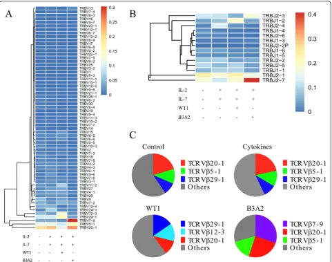
However, there were some differences in the usage of the Vβ genes and Jβ segments among the four groups (Fig. 1a, b). To obtain more detail regarding differences in the four groups, we further analyzed the data by the frequency of the Vβsubfamilies. We found that the top three Vβsubfamilies in the control group wereVβ20–1 (20.3%),Vβ5–1 (11.1%), and Vβ29–1 (9.2%), which was
Table 1Classification and counts for the sequencing results
Sample Read pairs Effective reads UniqueVβ UniqueJβ Unique VDJ Unique CDR3aa
Control 5227607 4297968 62 14 1499 17789
Cytokines 5110352 3254496 64 14 1448 13828
WT1 6591869 4968087 62 14 1437 14472
B3A2 5854623 3542234 64 14 1393 11747
Control: the untreated group; Cytokines: cells cultured with IL-2 and Il-7 in vitro for 21 days; WT1: cells treated with WT1 peptide (RMFPNAPYL, AA 126–134) and cultured with IL-2 and IL-7 in vitro for 21 days; B3A2: cells treated with 6 different BCR-ABL peptides and cultured with IL-2 and IL-7 in vitro for 21 days; An effective read is a read possessing the full CDR3 structure; UniqueVβ: types ofVβgene usage; UniqueJβ: types ofJβgene usage; Unique VDJ: unique combinations of V/D/J genes; Unique CDR3aa: unique CDR3 amino acid sequences
Fig. 1The expression frequency of theVβandJβgenes in the four groups.aThe expression frequency of theVβgene;bThe expression frequency
similar to that found in the cytokines-treated group (unspecific stimulation; Fig. 1c); however, in the WT1 group, the top three Vβsubfamilies changed toVβ29–1 (15.4%),Vβ12–3 (13.5%), and Vβ20–1 (10.8%), while in the B3A2 group,Vβ7–9 (30.3%), Vβ20–1 (24.8%), and
Vβ5–1 (15.4%) were the most frequent. These data dem-onstrate that stimulation with cytokines did not change the type of Vβ subfamilies used in normal T cells as expected, while stimulation with different peptides could induce the selective proliferation of differentVβsubfamily T cell clones.
Frequent usage pattern of VβVDJ rearrangement in T cells after stimulation with WT1 and BCR-ABL peptides
It is well known that VDJ recombination is an important event during the proliferation of the T cells, and CDR3 is an antigen recognition region that could be used as a specific marker for each T cell clone. Thus, we analyzed the usage frequency of VDJ recombination and the expression frequency of the predicted CDR3 amino acid
sequences in the four groups. As shown in Fig. 2a, the top three VDJ rearrangements in the control group were
Vβ20-1-Dβ2-Jβ2-1 (3.1%),Vβ9-Jβ1-6 (2.6%), andVβ
20-1-Dβ2-Jβ2-7 (1.7%), while in the cytokines group, it was
TCR Vβ29-1-Jβ2-4 (5.9%),Vβ20-1-Dβ1-Jβ2-4 (4.9%), and
Vβ24-1-Dβ2-Jβ2-1 (4.5%). The VDJ rearrangement usage pattern changed in the WT1 and B3A2 groups. The top three VDJ rearrangements in the WT1 group were
Vβ29-1-Dβ1-Jβ1-2 (12.2%), Vβ11-2-Dβ1-Jβ1-1 (4.7%), andV12-3-Dβ2-Jβ2-1 (4.2%). In the B3A2 group, they were
Vβ7-9-Dβ2-Jβ2-7 (29.0%),Vβ20-1-Dβ2-Jβ2-3 (12.4%), and
Vβ20-1-Dβ1-Jβ2-1 (6.7%). Similarly, the expression types and frequencies of the CDR3 amino acid sequences were different in the four groups (Fig.2b). We then analyzed the frequencies of the VDJ rearrangements corresponding to the CDR3 amino acids in the four groups. We found that the VDJ rearrangements corresponding to the highly ex-pressing CDR3 amino acids were also the patterns most highly used in each group. The highest expressed CDR3 amino acid sequence (CASSLAEREYPLEQYF; 28.8%) in
Fig. 2The predominant V (D) J rearrangements and expression levels of the CDR3 amino acids in the four groups.aThe ratios of
the B3A2 group was identified as aTCR Vβ7-9-Dβ2-Jβ2-7 rearrangement, while the second highest expressed CDR3 amino acid sequence (CASSSSGTGPNTEAFF) (4.7%) in
the WT1 group was identified as aTCR Vβ11-2-Dβ1-Jβ1-1 rearrangement (Fig.2c). Interestingly, this T cell clone was similar to the clonally expanded Vβ21 (different TCR V
Fig. 3GeneScan and Sanger sequencing results.aGeneScan results fromVβ21-amplified products in the WT1 group.bGeneScan results from theVβ
region naming system) in T cells from CML patients that we identified previously.
Identification of a WT1- or BCR-ABL-specific TCR Vβclone
We employed RT-PCR and GeneScan for further ana-lysis. Based on GeneScan analysis (Fig. 3a), we found that the Vβ21 clone in the WT1 group was biclonal (bi-peaks) and the size of the fragment was approximately 150 bp. In the B3A2 group (Fig. 3b), we found that the
Vβ6 clone was biclonal, and the control and cytokines groups were polyclonal (multi-peaks). The sequencing re-sults (Fig. 3c) indicated that the CDR3 sequence ofTCR
Vβ6 in the B3A2 group was from the TCR Vβ7-9-Dβ
2-Jβ2-7 clone, and the CDR3 sequence ofTCR Vβ21 in the WT1 group was from theTCR Vβ11-2-Dβ1-Jβ1-1 clone.
Discussion
Adoptive T cell transfusion using cancer antigen-specific T cells is the most effective immunotherapy [28]. How-ever, there are issues that limit the application of this ap-proach including difficulties in generating a sufficient number of cancer antigen-specific T cells for each pa-tient in vitro in a short period of time, and papa-tient antigen-specific T cells demonstrating low activation [29]. With the exception of chimeric antigene receptor (CAR)-T cells, TCR-engineered T cells have emerged from pre-clinical research to clinical trials and can over-come the low numbers of patient-derived CTLs [30–35]. Thus, identification of tumor antigen-specific TCRs is a key issue for such TCR-T cell generation and application. To design specific T cell immunotherapies for CML, the identification of common CML-specific TCRs might be focused on BCR-ABL or WT1 antigens [36–39]. Based on our previous finding that theTCR Vα13/β21 gene derived from CML patient-modified CD3+T cells can specifically target HLA-A11+ K562 cells [25], it would be interesting to further characterize the target of this TCR.
In this study, we first compared the frequent usage of the
TCR Vβ repertoire in CD3+ T cells treated with WT1 or BCR-ABL peptides by high-throughput TCRβ gene sequencing. The predominant TCR Vβ clone in WT1 peptide-induced T cells wasTCR Vβ11-2, whileTCR Vβ7-9 was predominant in BCR-ABL mixed peptide-induced T cells. As expected, the clonal response of theTCRVβ sub-family cells appeared to vary with different leukemia-associated antigen epitopes. To confirm this finding, we detected the expression of both Vβ11-2 and Vβ7-9 in treated CD3+ T cell samples by RT-PCR, and the PCR products were further analyzed by GeneScan to confirm the clonality of the T cells and direct Sanger sequencing to confirm the CDR3 rearrangement [40]. Significantly, clonal expansion of T cells expressingTCR Vβ11-2 orTCR Vβ7-9 was identified, and the CDR3 sequences were also con-firmed as Vβ11-2-Dβ1-Jβ1-1 and Vβ7-9-Dβ2-Jβ2-7,
respectively. Both are in-frame rearrangements, and their predicted CDR3 amino acid sequences were CASSSSGTGPNTEAFF (WT1 related) and CASSLAER-EYPLEQYF (BCR-ABL related). Therefore, both novel TCR clones might be responsible for BCR-ABL or WT1 epitopes, and whether they could be used to produce TCR-modified T cells requires further investigation. Inter-estingly, we found that the TCR Vβ11-2-Dβ1-Jβ1-1 se-quence is similar to the sese-quence in theVβ21 T cell clone (a different TCR V region naming system) in CML pa-tients that we previously identified, which could mediate specific cytotoxicity against CML with the TCR-modified T cell technique [25]. Whether these Vβ21 T cell clones from CML patients specifically recognize the WT1 pep-tide or cross respond based on different individuals re-quires more investigation. Previous studies have been showed that WT1 specific T cell clones were induced in AML patients by the same WT1126–134 peptide vaccines
which was used in this study, such T cell clone also expressedTCR Vβ11, however, the CDR3 sequence (ASS-DYNEQF) is different from the TCR Vβ11-2-Dβ1-Jβ1-1 (CASSSSGTGPNTEAFF) which we found in the study and the CML patients [20,41–43]. The reason that differ-ent TCR clone amplification induced by the same peptide, is due to the individual T cell response from different do-nors and patients. There were also studies showing differ-ent TCR clone (TCR Vβ5-1-Dβ2-Jβ2-5) inducted by WT1 (CMTWNQMNL) peptides [44]. Overall, the new iden-tified TCR Vβ11-2-Dβ1-Jβ1-1 clone in this study may provide new data of WT1 specific TCR clone bank. Moreover, on the other hand, all of this identified TCR clone may be thought as one of the immune biomarker of WT1 specific T cell clone in WT1 + malignancies.
Conclusion
In summary, we characterized the different usage pat-terns of theTCRVβrepertoire in T cells after WT1 and BCR-ABL peptide stimulation and identified two novel TCR clones (Vβ11-2-Dβ1-Jβ1-1 and Vβ7-9-Dβ2-Jβ2-7) related to both antigens. Functional studies will be per-formed to confirm their anti-CML cytotoxicity by pro-ducing TCR gene-modified T cells, it may be possible to provide a new TCR-T cell clone for WT1 + leukemia and maybe for WT1 + solid tumors immunotherapy.
Additional file
Additional file 1:Table S1.Sequences of the BCR-ABL antigen peptides. (DOCX 17 kb)
Abbreviations
AML:Acute myeloid leukemia; CAR: Chimeric Ag receptor;
IL: Interleukin; MDS: Myelodysplastic syndrome; PBMCs: Peripheral blood mononuclear cells; RACE: Rapid amplification of cDNA ends; SDs: Stable diseases; TKIs: Tyrosine kinase inhibitors; WT1: Wilms Tumor 1
Acknowledgements Not applicable
Authors’contributions
YQL contributed to the concept development and study design. YKZ, LX, SHC and XFZ performed the laboratory studies. YKZ participated in the manuscript and figure preparation. YKZ, LX, and YQL coordinated the study and helped draft the manuscript. All authors read and approved the final manuscript.
Funding
This study was supported by grants from the National Natural Science Foundation of China (Nos. 81770152 and 91642111), the Guangdong Provincial Applied Science and Technology Research & Development Program (No. 2016B020237006), and the Guangzhou Science and Technology Project (Nos. 201510010211, 20180710004, and 201803040017).
Availability of data and materials
The datasets used and/or analyzed during the current study are available from the corresponding author on reasonable request.
Ethics approval and consent to participate
All of the procedures were conducted according to the guidelines of the Medical Ethics Committees of the Health Bureau of the Guangdong Province of China. This study was approved by the Ethics Committee of the first affiliated hospital of Jinan University.
Consent for publication Not applicable.
Competing interests
The authors declare that they have no competing interests.
Author details
1Key Laboratory for Regenerative Medicine of Ministry of Education, Institute of Hematology, School of Medicine, Jinan University, 601 Huang Pu Da Dao Xi, 510632 Guangzhou, People’s Republic of China.2Department of Hematology, First Affiliated Hospital, Jinan University, Guangzhou 510632, China.3Department of Clinical Laboratory, First Affiliated Hospital, Jinan University, Guangzhou 510632, China.4Guangzhou Municipality Tianhe Nuoya Bio-engineering Co. Ltd, Guangzhou 510663, China.
Received: 7 May 2019 Accepted: 22 May 2019
References
1. Chopra R, Pu QQ, Elefanty AG. Biology of BCR-ABL. Blood Rev. 1999;13(4):211–29.
2. Druker BJ. Activity of a specific inhibitor of the BCR-ABL tyrosine kinase in the blast crisis of chronic myeloid leukemia and acute lymphoblastic leukemia with the Philadelphia chromosome (vol 344, pg 1038, 2001). N Engl J Med. 2001;345(3):232.
3. Jangamreddy JR, Panigrahi S, Lotfi K, Yadav M, Maddika S, Tripathi AK, et al. Mapping of Apoptin-interaction with BCR-ABL1, and development of apoptin-based targeted therapy. Oncotarget. 2014;5(16):7198–211. 4. Marin D, Milojkovic D, Olavarria E, Khorashad JS, de Lavallade H,
Reid AG, et al. European LeukemiaNet criteria for failure or suboptimal response reliably identify patients with CML in early chronic phase treated with imatinib whose eventual outcome is poor. Blood. 2008;112(12):4437–44.
5. Hehlmann R, Hochhous A, Baccarani M, European L. Chronic myeloid leukaemia. Lancet. 2007;370(9584):342–50.
6. Xu L, Chen H, Chen J, Han M, Huang H, Lai Y, et al. The consensus on indications, conditioning regimen, and donor selection of allogeneic hematopoietic cell transplantation for hematological diseases in China-recommendations from the Chinese Society of Hematology. J Hematol Oncol. 2018;11(1):33.
7. Collins RH Jr, Shpilberg O, Drobyski WR, Porter DL, Giralt S, Champlin R, et al. Donor leukocyte infusions in 140 patients with relapsed malignancy after allogeneic bone marrow transplantation. J Clin Oncol. 1997;15(2):433–44. 8. Kolb HJ, Schmid C, Barrett AJ, Schendel DJ. Graft-versus-leukemia
reactions in allogeneic chimeras. Blood. 2004;103(3):767–76.
9. Pinilla-Ibarz J, Shah B, Dubovsky JA. The biological basis for immunotherapy in patients with chronic myelogenous leukemia. Cancer Control. 2009;16(2):141–52.
10. Dazzi F, Goldman J. Donor lymphocyte infusions. Curr Opin Hematol. 1999;6(6):394–9.
11. Rosenfeld C, Cheever MA, Gaiger A. WT1 in acute leukemia, chronic myelogenous leukemia and myelodysplastic syndrome: therapeutic potential of WT1 targeted therapies. Leukemia. 2003;17(7):1301–12. 12. Yang L, Han Y, Saiz FS, Minden MD. A tumor suppressor and
oncogene: the WT1 story. Leukemia. 2007;21(5):868–76. 13. Sugiyama H. WT1 (Wilms’tumor gene 1): biology and Cancer
immunotherapy. Jpn J Clin Oncol. 2010;40(5):377–87.
14. Yoon JH, Kim HJ, Kwak DH, Park SS, Jeon YW, Lee SE, et al. High WT1 expression is an early predictor for relapse in patients with acute promyelocytic leukemia in first remission with negative PML-RARa after anthracycline-based chemotherapy: a single-center cohort study. J Hematol Oncol. 2017;10:4.
15. Cilloni D, Messa F, Gottardi E, Fava M, Arruga F, Defilippi L, et al. Sensitivity to imatinib therapy may be predicted by testing Wilms tumor gene expression and colony growth after a short in vitro incubation. Cancer. 2004;101(5):979–88.
16. Svensson E, Vidovic K, Lassen C, Richter J, Olofsson T, Fioretos T, et al. Deregulation of the Wilms’tumour gene 1 protein (WT1) by BCR/ABL1 mediates resistance to imatinib in human leukaemia cells. Leukemia. 2007;21(12):2485–94.
17. Otahalova E, Ullmannova-Benson V, Klamova H, Haskovec C. WT1 expression in peripheral leukocytes of patients with chronic myeloid leukemia serves for the prediction of Imatinib resistance. Neoplasma. 2009;56(5):393–7.
18. Vidovic K, Svensson E, Nilsson B, Thuresson B, Olofsson T, Lennartsson A, et al. Wilms’tumor gene 1 protein represses the expression of the tumor suppressor interferon regulatory factor 8 in human hematopoietic progenitors and in leukemic cells. Leukemia. 2010;24(5):992–1000. 19. Szanto A, Pap Z, Denes L, Lazar EB, Horvath A, Tunyogi AB, et al.
Real-time quantitative PCR detection of WT1 and M-BCR-ABL expressions in chronic myeloid leukemia. Romanian J Morphol Embryol. 2015;56(2):703–7.
20. Keilholz U, Letsch A, Busse A, Asemissen AM, Bauer S, Blau IW, et al. A clinical and immunologic phase 2 trial of Wilms tumor gene product 1 (WT1) peptide vaccination in patients with AML and MDS. Blood. 2009;113(26):6541–8.
21. Ochsenreither S, Fusi A, Busse A, Bauer S, Scheibenbogen C, Stather D, et al. “Wilms tumor protein 1”(WT1) peptide vaccination-induced complete remission in a patient with acute myeloid leukemia is accompanied by the emergence of a predominant T-cell clone both in blood and bone marrow. J Immunother. 2011;34(1):85–91.
22. Ochsenreither S, Fusi A, Geikowski A, Stather D, Busse A, Stroux A, et al. Wilms’tumor protein 1 (WT1) peptide vaccination in AML patients: predominant TCR CDR3beta sequence associated with remission in one patient is detectable in other vaccinated patients. Cancer Immunol Immunother. 2012;61(3):313–22.
23. Liu HT, Zha YY, Choudhury N, Malnassy G, Fulton N, Green M, et al. WT1 peptide vaccine in Montanide in contrast to poly ICLC, is able to induce WT1-specific immune response with TCR clonal enrichment in myeloid leukemia. Exp Hematol Oncol. 2018;7:11.
24. Zha X, Chen S, Yang L, Li B, Chen Y, Yan X, et al. Characterization of the CDR3 structure of the Vbeta21 T cell clone in patients with P210(BCR-ABL)-positive chronic myeloid leukemia and B-cell acute lymphoblastic leukemia. Hum Immunol. 2011;72(10):798–804.
25. Zha XF, Xu L, Chen SH, Yang LJ, Zhang YK, Lu YH, et al. Generation of V alpha 13/beta 21(+)T cell specific target CML cells by TCR gene transfer. Oncotarget. 2016;7(51):84246–57.
27. Li YQ, Geng SX, Du X, Chen SH, Yang LJ, Wu XL, et al. Restricted TRBV repertoire in CD4(+) and CD8(+) T-cell subsets from CML patients. Hematology. 2011;16(1):43–9.
28. Dossa RG, Cunningham T, Sommermeyer D, Medina-Rodriguez I, Biernacki MA, Foster K, et al. Development of T-cell immunotherapy for hematopoietic stem cell transplantation recipients at risk of leukemia relapse. Blood. 2018;131(1):108–20.
29. Sandri S, Bobisse S, Moxley K, Lamolinara A, De Sanctis F, Boschi F, et al. Feasibility of telomerase-specific adoptive T-cell therapy for B-cell chronic lymphocytic leukemia and solid malignancies. Cancer Res. 2016;76(9):2540–51.
30. Ochi T, Fujiwara H, Yasukawa A. Application of adoptive T-cell therapy using tumor antigen-specific T-cell receptor gene transfer for the treatment of human leukemia. J Biomed Biotechnol. 2010;2010:10.https://doi.org/10. 1155/2010/521248.
31. Im A, Pavletic SZ. Immunotherapy in hematologic malignancies: past, present, and future. J Hematol Oncol. 2017;10:10.
32. Tawara I, Kageyama S, Miyahara Y, Fujiwara H, Nishida T, Akatsuka Y, et al. Safety and persistence of WT1-specific T-cell receptor gene-transduced lymphocytes in patients with AML and MDS. Blood. 2017;130(18):1985–94.
33. Yu SN, Li AP, Liu Q, Li TF, Yuan X, Han XW, et al. Chimeric antigen receptor T cells: a novel therapy for solid tumors. J Hematol Oncol. 2017;10:13. 34. Li ZH, Song WR, Rubinstein M, Liu DL. Recent updates in cancer
immunotherapy: a comprehensive review and perspective of the 2018 China Cancer immunotherapy workshop in Beijing. J Hematol Oncol. 2018;11:15.
35. Zhang YK, Li YQ. T cell receptor-engineered T cells for leukemia immunotherapy. Cancer Cell Int. 2019;19:7.
36. Yamagami T, Sugiyama H, Inoue K, Ogawa H, Tatekawa T, Hirata M, et al. Growth inhibition of human leukemic cells by WT1 (Wilms tumor gene) antisense oligodeoxynucleotides: implications for the involvement of WT1 in leukemogenesis. Blood. 1996;87(7):2878–84.
37. Anuchapreeda S, Thanarattanakorn P, Sittipreechacharn S, Chanarat P, Limtrakul P. Curcumin inhibits WT1 gene expression in human leukemic K562 cells. Acta Pharmacol Sin. 2006;27(3):360–6.
38. Kerst G, Bergold N, Viebahn S, Gieseke F, Kalinova M, Trka J, et al. WT1 protein expression in slowly proliferating myeloid leukemic cell lines is scarce throughout the cell cycle with a minimum in G (0)/G (1) phase. Leuk Res. 2008;32(9):1393–9.
39. Semsri S, Krig SR, Kotelawala L, Sweeney CA, Anuchapreeda S. Inhibitory mechanism of pure curcumin on Wilms’tumor 1 (WT1) gene expression through the PKCalpha signaling pathway in leukemic K562 cells. FEBS Lett. 2011;585(14):2235–42.
40. Chen SH, Huang X, Zheng HT, Geng SX, Wu XL, Yang LJ, et al. The evolution of malignant and reactive gamma delta plus T cell clones in a relapse T-ALL case after allogeneic stem cell transplantation. Mol Cancer. 2013;12:7.
41. Mailander V, Scheibenbogen C, Thiel E, Letsch A, Blau IW, Keilholz U. Complete remission in a patient with recurrent acute myeloid leukemia induced by vaccination with WT1 peptide in the absence of hematological or renal toxicity. Leukemia. 2004;18(1):165–6.
42. Zhao Q, Ahmed M, Tassev DV, Hasan A, Kuo TY, Guo HF, et al. Affinity maturation of T-cell receptor-like antibodies for Wilms tumor 1 peptide greatly enhances therapeutic potential. Leukemia. 2015;29(11):2238–47.
43. Nguyen THO, Tan ACL, Xiang SD, Goubier A, Harland KL, Clemens EB, et al. Understanding CD8(+) T-cell responses toward the native and alternate HLA-A*02:01-restricted WT1 epitope. Clin Transl Immunology. 2017;6:12.
44. Watanabe K, Toji S, Ohtake J, Nakano K, Satoh T, Kitamura H, et al. Establishment of a stable T lymphoma cell line transduced with HLA-A*24: 02-restricted WT1-specific TCR genes and its application to antigen-specific immunomonitoring. Biomed Res Tokyo. 2013;34(1):41–50.
Publisher’s Note