warwick.ac.uk/lib-publications
http://wrap.warwick.ac.uk/78919
Copyright and reuse:
The Warwick Research Archive Portal (WRAP) makes this work of researchers of the
University of Warwick available open access under the following conditions.
This article is made available under the Creative Commons Attribution 4.0 International
license (CC BY 4.0) and may be reused according to the conditions of the license. For more
details see:
http://creativecommons.org/licenses/by/4.0/
A note on versions:
The version presented in WRAP is the published version, or, version of record, and may be
cited as it appears here.
www.nature.com/scientificreports
High-resolution analytical imaging
and electron holography of
magnetite particles in amyloid
cores of Alzheimer’s disease
Germán Plascencia-Villa
1, Arturo Ponce
1, Joanna F. Collingwood
2, M. Josefina
Arellano-Jiménez
1, Xiongwei Zhu
3, Jack T. Rogers
4, Israel Betancourt
1, Miguel José-Yacamán
1&
George Perry
5Abnormal accumulation of brain metals is a key feature of Alzheimer’s disease (AD). Formation of amyloid-β plaque cores (APC) is related to interactions with biometals, especially Fe, Cu and Zn, but their particular structural associations and roles remain unclear. Using an integrative set of advanced transmission electron microscopy (TEM) techniques, including spherical aberration-corrected scanning transmission electron microscopy (Cs-STEM), nano-beam electron diffraction, electron holography and analytical spectroscopy techniques (EDX and EELS), we demonstrate that Fe in APC is present as iron oxide (Fe3O4) magnetite nanoparticles. Here we show that Fe was accumulated primarily as nanostructured particles within APC, whereas Cu and Zn were distributed through the amyloid fibers. Remarkably, these highly organized crystalline magnetite nanostructures directly bound into fibrillar Aβ showed characteristic superparamagnetic responses with saturated magnetization with circular contours, as observed for the first time by off-axis electron holography of nanometer scale particles.
Alzheimer’s disease (AD) is the most common cause of irreversible dementia among older people, affecting more than 5 million patients older than 65 years in the USA alone1. The exact mechanisms that trigger AD are not completely understood. Evidence suggests that AD is caused by a complex synergism between genetic predispo-sition, ageing, environmental, occupational, metal over-exposure and ancillary factors over a long period of time. Particularly, in advanced stages of AD it is evident there is a severe loss of neuronal connections as well as a high degree of formation and spread of abnormal filaments of amyloid-β (Aβ , forming amyloid plaques cores, APC), and tau protein (neurofibrillary tangles NFT) and altered levels of biometals and neurotransmitters1,2. Specific factors that promote accumulation and aggregation of amyloid-β into APC remain unclear. Several studies found connections with metal-catalyzed oxidative stress, high production of free radicals, reactive oxygen species and mitochondrial dysfunction3,4; genetic predisposition5, epigenetic factors6,7, and also activation of inflammatory pathways8, and other processes related to ageing.
Aβ is formed through the amyloidogenic pathway (sequential cleavage by β - and γ -secretase) of Aβ PP (amyloid-β protein precursor). The expression, processing and aggregation of Aβ are partially regulated by neurocortical metal ions (Zn, Fe and Cu) by modulating the translational and transcriptional control of Aβ PP, enzymatic activities of cell surface metalloproteinase family of desintegrin and metalloprotease (ADAM, which includes secretases), homodimerization of Aβ , and modulating the expression and activity of BACE1 and SOD1 through oxidative stress signaling2,9,10. Aβ
1−42 possesses binding sites for Cu2+ and Zn2+ at physiological pH, but
under acidic conditions Fe ions bind to Aβ , whereas Cu displaces Zn9. The dyshomeostasis in Cu, Zn and Fe levels and molecular interactions with Aβ may trigger its aggregation into fibrillar forms in APC2,9,11,12. Metabolism and transport of Fe is highly regulated at the translational level, controlling expression of corresponding Aβ , ferritin,
1Department of Physics and Astronomy, The University of Texas at San Antonio (UTSA), San Antonio, TX, 78249,
USA. 2School of Engineering, University of Warwick, Coventry, CV4 7AL, UK. 3Department of Pathology, Case
Western Reserve University, Cleveland, OH, 44106, USA. 4Department of Psychiatry, Harvard Medical School,
Boston, MA 02115, USA. 5Department of Biology, The University of Texas at San Antonio (UTSA), San Antonio,
TX, 78249, USA. Correspondence and requests for materials should be addressed to G.P.-V. (email: german. [email protected])
received: 04 March 2016
Accepted: 31 March 2016
Published: 28 April 2016
transferrin receptor and ferroportin genes; nevertheless ageing and AD promote over-expression of ferritin and under-expression of transferrin, that in consequence may increase levels of Fe associated with neurofibrillary tangles and APC9,10,13. Interaction of metal ions with Aβ has been related to production or reduction of reactive oxygen species (ROS), causing a misbalance in neuronal redox potential and induction of oxidative stress with a significant role in progression of AD, whereas Aβ PP may have ferroxidase activity and interaction with ferro-portin14. Consequently, alterations of homeostasis, transport and regulatory proteins of Fe, Cu and Zn ions are closely related with the progression and pathogenesis of AD, yet with all these changes the locations of metals in end-stage AD have not been clearly established.
Analyses and characterization of hybrid samples, in this case metallo-proteins, requires use of new advanced imaging techniques. By their size range, the interactions between proteins and metals occur at the nano-bio interface, comprising the dynamic physicochemical interactions between inorganic materials and biological com-ponents15. In particular, ultra-high resolution field-emission scanning electron microscopy (UHR FE-SEM) and spherical aberration-corrected scanning transmission electron microscopy (Cs-STEM) allow higher resolution and sensitivity than conventional imaging techniques16,17. Specifically, by using cold field-emission electron guns and improved electron column design STEM allows efficient collection of multiple elastic and inelastic signals (HAADF, EDX, EELS, holography) to obtain an advanced analytical imaging and quantitative characterization with atomic resolution of sensitive biological samples.
In this work, we present the analysis of amyloid plaque cores isolated from Alzheimer disease patients through an integrative set of advanced analytical electron microscopy techniques. Particularly, to determine the presence and chemical identity of inorganic materials associated with APC, confirming the presence of nanostructured aggre-gates of iron oxide, whereas other metals such as Cu and Zn were associated with Aβ fibers. Atomic-resolution aberration-corrected STEM imaging, coupled with energy dispersive X-ray spectroscopy (EDX) and electron energy loss spectroscopy (EELS) detectors allowed a precise identification and quantification of metals, confirm-ing that iron-rich aggregates effectively had properties consistent with magnetite (Fe3O4). Magnetite nanoparti-cles located with APC showed a size range of 8–50 nm diameter with characteristic superparamagnetic responses as observed by off-axis electron holography. Particularly, holography of Fe3O4 particles revealed the phase con-tribution of the ferrite mean inner potential associated with the composition and density as circular contours. In conclusion, advanced electron analytical microscopy confirmed that the predominant form of iron within APC as nanostructured particles, and revealed their ultra-structural location, chemical identity and functional properties with high spatial sub-nanometer resolution, providing evidence of the metallo-biology implicated in iron accu-mulation and Aβ aggregation in Alzheimer’s disease and its connection to a dysregulated processing/transport of metal ions in human neurons.
Results
[image:3.595.157.397.47.176.2]Isolation and microscopic detection of amyloid plaque cores.
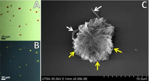
A definitive diagnosis of Alzheimer’s disease is made by neuropathological evaluation of postmortem autopsy of the most affected areas: the frontal and temporal lobes, and hippocampus18. We employed frozen brain tissues collected from patients that showed evidence for the presence of APC, demonstrated by pathological analysis with immunocytochemistry (Braak stage VI). Amyloid plaque cores were isolated from homogenized gray matter tissue by gradient ultracentrifuga-tion19,20. The histopathological dyes Congo red and thioflavin S provided qualitative evidence for presence of APC in the gradient fractions. Figure 1A,B shows APC with Congo red staining under transmitted and polarized light, respectively. Additionally, enriched fractions of APC were sorted by size through FACS. Small fragments, cell debris and oversized aggregates were mostly removed, recovering principally APC of 5–30 μ m by using a 100 μ m nozzle for further analysis (Supplementary Fig. S1). This extensive isolation process ensured that only complete APC were employed during the high resolution analytical imaging.www.nature.com/scientificreports/
Imaging of APC by UHR FE-SEM and STEM.
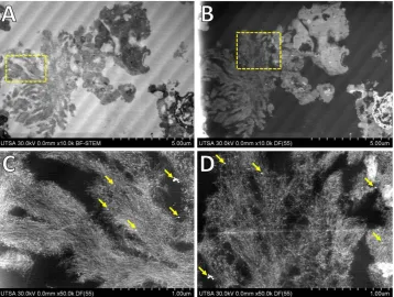
Besides histological identification of APC with polar-ized light, the amyloid cores are characterpolar-ized by their fibrillar morphology. Aβ plaques have variable diameters from 1 μ m up to 50 μ m which are fibrillar or dense-cored21–23. Imaging of isolated APC through UHR FE-SEM revealed a compact dense plaque with fibrillar morphology (Fig. 1C). APC are described as pompoms showing protruding spikes or tufts, details of the fibrillar structures formed by Aβ from different sections of the APC are indicated with yellow arrows in Fig. 1C. APC were of 8–15 μ m in diameter showing pompom-like morphology with elongated compact arrangement of filaments of 7–12 nm in diameter, with presence of some fractions of blood capillaries along the dense clump (white arrows in Fig. 1C). The presence of APC and tangles in AD cause a progressive neurodegenerative disease resulting in synaptic failure and neuronal death24–27. Also, a compro-mised integrity of intracellular vesicles is an early sign of Aβ pathogenesis which eventually may lead to neuronal cell death28. Altered levels of biometals have been related with neurogenerative diseases, especially in AD with accumulation of highly active iron ions (Fe2+). Particularly, this excess iron by interaction with Aβ cause itspre-cipitation and also may lead formation of inorganic aggregates of recognized iron oxides observed in the human brain with variable arrangements: ferrihydrite (5Fe2O3 • 9H2O), haematite (Fe2O3), magnetite (Fe3O4), maghemite (γ -Fe2O3), wüstite (FeO) and goethite (α -FeO(OH))29. Ultra-thin sections of isolated APC showed presence of electrodense aggregates coupled to compact fibrillar arrangements (Fig. 2). The morphological phenotype of APC most present in advanced AD are dense-cored, with a compact central mass encircled by an outer com-pact layer of Aβ protruding as spikes22. Similar iron-based particles were previously observed in frozen, pelleted or homogenized human brain tissue30,31, but their association with cellular components remained unclear. Due to their nanometer-scale size, electron beam imaging of these iron-based materials is not possible in complete plaques, but using the ADF detector in STEM allowed us to locate these aggregates that presented higher con-trast (due to Z2-contrast, Z is the atomic number) in comparison with the proteinaceous matrix formed by Aβ . Negative-staining enhanced the contrast and clearly revealed that the aggregates were linked to the Aβ fibers, identified by their characteristic fibrillary crossed structure of 7–12 nm in diameter.
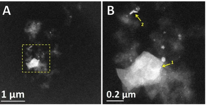
[image:4.595.157.516.47.317.2]originated only from scattering of elements present in APC. Among the cores, some round and elongated struc-tures provided an extraordinary contrast (arrows in Fig. 3B), similar electron dense regions in APC have previ-ously been observed and identified to contain Fe, Al, Si, Cu, and trace amounts of other inorganic elements26,32,33. Previously we showed the affinity and metal binding activity of APC by Raman spectroscopy, showing Zn2+ and
Cu2+ coordinated to His side chains of Aβ34. Additional evidence of the high affinity of Aβ for metals (Cu2+) arises
from in vitro fluorescence and electron paramagnetic resonance spectroscopies35. Aggregation of Aβ increases in a significant way when metal ions are present, causing precipitation and ultimately formation of synthetic Aβ fibers highly enriched in metals36,37.
Atomic resolution STEM of APC-metal particles.
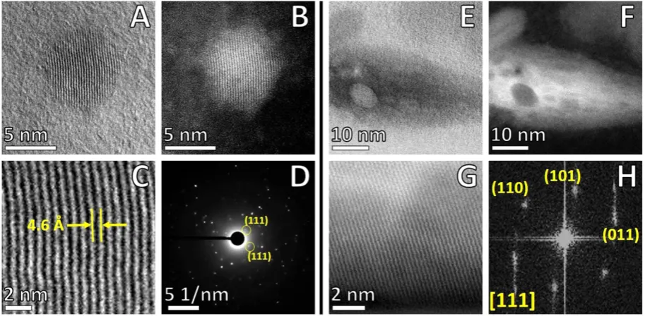
HAADF-STEM revealed details of size, shape and arrangements of inorganic materials into APC. Figure 4A,B shows high magnification BF/HAADF-STEM of a spherical particle with a diameter of 8 nm, where it is possible to distinguish atomic columns with high arrange-ment into a crystalline nanostructure. Detail of HAADF-STEM at atomic resolution in Fig. 4C revealed an interplanar spacing of 0.46 nm, which is close to the cubic crystal structure of magnetite (Fe3O4). Nano-beam diffraction and FFT pattern of the selected area showed a bcc (body-centered cubic) system oriented in the (011) beam direction. Besides individually spherical particles, we identified the presence of elongated nanostructures, which showed similar bcc crystalline structures and interplanar spacing. High magnification STEM revealed that these metal-rich aggregates could be formed by several small particles (Fig. 4E,F). These irregular morphologies can be derived from an incomplete crystallization and agglomeration effects of iron oxide particles. Magnetite (Fe3O4) is the most stable iron oxide formed by hydrolysis of Fe2+ and Fe3+ ions, in the presence of OH− precip-itating by partial reduction from ferric to ferrous ions at room temperature38,39, even though the mineralization mechanism of magnetite formation in human brain is still unknown. Synthetic magnetite-Aβ caused severe dete-rioration of in vitro clustered neuronal circuits from rat, by observing alterations in functional connectivity and damage39. Fe ions and high levels of OH• radicals have been associated with increased oxidative stress byover-production of reactive oxygen species (ROS: O2−, OH• and H2O2) which may trigger neuronal damage in AD40. The development of AD occurs over a long period, our findings suggest Fe does not bind to Aβ in plaques, it was accumulated preferentially into aggregates.
EELS and EDX of APC.
Cs-STEM in combination with electron energy loss spectroscopy (EELS) and energy dispersive X-ray spectroscopy (EDX) provided chemical and electronic state information of elements present within APC. Figure 5 shows BF/HAADF-STEM and the corresponding EELS spectra of four different regions of the APC. Areas 1, 2 and 3 were selected due to their higher contrast in the area under analysis, whereas STEM revealed the crystallinity and arrangement. Corresponding EELS spectra clearly revealed presence of peaks located at 530 and 708–710 eV, analysis and integration with DeConvEELS (GATAN) identified that peaks cor-responded to OK and FeL3 with characteristic peaks centered at 532 and 708 eV, respectively. In contrast, EELSspectra of area 4 adjacent to the nanoparticles, did not present Fe nor O peaks even though it presented contrast in STEM imaging. Background substracted spectrum showed presence of FeL3 and FeL2 peaks in areas 1–3 but not
in 4. Previously, EELS of complete APC confirmed the presence of Fe, but high scattering signals from protein matrix and carbon support prevented clear identification of Fe2+ and/or Fe3+ which would have permitted
[image:5.595.159.516.45.227.2]dis-tinction between mixed valence magnetite and maghemite26. Complementary to EELS, the corresponding EDX spectra for the same areas under analysis were acquired. Corresponding characteristic peaks for O at 0.523 keV Figure 3. Aberration corrected-BF/HAADF-STEM imaging. APC were cut in ultra-thin sections of
www.nature.com/scientificreports/
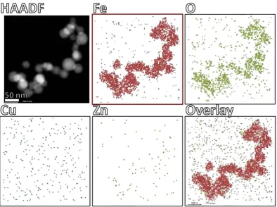
[image:6.595.98.555.48.272.2]and Fe (0.705 and 6.403 keV) were identified in the spectra, confirming that elements under analysis effectively were iron oxide particles as observed by HAADF-STEM and nano-diffraction. Control area (#4) did not show sufficient counts for Fe, but clearly showed presence of small amounts of Cu 1.30%w (0.30%at) and 0.19%w (0.04%at) of Zn (Supplementary Fig. S2). Both EELS and EDX are coupled with a scanning focused electron probe obtaining analytical information for chemical identification with high spatial resolution. The size of the probe determined the quality of spectra obtained, under the conditions employed it was 0.8 Å. Figure S2 shows BF/HAADF-STEM of the areas analyzed where it was possible to see the points where the electron probe was focused during EELS and EDX acquisition after a long period of exposure, confirming that EELS and EDX spectra acquired corresponded to signals from the nanostructured particles. EDX acquired under scanning mode gen-erated a map of the spatial distribution of the inorganic elements in the APC core. Spectral mapping of Fe and O clearly co-localized with high-contrast particles observed by HAADF-STEM (Fig. 6). In contrast, maps of Cu and Figure 4. Atomic resolution Cs-BF/HAADF-STEM imaging. High-contrast spots corresponding to iron oxide particles within APC (yellow arrows, labeled as 1 and 2 in Fig. 2). (A) BF-STEM spherical Fe nanoparticle. (B) HAADF-STEM spherical Fe nanoparticle. (C) Detail of surface of Fe nanoparticle. (D) Nano-beam electron diffraction. Elongated particles: (E) BF-STEM non-spherical Fe nanoparticle. (F) HAADF-STEM non-spherical Fe nanoparticle. (G) Detail of periodic surface of Fe nanoparticle. (H) Fast Fourier Transform (FFT) pattern of selected area in (G). Imaging with JEOL ARM-200F, operated in STEM mode at accelerating voltage of 80 kV.
[image:6.595.155.550.372.552.2]Zn showed a more disperse distribution throughout the thin-section, indicating that the nanoparticulate material is adjacent (rather than coincident) with Cu and Zn and exclusively iron oxide in the Aβ compact matrix within the limits of detection. Integration of characteristic peaks showed that sample contained 24.25% of C, 67.54% of O, 4.82% of Fe, 2.98% of Cu and 0.42% of Zn, expressed as normalized weight percentage (Supplementary Fig. S3). At the molecular level the binding of oxidized and reduced ionic forms of Cu, Fe and Zn to Aβ occurs through side chains of Asp1, Ala2, Arg5, His6, Ser8, Tyr10, Glu11, His13 and His149,41. Residues 1–17 of Aβ are disordered, while 18–42 domain form a β -strand-turn-β -strand motif which through intermolecular interactions assemble into protofilaments42. NMR spectroscopy and circular dichroism in combination with MD simulations previously revealed that the N-terminal of Aβ adopts different conformational steps depending on surround-ing media, whereas the C-terminal turns from α -helix to beta conformation43. The dynamic structure and high mobility of the Aβ C-terminal explains the versatility to bind Cu, Fe and Zn ions under different physicochemical conditions (neutral or acidic), which ultimately may adopt a stable conformation upon binding with metallic pre-cursors to assemble into protofibrillar structures forming APC. One of the most striking findings here is that the majority of the iron is not bound to Aβ fibers isolated from brain, consistent with our previous Raman scattering and electron tomography analysis of APC26,34.
[image:7.595.157.552.46.340.2]www.nature.com/scientificreports/
study demonstrated the first direct observation of magnetism of individual Fe3O4 nanoparticles directly bound within APC, in comparison with previous studies that employed SQUID magnetometry of intact or powdered and pelleted brain tissues from AD where the spatial relationship of the magnetite with APC could not be tested. In conjunction with STEM, we can confirm that the magnetic responses arise exclusively from magnetite embedded into APC, even though presence of iron biomineralization has been observed in other brain areas.
Discussion
Amyloid proteins are identified by their high affinity to Congo red and the resulting characteristic green bire-fringence by polarizing microscopy, which is an anisotropic optical effect produced when bound to amyloid pro-teins21,48. These dyes bind with high specificity by interactions with positively charged residues (His, Arg and Lys) located on the vicinity groove of stacked β -sheets, without conformational alteration of 3D structure of protein fibers. Extracellular amyloid plaques and neurofibrillary tangles are both present in AD. The precise mechanisms of pathogenesis of APC is not clearly established, but in some steps it involves sorting to multivesicular bodies with intracellular growth of Aβ fibrils which over time infiltrate to multivesicular bodies promoting neuronal death, and are finally released to the extracellular space28,49. APC isolated from AD patients showed pompom-like structure of 5–15 μ m with fibers protruding in all directions, their high stability notably resisted all purification steps and storage before and during analysis, some portions of vesicular bodies were also observed in APC.
[image:8.595.159.549.48.352.2]metal distribution in APC sections, with additional versatility from the use of low-voltage doses to reduce radia-tion damage to the proteins but keeping high spatial resoluradia-tion of the magnetite nanostructures. By the size range of Fe3O4 particles (8–50 nm in diameter) and association with Aβ , this indicated that the iron oxide nanoparti-cles present in APC cannot be cleared by neurons or microglia cells, which may promote their accumulation. The origin of these magnetite particles might be related to ferritin with a 8 nm core54, that afterwards aggregate forming larger aggregates or could be formed by the redox properties of Aβ . Also, APC are tightly enveloped by microglia and astrocytes forming a barrier influencing plaque composition, axonal dystrophy and neurotoxic-ity27. The cytotoxic effects of iron oxide nanoparticles have been extensively studied. Upon exposure to iron oxide nanoparticles, cells may suffer membrane damage, impaired mitochondrial function, inflammation and apoptotic responses, chromosome condensation, DNA damage, and generation of ROS55. The Fe nanoparticles tested were coated with inorganic surfactants; bio-compatible compounds (proteins, peptides, carbohydrates or zwitterionic agents) can effectively reduce their toxicity. Like in the case of poly-lysine coated magnetite particles, they show no observable toxic effects after 24 h dose on primary Schwann cells of the peripheral nervous system and human neuroblastoma cells (SH-SY5Y)56. Some cytotoxic effects of magnetic nanoparticles can be attributed to chemical coating and by-products and not directly to the metal core, although long term effects in neurons and brain are unknown. Similarly, magnetite particles interacting with in vitro neuronal networks showed no side effects, but when combined with Aβ caused significant deterioration in neuronal functions and connectivity39.
In a previous study, we showed iron aggregates in complete APC were magnetite (Fe3O4) or maghemite (γ -phase, Fe2O3), but specific discrimination between the two Fe phases was not possible due to high similarities in their crystalline structure26. Also, EELS on complete APC precluded determining the chemical state of mag-netite/maghemite nanoparticulate inclusions. Synchrotron X-ray microscopy was able to effectively identify the presence of iron foci in the form of magnetite and ferrihydrite-like phases in APC-rich superior temporal gyrus, but with a limitation of 5 μm spatial resolution and an embedding method that did not facilitate direct correlation with the APCs.
In this study, EELS and EDX performed in STEM mode on transversal ultrathin-sections allowed us to focus the coherent electron beam to a single point (0.8 Å), obtaining specific information about the elemental composi-tion with characteristic peaks for Fe and O generated from the specific posicomposi-tion selected with the electron beam, confirming that high-contrast Fe particles in APC are consistent with the magnetite phase. Selected area EELS analysis in the region 700–740 revealed the Fe 2p L2,3 edge with high resolution and sensitivity (Fig. 5). Spectra excluded Fe as a major component bound to Aβ , showing typical shape for a mixed valence (Fe2+/Fe3+) with Fe
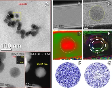
L2 around 708 eV and Fe L3 at 720–723 eV57, confirming identity of Aβ magnetite (Fe3O4) particles in compar-ison with α -Fe2O3 and γ -Fe2O4 that present an additional edge before L3. Adjustments to the configuration of the TEM to obtain electron holograms at specific voltages allowed fine-tuning of the field of view from μ m to nm scale46. Magnetite particles showed superparamagnetic behavior, with vanishing influence of the magnetic contribution on the reconstructed phase, which allowed the mean inner potential to work as the prevailing signal contribution in the form of circular contours (Fig. 7E). This observation indicated a phase shifting proportional to thickness variations along the nanoparticle direction parallel to the incident electron beam. Additionally, since electromagnetic fields are present in the phase shift, the electrostatic and magnetic contributions must be sep-arated; holograms with constant thickness do not produce fringes within nanoparticles whereas observations of fringes clearly reveal magnetic lines of force45. The reconstructed phase showed fringe contours (Fig. 7D,E), which are produced mainly by contributions from the mean inner potential with a vanishing contribution from the magnetic response of the Fe3O4 nanoparticles. The magnetic response of iron oxide is related to nanoparti-cle size; specifically the critical diameter for single domain partinanoparti-cle formation (Dsd) of Fe3O4 is ~80 nm at room temperature58. Whereas, particles of smaller size (25–30 nm) approach the lower critical diameter for super-paramagnetic response (Dsp) characterized by a fast flipping of the magnetization vectors presenting vanishing magnetization values58,59. Micromagnetic simulations of the equilibrium magnetization of Fe
www.nature.com/scientificreports/
by using superparamagnetic properties of Fe3O4 particles as imaging markers for early diagnosis and monitoring in AD patients61–63.
Conclusions
Metals play a significant role in neurobiology, particularly in neurodegenerative diseases. Iron, copper and zinc bind to Aβ , these interactions result in aggregation and precipitation into APC. High affinity of Aβ for metal ions is correlated with accumulation of Fe and their eventual aggregation into more stable particulate forms. These events are a possible mechanism of neurons associated to mitigate increased oxidative stress and misbalance of brain metals.
Using an integrative set of advanced analytical electron microscopy techniques allowed identification and quantification of the amounts of Fe, Cu and Zn present within APC. Cu and Zn were directly associated with Aβ fibers, while the majority of Fe was essentially independent from the fibers. Remarkably, iron oxide nanoparticles were highly structured and crystalline with 8–50 nm in diameter within the APC but separate from the fibers. Ultrastructural location, atomic arrangement and chemical composition of Fe aggregates as magnetite (Fe3O4) were confirmed by aberration-corrected STEM, electron nano-diffraction and in situ spectroscopy techniques (EELS and EDX). Interestingly, Fe3O4 present within APC showed a superparamagnetic response as observed by off-axis electron holography. The magnetic contours of magnetite corresponded to the mean inner potential and no lines of magnetic force were observed, which in fact is in agreement with the lower critical size for the superparamagnetic response and consistent with our previous observations from the collective content of APC. The presence of magnetic nanoparticles associated with AD reveals the importance of metallobiology of human neurons, as a key point for identification of misbalanced processing/transport of metal ions in neurodegenerative diseases.
Methods
Tissue source.
Human brain tissue was obtained, with informed written consent of relatives from autopsy AD patients for neuropathology examination. All protocols used in this study were approved by the Bioethics Committee (Department of Pathology, Case Western Reserve University). All methods were carried out in accordance with the approved guidelines and regulations.Isolation of Amyloid Plaque Cores (APC).
All chemicals were of ACS or molecular biology grade and buffers prepared with fresh ultrapure water (MilliQ, Millipore). APC were isolated using a modified reported method19,20. Brains of patients diagnosed with Alzheimer (78 and 89 years old, Braak stage VI) were removed at autopsy (5 h postmortem), divided in half and cut into 1 cm slices (one hemisphere fixed with 10% para-formaldehyde and the other was stored at − 70 °C). Sections of frozen tissue were thawed, and then grey matter from frontal and temporal lobes was cleaned by removing blood vessels, meninges and white matter. Grey matter was fined minced with a razor blade and dissolved for 2 h in homogenization buffer (2% SDS, 50 mM Tris, pH 7.6) adding a 5× v/w ratio of buffer. Tissue was homogenized in a Dounce glass homogenizer and then heated at 95 °C for 10 min. Homogenate was filtered through nylon mesh of 110 μ m removing cell debris and immedi-ately centrifuged at 1000 rpm for 30 min. Pellets were resuspended in wash buffer (1% SDS, 150 mM NaCl, 0.02% NaN3) and centrifuged again at 1000 rpm for 15 min. Supernatant was removed and pellets resuspended in small volume of wash buffer and passed through nylon mesh of 35 μ m. A non-continuous gradient (1.2, 1.4, 1.6 and 1.8 M sucrose in buffer 1% SDS, 50 mM Tris, pH 7.6) was prepared in 32 ml thickwall polycarbonate tubes and adding 1–2 ml of concentrated sample. Tubes centrifuged at 72,000 g for 1 h at 10 °C using a Beckman L8–80 M ultracentrifuge and SW28 Ti rotor. All interfaces were collected, diluted 5× in wash buffer and centrifuged at 1500 rpm for 30 min. Finally pellets were resuspended in wash buffer and stored at 4 °C for further analysis. APC were sorted with BD FACS-Aria II by forward angle light-scattering and auto-fluorescence, using a 100 μ m nozzle to recover 5–30 μ m particles and removing small debris and large aggregates. Finally, APC were concentrated by centrifugation.Optical microscopy.
Microscopy detection of isolated APC was performed by Congo red staining. 2–3 μ l of each sample were placed on a positively charged microscope slide and dried at 100 °C for 10 min. Slides were stained for 30 min with Congo red solution (1% w/v in 80% ethanol, with 0.01 M NaOH), rinsed with 70% eth-anol, 95% etheth-anol, xylenes and air dried. Coverslip was placed with help of water based mounting media. Slides were analyzed by polarization microscopy in a ZEISS AXIO A1 Imager to observe birefringence (green color) characteristic of APC stained with Congo red.of 7.98 V with field of view of 65 × 65 nm at 300 K. Processing and reconstructing of the interference fringes and reference waves with HoloWorks v.5.0 (Gatan Microscopy Suite, DigitalMicrograph) using a fringe contrast of 26%, fringes width (W) of 52 nm and fringes spacing (σ ) of 0.36 nm.
References
1. Alzheimer’s Disease Education and Referral (ADEAR) Center. Alzheimer’s Disease Fact Sheet - National Institute on Aging. NIH Publication 15–6423, 1–8 (2015), Available at: https://www.nia.nih.gov/alzheimers/publication/alzheimers-disease-fact-sheet (Update: 07/01/2016) (Access: 03/03/2016).
2. Bush, A. I. The metallobiology of Alzheimer’s disease. Trends Neurosci 26, 207–214 (2003), 10.1016/S0166-2236(03)00067-5. 3. Moreira, P. I., Carvalho, C., Zhu, X., Smith, M. A. & Perry, G. Mitochondrial dysfunction is a trigger of Alzheimer’s disease
pathophysiology. Biochim Biophys Acta 1802, 2–10 (2010), 10.1016/j.bbadis.2009.10.006.
4. Wang, X. et al. Oxidative stress and mitochondrial dysfunction in Alzheimer’s disease. Biochim Biophys Acta 1842, 1240–1247 (2014), 10.1016/j.bbadis.2013.10.015.
5. Bird, T. D. Alzheimer Disease Overview. GeneReviews (2015), Available at: http://www.ncbi.nlm.nih.gov/books/NBK116/(Update: 24/09/2015) (Access: 03/03/2016).
6. De Jager, P. L. et al. Alzheimer’s disease: early alterations in brain DNA methylation at ANK1, BIN1, RHBDF2 and other loci. Nat Neurosci 17, 1156–1163 (2014), 10.1038/nn.3786.
7. Lord, J. & Cruchaga, C. The epigenetic landscape of Alzheimer’s disease. Nat Neurosci 17, 1138–1140 (2014), 10.1038/nn.3792. 8. Wyss-Coray, T. & Rogers, J. Inflammation in Alzheimer disease-a brief review of the basic science and clinical literature. Cold Spring
Harb Perspect Med 2, a006346 (2012), 10.1101/cshperspect.a006346.
9. Roberts, B. R., Ryan, T. M., Bush, A. I., Masters, C. L. & Duce, J. A. The role of metallobiology and amyloid-beta peptides in Alzheimer’s disease. J Neurochem 120, Suppl 1, 149–166 (2012), 10.1111/j.1471-4159.2011.07500.x.
10. Cho, H. H. et al. Selective translational control of the Alzheimer amyloid precursor protein transcript by iron regulatory protein-1.
J Biol Chem 285, 31217–31232 (2010), 10.1074/jbc.M110.149161.
11. Greenough, M. A., Camakaris, J. & Bush, A. I. Metal dyshomeostasis and oxidative stress in Alzheimer’s disease. Neurochem Int 62, 540–555 (2013), 10.1016/j.neuint.2012.08.014.
12. Pal, A., Siotto, M., Prasad, R. & Squitti, R. Towards a unified vision of copper involvement in Alzheimer’s disease: a review connecting basic, experimental, and clinical research. J Alzheimers Dis 44, 343–354 (2015), 10.3233/JAD-141194.
13. Rogers, J. & Munro, H. Translation of ferritin light and heavy subunit mRNAs is regulated by intracellular chelatable iron levels in rat hepatoma cells. Proc Natl Acad Sci USA 84, 2277–2281 (1987), 10.1073/pnas.84.8.2277.
14. Duce, J. A. et al. Iron-export ferroxidase activity of beta-amyloid precursor protein is inhibited by zinc in Alzheimer’s disease. Cell
142, 857–867 (2010), 10.1016/j.cell.2010.08.014.
15. Nel, A. E. et al. Understanding biophysicochemical interactions at the nano-bio interface. Nat Mater 8, 543–557 (2009), 10.1038/ nmat2442.
16. Pennycook, S. J. Scanning transmission electron microscopy: Seeing the atoms more clearly. MRS Bulletin 37, 943–951 (2012), 10.1557/mrs.2012.239.
17. Pennycook, S. J. Seeing the atoms more clearly: STEM imaging from the Crewe era to today. Ultramicroscopy 123, 28–37 (2012), 10.1016/j.ultramic.2012.05.005.
18. Nowotny, P., Kwon, J. M. & Goate, A. M. Alzheimer disease. eLS 1–6 (2001), 10.1038.npg.els.0000228.
19. Selkoe, D. J., Abraham, C. R., Podlisny, M. B. & Duffy, L. K. Isolation of low-molecular-weight proteins from amyloid plaque fibers in Alzheimer’s disease. J Neurochem 46, 1820–1834 (1986), 10.1111/j.1471-4159.1986.tb08501.x.
20. Rostagno, A. & Ghiso, J. Isolation and biochemical characterization of amyloid plaques and paired helical filaments. Curr Protoc Cell Biol 3, 1–33 (2009), 10.1002/0471143030.cb0333s44.
21. Hayne, D. J., Lim, S. & Donnelly, P. S. Metal complexes designed to bind to amyloid-beta for the diagnosis and treatment of Alzheimer’s disease. Chem Soc Rev 43, 6701–6715 (2014), 10.1039/c4cs00026a.
22. Dickson, T. C. & Vickers, J. C. The morphological phenotype of beta-amyloid plaques and associated neuritic changes in Alzheimer’s disease. Neuroscience 105, 99–107 (2001), 10.1016/S0306-4522(01)00169-5.
23. Kawai, M., Cras, P. & Perry, G. Serial reconstruction of β -protein amyloid plaques: relationship to microvessels and size distribution.
Brain Res 592, 278–282 (1992), 10.1016/0006-8993(92)91686-9.
24. Kuo, Y. M. et al. Comparative analysis of amyloid-beta chemical structure and amyloid plaque morphology of transgenic mouse and Alzheimer’s disease brains. J Biol Chem 276, 12991–12998 (2001), 10.1074/jbc.M007859200.
25. Colletier, J. P. et al. Molecular basis for amyloid-beta polymorphism. Proc Natl Acad Sci USA 108, 16938–16943 (2011), 10.1073/ pnas.1112600108.
26. Collingwood, J. F. et al. Three-dimensional tomographic imaging and characterization of iron compounds within Alzheimer’s plaque core material. J Alzheimers Dis 14, 235–245 (2008), 10.3233/JAD-18560134.
27. Condello, C., Yuan, P., Schain, A. & Grutzendler, J. Microglia constitute a barrier that prevents neurotoxic protofibrillar Abeta42 hotspots around plaques. Nat Commun 6, 6176 (2015), 10.1038/ncomms7176.
28. Friedrich, R. P. et al. Mechanism of amyloid plaque formation suggests an intracellular basis of Abeta pathogenicity. Proc Natl Acad Sci USA 107, 1942–1947 (2010), 10.1073/pnas.0904532106.
29. Collingwood, J. & Telling, N. D. Iron oxides in the human brain in Iron oxides: from nature to applications (ed. Faivre, D.) ISBN 978-3-527-33882-5 (Wiley-VCH, 2016).
30. Hautot, D., Pankhurst, Q. A., Khan, N. & Dobson, J. Preliminary evaluation of nanoscale biogenic magnetite in Alzheimer’s disease brain tissue. Proc Biol Sci 270, Suppl 1, S62–S64 (2003), 10.1098/rsbl.2003.0012.
www.nature.com/scientificreports/
32. Collingwood, J. & Dobson, J. Mapping and characterization of iron compounds in Alzheimer’s tissue. J Alzheimers Dis 10, 215–222 (2006).
33. Collingwood, J. F. et al.In situ characterization and mapping of iron compounds in Alzheimer’s disease tissue. J Alzheimers Dis 7, 267–272 (2005).
34. Dong, J. et al. Metal binding and oxidation of amyloid-beta within isolated senile plaque cores: Raman microscopic evidence.
Biochemistry 42, 2768–2773 (2003), 10.1021/bi0272151.
35. Jiang, D. et al. The elevated copper binding strength of amyloid-β aggregates allows the sequestration of copper from albumin: a pathway to accumulation of copper in senile plaques. Biochemistry 52, 547–556 (2013), 10.1021/bi301053h.
36. Masters, C. L. et al. Amyloid plaque core protein in Alzheimer disease and Down syndrome. Proc Natl Acad Sci USA 82, 4245–4249 (1985), 10.1097/00005072-198505000-00178.
37. Everett, J. et al. Evidence of redox-active iron formation following aggregation of ferrihydrite and the Alzheimer’s disease peptide beta-amyloid. Inorg Chem 53, 2803–2809 (2014), 10.1021/ic402406g.
38. Mascolo, M., Pei, Y. & Ring, T. Room temperature co-precipitation synthesis of magnetite nanoparticles in a large ph window with different bases. Materials 6, 5549–5567 (2013), 10.3390/ma6125549.
39. Teller, S., Tahirbegi, I. B., Mir, M., Samitier, J. & Soriano, J. Magnetite-Amyloid-beta deteriorates activity and functional organization in an in vitro model for Alzheimer’s disease. Sci Rep 5, 17261 (2015), 10.1038/srep17261.
40. Smith, M. A., Harris, P. L., Sayre, L. M. & Perry, G. Iron accumulation in Alzheimer disease is a source of redox-generated free radicals. Proc Natl Acad Sci USA 94, 9866–9868 (1997), 10.1073/pnas.94.18.9866.
41. Faller, P. & Hureau, C. Bioinorganic chemistry of copper and zinc ions coordinated to amyloid-beta peptide. Dalton Trans 21, 1080–1094 (2009), 10.1039/b813398k.
42. Lührs, T. et al. 3D structure of Alzheimer’s amyloid-β (1–42) fibrils. Proc Natl Acad Sci USA 102, 17342–17347 (2005), 10.1073/ pnas.0506723102.
43. Tomaselli, S. et al. The alpha-to-beta conformational transition of Alzheimer’s Abeta-(1–42) peptide in aqueous media is reversible: a step by step conformational analysis suggests the location of beta conformation seeding. Chembiochem 7, 257–267 (2006), 10.1002/ cbic.200500223.
44. Midgley, P. A. & Dunin-Borkowski, R. E. Electron tomography and holography in materials science. Nat Mater 8, 271–280 (2009), 10.1038/nmat2406.
45. Tonomura, A. Applications of electron holography. Rev Mod Phys 59, 639–669 (1987), 10.1103/RevModPhys.59.639.
46. Cantu-Valle, J., Ruiz-Zepeda, F., Mendoza-Santoyo, F., Jose-Yacaman, M. & Ponce, A. Calibration for medium resolution off-axis electron holography using a flexible dual-lens imaging system in a JEOL ARM 200F microscope. Ultramicroscopy 147, 44–50 (2014), 10.1016/j.ultramic.2014.06.003.
47. Völkl, E., Allard, L. F. & Frost, B. A software package for the processing and reconstruction of electron holograms. J Microsc 180, 39–50 (1995), 10.111/j.1365-2818.tb03655.x.
48. Schutz, A. K. et al. The amyloid-Congo red interface at atomic resolution. Angew Chem Int Ed Engl 50, 5956–5960 (2011), 10.1002/ anie.201008276.
49. Meyer, E. P., Ulmann-Schuler, A., Staufenbiel, M. & Krucker, T. Altered morphology and 3D architecture of brain vasculature in a mouse model for Alzheimer’s disease. Proc Natl Acad Sci USA 105, 3587–3592 (2008), 10.1073/pnas.0709788105.
50. Quintana, C. & Gutierrez, L. Could a dysfunction of ferritin be a determinant factor in the aetiology of some neurodegenerative diseases? Biochim Biophys Acta 1800, 770–782 (2010), 10.1016/j.bbagen.2010.04.012.
51. Rouault, T. A. Iron metabolism in the CNS: implications for neurodegenerative diseases. Nat Rev Neurosci 14, 551–564 (2013), 10.1038/nrn3453.
52. Everett, J. et al. Ferrous iron formation following the co-aggregation of ferric iron and the Alzheimer’s disease peptide beta-amyloid (1–42). J R Soc Interface 11, 20140165 (2014), 10.1098/rsif.2014.0165.
53. Yang, H. et al. Upregulation of mitochondrial ferritin by proinflammatory cytokines: implications for a role in Alzheimer’s disease.
J Alzheimers Dis 45, 797–811 (2015), 10.3233/JAD-142595.
54. Pankhurst, Q., Hautot, D., Khan, N. & Dobson, J. Increased levels of magnetic iron compounds in Alzheimer’s disease. J Alzheimers Dis 13, 49–52 (2008).
55. Singh, N., Jenkins, G. J. S., Asadi, R. & Doak, S. H. Potential toxicity of superparamagnetic iron oxide nanoparticles (SPION). Nano Rev 1, 5358 (2010), 10.3402/nano.v1i0.5358.
56. Riggio, C. et al. Poly-l-lysine-coated magnetic nanoparticles as intracellular actuators for neural guidance. Int J Nanomedicine 7, 3155–3166 (2012), 10.2147/ijn.s28460.
57. Almeida, T. P. et al. Visualized effect of oxidation on magnetic recording fidelity in pseudo-single-domain magnetite particles. Nat Commun 5, 5154 (2014), 10.1038/ncomms6154.
58. Singamaneni, S., Bliznyuk, V. N., Binek, C. & Tsymbal, E. Y. Magnetic nanoparticles: recent advances in synthesis, self-assembly and applications. J Mater Chem 21, 16819–16845 (2011), 10.1039/C1JM11845E.
59. Vikram, S. et al. Tuning the magnetic properties of iron oxide nanoparticles by a room-temperature air-atmosphere (RTAA) co-precipitation method. J Nanosci Nanotechnol 15, 3870–3878 (2015), 10.1166/jnn.2015.9544.
60. Tao, Y., Wang, Y., Rogers, J. T. & Wang, F. Perturbed iron distribution in Alzheimer’s disease serum, cerebrospinal fluid, and selected brain regions: a systematic review and meta-analysis. J Alzheimers Dis 42, 679–690 (2014), 10.3233/JAD-140396.
61. Filippi, M. et al. Magnetic resonance imaging in Alzheimer’s disease: from diagnosis to monitoring treatment effect. Curr Alzheimer Res 9, 1198–1209 (2012), 10.2174/156720512804142949.
62. Frisoni, G. B., Fox, N. C., Jack, C. R., Scheltens, P. & Thompson, P. M. The clinical use of structural MRI in Alzheimer disease. Nat Rev Neurol 6, 67–77 (2010), 10.1038/nrneurol.2009.215.
63. Antharam, V. et al. High field magnetic resonance microscopy of the human hippocampus in Alzheimer’s disease: quantitative imaging and correlation with iron. Neuroimage 59, 1249–1260 (2012), 10.1016/j.neuroimage.2011.08.019.
Acknowledgements
This work was supported by Welch Foundation (AX-1615), NSF (DMR-1103730), NIH RCMI (5G12RR013646-12 and G(5G12RR013646-12MD007591) and Semmes Foundation. Technical assistance of Sandra Siedlak. Facilities of Department of Pathology, Case Western Reserve University. Facilities of Kleberg Advanced Microscopy Center (KAMiC), NIH RCMI Nanotechnology and Human Health Core and NIH RCMI Biophotonics Core at UTSA. We dedicate this paper to Mark A. Smith for first noting the magnetic properties of APC.