Received 6 November 2019 Accepted 13 November 2019
Edited by H. Stoeckli-Evans, University of Neuchaˆtel, Switzerland
Keywords:crystal structure; hydrogen bonding; DFT; Hirshfeld surface analysis; HOMO–LUMO calculations.
CCDC reference:1965448
Supporting information:this article has supporting information at journals.iucr.org/e
Crystal structure, Hirshfeld surface analysis and
DFT studies of
2-[5-(4-methylbenzyl)-6-oxo-3-phenyl-1,6-dihydropyridazin-1-yl]acetic acid
Said Daoui,aCemile Baydere,b* Fouad El Kalai,a* Lhassane Mahi,cNecmi Dege,b Khalid Karrouchidand Noureddine Benchata
aLaboratory of Applied Chemistry and Environment (LCAE), Faculty of Sciences, Mohamed I University, 60000 Oujda,
Morocco,bDepartment of Physics, Faculty of Arts and Sciences, Ondokuz Mayıs University, 55139-Samsun, Turkey, cMoroccan Foundation for Advanced Science, Innovation and Research (Mascir), Department of Nanotechnology, Rabat
Design Center, Rue Mohamed Al Jazouli-Madinat Al Irfane, Rabat 10 100, Morocco, anddLaboratory of Plant Chemistry,
Organic and Bioorganic Synthesis, URAC23, Faculty of Science, B.P. 1014, GEOPAC Research Center, Mohammed V University, Rabat, Morocco. *Correspondence e-mail: [email protected], [email protected]
The title pyridazinone derivative, C20H18N2O3, is not planar. The phenyl ring
and the pyridazine ring are inclined to each other by 10.55 (12), whereas the
4-methylbenzyl ring is nearly orthogonal to the pyridazine ring, with a dihedral angle of 72.97 (10). In the crystal, molecules are linked by pairs of O—H O
hydrogen bonds, forming inversion dimers with anR2 2
(14) ring motif. The dimers are linked by C—H O hydrogen bonds, generating ribbons propagating along the c-axis direction. The intermolecular interactions were additionally investigated using Hirshfeld surface analysis and two-dimensional fingerprint plots. They revealed that the most significant contributions to the crystal packing are from H H (48.4%), H O/O H (21.8%) and H C/C H (20.4%) contacts. Molecular orbital calculations providing electron-density plots of HOMO and LUMO molecular orbitals and molecular electrostatic potentials (MEP) were also computed, both with the DFT/B3LYP/6–311 G++(d,p) basis set.
1. Chemical context
In a continuation of our investigations of the molecular structures and Hirshfeld surfaces of new pyridazinone deriv-atives (Daouiet al., 2019a,b), we report herein on the synthesis and crystal and molecular structures of the title compound, 2-[5-(4-methylbenzyl)-6-oxo-3-phenyl-1,6-dihydropyridazin-1-yl]acetic acid, as well as the analysis of the Hirshfeld surfaces.
2. Structural commentary
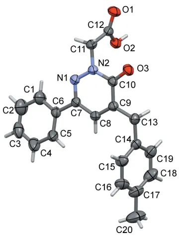
The molecule structure of the title compound is shown in Fig. 1. The phenyl (C1–C6) and pyridazine (C7–C10/N1/N2) rings are twisted relative to each other, making a dihedral
angle of 10.55 (12). The 4-methylbenzl ring (C14–C19) is inclined to the pyridazine ring by 72.97 (10). Atoms C9 and
C10 of the pyridazine ring show the largest deviations from planarity (r.m.s. deviation = 0.0075 A˚ ) in positive and negative directions [C10 = 0.0127 (11) A˚ and C9 =
0.0090 (11) A˚ ]. The O3 C10 bond length of the pyridaz-inone carbonyl function is 1.2433 (19) A˚ and the N1—N2 bond length in the pyridazine ring is 1.3516 (19) A˚ , both in accordance with values reported for related pyridazinones (El Kalaiet al., 2019a; Xuet al., 2005).
3. Supramolecular features
In the crystal, molecules are linked by pairs of O—H O hydrogen bonds, forming inversion dimers with anR22(14) ring
motif (Table 1 and Fig. 2). The dimers are linked by C—H O hydrogen bonds, forming ribbons that extend along thec-axis direction (Table 1 and Fig. 2). There are no other significant intermolecular interactions present.
4. Database survey
[image:2.610.82.263.67.304.2] [image:2.610.313.565.87.133.2]A search of the Cambridge Structural Database (CSD, version 5.40, update August 2019; Groom et al., 2016) using 2-[6-oxopyridazin-1(6H)-yl]acetic acid as the main skeleton revealed the presence of six structures similar to the title compound, but with different substituents on the pyridazine ring. Two of these structures are similar to the title compound, Table 1
Hydrogen-bond geometry (A˚ ,).
D—H A D—H H A D A D—H A
O2—H2 O3i 0.82 1.83 2.6358 (16) 167 C3—H3 O1ii 0.93 2.51 3.430 (3) 172
[image:2.610.70.524.533.721.2]Symmetry codes: (i)xþ1;yþ2;z; (ii)x;y;zþ1.
Figure 2
A view along theaaxis of the crystal packing of the title compound. The O—H O and C—H O hydrogen bonds (see Table 1) are shown as dashed lines. For clarity, only H atoms H2 and H3 (grey balls) have been included.
Figure 1
viz. ethyl {5-[(3-chlorophenyl)methyl]-6-oxo-3-phenylpyrid-azin-1(6H)-yl}acetate (FODQUN; El Kalaiet al., 2019a) and ethyl 3-methyl-6-oxo-5-[3-(trifluoromethyl)phenyl]-1,6-di-hydro-1-pyridazineacetate (QANVOR; Xuet al., 2005).
In FODQUN, the phenyl ring and the pyridazine ring are inclined to each other by 17.41 (13), whereas the
3-chloro-phenyl ring is nearly orthogonal to the pyridazine ring with a dihedral angle of 88.19 (13). In the crystal, C—H O hydrogen bonds generate inversion dimers with anR2
2(10) ring
motif. The dimers are linked by further C—H O hydrogen bonds, enclosingR2
2
(20) ring motifs, forming ribbons, similar to the situation in the crystal of the title compound. Weak intermolecular C—H interactions and – interactions are also observed in the crystal structure.
In QANVOR, the phenyl and pyridazinone rings are approximately coplanar with a dihedral angle of 4.84 (14). In
the crystal, inversion-related molecules form dimers through non-classical C—H O hydrogen bonds. The dimers are linked by a number of C–H F hydrogen bonds, forming a three-dimensional structure.
5. Hirshfeld surface analysis
Hirshfeld surface analysis was used to quantify the inter-molecular contacts of the title compounds, using the software
CrystalExplorer17.5 (Turner et al., 2017). The Hirshfeld surfaces were calculated using a standard (high) surface resolution with the three-dimensional dnorm surfaces plotted
over a fixed colour scale of0.7290 (red) to 1.4764 (blue) a.u.. The Hirshfeld surfaces of the title compound were mapped over dnorm, shape index and curvedness, and are shown in
Fig. 3a–c.
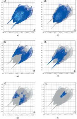
The overall two-dimensional fingerprint plot and those delineated into H H, H C/ C H, H O/O H, H N/ N H and C C contacts are illustrated in Fig. 4a–f, respectively. The H H interaction makes the largest contri-bution (48.4%) to the overall crystal packing. The pair of wings in the fingerprint plot delineated into H C/C H contacts, which contribute 20.4% to the Hirshfeld surface, have a nearly symmetrical distribution of points with the tips at de + di 2.70 A˚ . H O/O H contacts make a 21.8%
contribution to the Hirshfeld surface. The contacts are
represented by a pair of sharp spikes in the regionde + di
1.64 A˚ in the fingerprint plot, Fig. 4d. The H O/O H contacts arise from intermolecular O—H O and C—H O hydrogen bonding (Table 2). The contributions of the other contacts to the Hirshfeld surface are negligible, i.e. H N/ N H of 4.1% and C C of 4.0%.
6. Frontier molecular orbital analysis
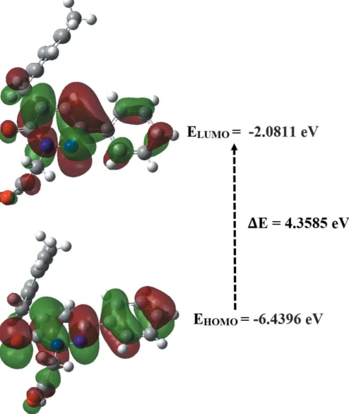
The energy levels for the title compound were computed theoretically via density functional theory (DFT) using the standard B3LYP functional and 6–311 G++ (d,p) basis-set calculations (Becke, 1993) as implemented inGAUSSIAN 09
[image:3.610.312.569.73.470.2](Frischet al., 2009). The HOMO (highest occupied molecular orbital) acts as an electron donor and the LUMO (lowest occupied molecular orbital) as an electron acceptor. When the energy gap is small, the molecule is highly polarizable and has high chemical reactivity. The energy levels, energy gaps, Figure 3
(a) The Hirshfeld surface of the title compound mapped overdnorm, and
plotted in the range0.7290 to 1.4764 a.u.. (b) the Hirshfeld surface mapped over shape-index, (c) the Hirshfeld surface mapped over curvedness.
Figure 4
[image:3.610.46.296.594.697.2]hardness (), softness () and electronegativity () are given in Table 2. The electron transition from the HOMO to the LUMO energy level is shown in Fig. 5. The chemical hardness and softness of a molecule is a sign of its chemical stability. From the HOMO–LUMO energy gap, we can see whether or not the molecule is hard or soft. If the energy gap is large, the molecule is hard and if small the molecule is soft. Soft mol-ecules are more polarizable than hard ones because they need less energy for excitation. Therefore, from Table 2 we conclude that the title compound can be classified as a hard material with a HOMO–LUMO energy gap of 4.3585 eV.
7. Molecular electrostatic potentials
Molecular electrostatic potential (MEP) displays molecular size and shape as well as positive, negative and neutral elec-trostatic potential regions in terms of colour grading and is useful in investigating relationships between molecular
structure and physicochemical properties (Murray & Sen, 1996; Scrocco & Tomasi, 1978). The MEP map (Fig. 6) was calculated at the B3LYP/6-311 G++ (d,p) level of theory. The red and blue-coloured regions indicate nucleophiles (electron rich) and electrophile regions (electron poor), respectively. The white regions indicate neutral atoms. In the title molecule, the red regions are concentrated at the carbonyl group. It possesses the most negative potential and is thus the strongest repulsion site (electrophilic attack). The blue regions indicate the strongest attraction regions, which are occupied mostly by hydrogen atoms.
8. Synthesis and crystallization
A suspension of ethyl 2-[5-(4-methylbenzyl)-6-oxo-3-phenyl-pyridazin-1(6H)-yl]acetate (3.6 mmol), and 6 N NaOH (14.4 mmol) in ethanol (50 ml) was stirred at 353 K for 4 h. The mixture was then concentratedin vacuo, diluted with cold water, and acidified with 6 N HCl. The final product was filtered off with suction and recrystallized from ethanol. Yellow prismatic crystals were obtained by slow evaporation of the solvent at room temperature.
9. Refinement
Crystal data, data collection and structure refinement details are summarized in Table 3. The hydrogen atoms were fixed geometrically (O—H = 0.82 A˚ , C—H = 0.93–0.96 A˚) and allowed to ride on their parent atoms withUiso(H) = 1.5Ueq(O,
C-methyl) and 1.2Ueq(C) for other H atoms. For atoms C17–
C20, SIMU, DELU and ISOR commands were used (SHELXL; Sheldrick, 2015b).
Acknowledgements
[image:4.610.316.564.71.249.2]The authors acknowledge the Faculty of Arts and Sciences, Ondokuz Mayıs University, Turkey, for the use of the Stoe Figure 6
[image:4.610.43.296.93.170.2]Theoretical molecular electrostatic potential surface for the title compound, calculated using the DFT/B3LYP/6–311 G++ (d,p) basis set. Table 2
Calculated frontier molecular orbital energies (eV).
FMO Energy
E(HOMO) –6.4396
E(LUMO) –2.0811
E(HOMO–LUMO) 4.3585
Hardness, 2.1792
Softness, 0.4589
Electronegativity, 4.2603
Figure 5
[image:4.610.48.295.432.725.2]IPDS 2 diffractometer (purchased under grant F.279 of the University Research Fund).
References
Akhtar, W., Shaquiquzzaman, M., Akhter, M., Verma, G., Khan, M. F. & Alam, M. M. (2016).Eur. J. Med. Chem.123, 256–281. Asif, M. (2013).Mini-Rev. Org. Chem.10, 113–122.
Barberot, C., Moniot, A., Allart-Simon, I., Malleret, L., Yegorova, T., Laronze-Cochard, M., Bentaher, A., Me´debielle, M., Bouillon, J. P., He´non, E., Sapi, J., Velard, F. & Ge´rard, S. (2018). Eur. J. Med. Chem.146, 139–146.
Becke, A. D. (1993).J. Chem. Phys.98, 5648–5652.
Boukharsa, Y., Meddah, B., Tiendrebeogo, R. Y., Ibrahimi, A., Taoufik, J., Cherrah, Y., Benomar, A., Faouzi, M. E. A. & Ansar, M. (2016).Med. Chem. Res.25, 494–500.
Chetouani, A., Aouniti, A., Hammouti, B., Benchat, N., Benhadda, T. & Kertit, S. (2003).Corros. Sci.45, 1675–1684.
Daoui, S., Baydere, C., El Kalai, F., Saddik, R., Dege, N., Karrouchi, K. & Benchat, N. (2019a).Acta Cryst.E75, 1734–1737.
Daoui, S., Cinar, E. B., El Kalai, F., Saddik, R., Karrouchi, K., Benchat, N., Baydere, C. & Dege, N. (2019b). Acta Cryst. E75, 1352–1356.
Demirayak, S., Karaburun, A. C. & Beis, R. (2004). Eur. J. Med. Chem.39, 1089–1095.
Dubey, S. & Bhosle, P. A. (2015).Med. Chem. Res.24, 3579–3598. El-Hashash, M. A., Guirguis, D. B., AbdEl-Wahed, N. A. M. &
Kadhim, M. A. (2014).J. Chem. Eng. Process Technol,5, 1000191– 1000196.
El Kalai, F., Baydere, C., Daoui, S., Saddik, R., Dege, N., Karrouchi, K. & Benchat, N. (2019a).Acta Cryst.E75, 892–895.
El Kalai, F., Chelfi, T., Benchat, N., Hacht, B., Bouklah, M., Elaatiaoui, A., Daoui, S., Allali, M., Ben Hadda, T. & Almalki, F. (2019b).J. Mol. Struct.1191, 24–31.
Farrugia, L. J. (2012).J. Appl. Cryst.45, 849–854.
Frisch, M. J., Trucks, G. W., Schlegel, H. B., Scuseria, G. E., Robb, M. A., Cheeseman, J. R.,et al.(2009).GAUSSIAN09. Gaussian Inc., Wallingford, CT, USA.
Groom, C. R., Bruno, I. J., Lightfoot, M. P. & Ward, S. C. (2016).Acta Cryst.B72, 171–179.
Li, D., Zhan, P., Liu, H., Pannecouque, C., Balzarini, J., De Clercq, E. & Liu, X. (2013).Bioorg. Med. Chem.21, 2128–2134.
Macrae, C. F., Bruno, I. J., Chisholm, J. A., Edgington, P. R., McCabe, P., Pidcock, E., Rodriguez-Monge, L., Taylor, R., van de Streek, J. & Wood, P. A. (2008).J. Appl. Cryst.41, 466–470.
Murray, J. S. & Sen, K. (1996). Molecular Electrostatic Potentials: Concepts and Applications. Amsterdam: Elsevier.
Partap, S., Akhtar, M. J., Yar, M. S., Hassan, M. Z. & Siddiqui, A. A. (2018).Bioorg. Chem.77, 74–83.
Scrocco, E. & Tomasi, J. (1978). Advances in Quantum Chemistry. New York: Academic Press.
Sheldrick, G. M. (2015a).Acta Cryst.A71, 3–8. Sheldrick, G. M. (2015b).Acta Cryst.C71, 3–8. Spek, A. L. (2009).Acta Cryst.D65, 148–155.
Stoe & Cie (2002). X-AREAand X-RED32. Stoe & Cie GmbH, Darmstadt, Germany.
Turner, M. J., McKinnon, J. J., Wolff, S. K., Grimwood, D. J., Spackman, P. R., Jayatilaka, D. & Spackman, M. A. (2017). CrystalExplorer17. University of Western Australia. http://hirsh-feldsurface.net.
Westrip, S. P. (2010).J. Appl. Cryst.43, 920–925.
[image:5.610.312.566.90.367.2]Xu, H., Song, H.-B., Yao, C.-S., Zhu, Y.-Q., Hu, F.-Z., Zou, X.-M. & Yang, H.-Z. (2005).Acta Cryst.E61, o1561–o1563.
Table 3
Experimental details.
Crystal data
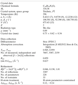
Chemical formula C20H18N2O3
Mr 334.36
Crystal system, space group Triclinic,P1
Temperature (K) 296
a,b,c(A˚ ) 8.4213 (7), 9.0739 (9), 12.2238 (12) ,,() 106.501 (8), 92.390 (8), 100.750 (8)
V(A˚3) 875.43 (15)
Z 2
Radiation type MoK
(mm1) 0.09
Crystal size (mm) 0.750.620.34
Data collection
Diffractometer StoeIPDS2
Absorption correction Integration (X-RED32; Stoe & Cie, 2002)
Tmin,Tmax 0.945, 0.959 No. of measured, independent and
observed [I> 2(I)] reflections
7687, 3387, 2159
Rint 0.029
(sin/)max(A˚1) 0.617
Refinement
R[F2> 2(F2)],wR(F2),S 0.046, 0.130, 1.01 No. of reflections 3387
No. of parameters 228
No. of restraints 33
H-atom treatment H-atom parameters constrained
max,min(e A˚3) 0.12,0.14
sup-1
Acta Cryst. (2019). E75, 1925-1929
supporting information
Acta Cryst. (2019). E75, 1925-1929 [https://doi.org/10.1107/S2056989019015317]
Crystal structure, Hirshfeld surface analysis and DFT studies of
2-[5-(4-methyl-benzyl)-6-oxo-3-phenyl-1,6-dihydropyridazin-1-yl]acetic acid
Said Daoui, Cemile Baydere, Fouad El Kalai, Lhassane Mahi, Necmi Dege, Khalid Karrouchi and
Noureddine Benchat
Computing details
Data collection: X-AREA (Stoe & Cie, 2002); cell refinement: X-AREA (Stoe & Cie, 2002); data reduction: X-RED32
(Stoe & Cie, 2002); program(s) used to solve structure: SHELXT2017 (Sheldrick, 2015a); program(s) used to refine
structure: SHELXL2018 (Sheldrick, 2015b); molecular graphics: Mercury (Macrae et al., 2008) and PLATON (Spek,
2009); software used to prepare material for publication: WinGX (Farrugia, 2012), SHELXL2018 (Sheldrick, 2015b),
PLATON (Spek, 2009) and publCIF (Westrip, 2010).
2-[5-(4-Methylbenzyl)-6-oxo-3-phenyl-1,6-dihydropyridazin-1-yl]acetic acid
Crystal data
C20H18N2O3
Mr = 334.36
Triclinic, P1 a = 8.4213 (7) Å b = 9.0739 (9) Å c = 12.2238 (12) Å α = 106.501 (8)° β = 92.390 (8)° γ = 100.750 (8)° V = 875.43 (15) Å3
Z = 2 F(000) = 352
Dx = 1.268 Mg m−3
Mo Kα radiation, λ = 0.71073 Å Cell parameters from 9277 reflections θ = 2.4–30.5°
µ = 0.09 mm−1
T = 296 K Prism, yellow
0.75 × 0.62 × 0.34 mm
Data collection
Stoe IPDS 2 diffractometer
Radiation source: sealed X-ray tube, 12 x 0.4 mm long-fine focus
Plane graphite monochromator
Detector resolution: 6.67 pixels mm-1
rotation method scans
Absorption correction: integration (X-RED32; Stoe & Cie, 2002)
Tmin = 0.945, Tmax = 0.959
7687 measured reflections 3387 independent reflections 2159 reflections with I > 2σ(I)
Rint = 0.029
θmax = 26.0°, θmin = 2.5°
h = −10→10 k = −10→11 l = −15→15
Refinement
Refinement on F2
Least-squares matrix: full
R[F2 > 2σ(F2)] = 0.046
wR(F2) = 0.130
S = 1.01
3387 reflections 228 parameters 33 restraints
sup-2
Acta Cryst. (2019). E75, 1925-1929
Secondary atom site location: difference Fourier map
Hydrogen site location: inferred from neighbouring sites
H-atom parameters constrained
w = 1/[σ(Fo) + (0.0706P)]
where P = (Fo2 + 2Fc2)/3
(Δ/σ)max < 0.001
Δρmax = 0.12 e Å−3
Δρmin = −0.14 e Å−3
Special details
Geometry. All esds (except the esd in the dihedral angle between two l.s. planes) are estimated using the full covariance matrix. The cell esds are taken into account individually in the estimation of esds in distances, angles and torsion angles; correlations between esds in cell parameters are only used when they are defined by crystal symmetry. An approximate (isotropic) treatment of cell esds is used for estimating esds involving l.s. planes.
Fractional atomic coordinates and isotropic or equivalent isotropic displacement parameters (Å2)
x y z Uiso*/Ueq
O2 0.63260 (15) 1.10394 (16) 0.10937 (10) 0.0735 (4)
H2 0.597450 1.162100 0.078353 0.110*
O3 0.52647 (15) 0.73205 (16) −0.01452 (10) 0.0764 (4)
O1 0.80943 (17) 1.09371 (19) −0.02162 (12) 0.0945 (5)
N2 0.68471 (16) 0.84641 (18) 0.15268 (12) 0.0669 (4)
N1 0.72008 (16) 0.88379 (18) 0.26737 (12) 0.0668 (4)
C8 0.46392 (19) 0.7210 (2) 0.26864 (14) 0.0620 (4)
H8 0.389598 0.679405 0.311801 0.074*
C9 0.4288 (2) 0.6836 (2) 0.15452 (14) 0.0620 (4)
C7 0.61225 (19) 0.8228 (2) 0.32473 (14) 0.0613 (4)
C14 0.1506 (2) 0.5207 (2) 0.15768 (15) 0.0664 (4)
C6 0.6544 (2) 0.8672 (2) 0.45037 (15) 0.0680 (5)
C10 0.5447 (2) 0.7524 (2) 0.09055 (14) 0.0634 (4)
C12 0.7494 (2) 1.0492 (2) 0.05303 (14) 0.0670 (5)
C13 0.2753 (2) 0.5779 (2) 0.08692 (16) 0.0734 (5)
H13A 0.226930 0.634241 0.042417 0.088*
H13B 0.303827 0.487485 0.033334 0.088*
C11 0.8048 (2) 0.9243 (3) 0.09364 (17) 0.0773 (5)
H11A 0.830690 0.846172 0.028180 0.093*
H11B 0.903514 0.971292 0.145133 0.093*
C15 0.1700 (3) 0.4072 (2) 0.20852 (19) 0.0832 (6)
H15 0.258956 0.359741 0.194728 0.100*
C19 0.0148 (3) 0.5824 (2) 0.1768 (2) 0.0885 (6)
H19 −0.003886 0.657256 0.142058 0.106*
C17 −0.0729 (3) 0.4267 (3) 0.3010 (2) 0.1019 (7)
C18 −0.0948 (3) 0.5352 (3) 0.2469 (2) 0.1060 (7)
H18 −0.186687 0.578672 0.257801 0.127*
C16 0.0607 (3) 0.3626 (3) 0.2792 (2) 0.0992 (7)
H16 0.078355 0.286573 0.313098 0.119*
C3 0.7361 (4) 0.9592 (4) 0.6852 (2) 0.1101 (9)
H3 0.763972 0.989204 0.763999 0.132*
C1 0.7831 (3) 0.9861 (3) 0.5033 (2) 0.1070 (8)
H1 0.844994 1.038827 0.459306 0.128*
sup-3
Acta Cryst. (2019). E75, 1925-1929
H5 0.478402 0.715558 0.486763 0.134*
C4 0.6081 (3) 0.8448 (5) 0.6369 (2) 0.1338 (11)
H4 0.545236 0.796070 0.682551 0.161*
C2 0.8236 (4) 1.0299 (4) 0.6197 (2) 0.1281 (10)
H2A 0.913185 1.110017 0.653001 0.154*
C20 −0.1911 (4) 0.3793 (4) 0.3811 (3) 0.1591 (13)
H20A −0.271221 0.443328 0.391357 0.239*
H20B −0.133254 0.393504 0.453929 0.239*
H20C −0.243577 0.270862 0.348701 0.239*
Atomic displacement parameters (Å2)
U11 U22 U33 U12 U13 U23
O2 0.0725 (8) 0.0961 (10) 0.0601 (7) 0.0214 (7) 0.0150 (6) 0.0325 (6)
O3 0.0785 (8) 0.1010 (10) 0.0580 (7) 0.0288 (7) 0.0140 (6) 0.0293 (7)
O1 0.0939 (10) 0.1310 (12) 0.0796 (9) 0.0267 (9) 0.0331 (8) 0.0589 (9)
N2 0.0532 (8) 0.0909 (10) 0.0664 (9) 0.0154 (7) 0.0119 (7) 0.0379 (8)
N1 0.0535 (8) 0.0864 (10) 0.0657 (9) 0.0138 (7) 0.0053 (7) 0.0312 (8)
C8 0.0572 (9) 0.0719 (11) 0.0606 (10) 0.0117 (8) 0.0105 (8) 0.0260 (8)
C9 0.0593 (9) 0.0686 (10) 0.0612 (10) 0.0155 (8) 0.0085 (8) 0.0226 (8)
C7 0.0534 (9) 0.0744 (11) 0.0620 (10) 0.0180 (8) 0.0085 (8) 0.0263 (8)
C14 0.0630 (10) 0.0584 (10) 0.0684 (10) 0.0029 (8) 0.0007 (8) 0.0111 (8)
C6 0.0595 (10) 0.0875 (12) 0.0599 (10) 0.0225 (9) 0.0044 (8) 0.0219 (9)
C10 0.0600 (10) 0.0788 (11) 0.0586 (10) 0.0231 (8) 0.0098 (8) 0.0257 (8)
C12 0.0584 (10) 0.0908 (13) 0.0515 (9) 0.0060 (9) 0.0068 (8) 0.0267 (9)
C13 0.0737 (11) 0.0748 (11) 0.0642 (10) 0.0050 (9) −0.0002 (9) 0.0165 (9)
C11 0.0556 (9) 0.1085 (15) 0.0800 (12) 0.0164 (10) 0.0190 (9) 0.0462 (11)
C15 0.0817 (13) 0.0735 (12) 0.0977 (14) 0.0177 (10) 0.0199 (11) 0.0284 (11)
C19 0.0741 (13) 0.0770 (13) 0.1100 (17) 0.0140 (10) 0.0034 (12) 0.0228 (12)
C17 0.0866 (14) 0.0909 (14) 0.1061 (16) −0.0089 (12) 0.0287 (11) 0.0092 (10)
C18 0.0733 (12) 0.1004 (16) 0.1320 (19) 0.0157 (12) 0.0256 (13) 0.0142 (12)
C16 0.1108 (18) 0.0792 (14) 0.1074 (17) 0.0029 (13) 0.0224 (14) 0.0366 (13)
C3 0.1037 (19) 0.164 (3) 0.0617 (13) 0.0585 (19) 0.0004 (14) 0.0139 (16)
C1 0.1183 (19) 0.1081 (17) 0.0751 (14) −0.0077 (15) −0.0081 (13) 0.0191 (12)
C5 0.0720 (13) 0.189 (3) 0.0717 (13) −0.0062 (14) −0.0022 (11) 0.0583 (15)
C4 0.0910 (17) 0.243 (4) 0.0762 (15) 0.023 (2) 0.0094 (14) 0.068 (2)
C2 0.143 (2) 0.133 (2) 0.0775 (17) 0.0047 (19) −0.0182 (17) 0.0019 (16)
C20 0.138 (2) 0.158 (3) 0.145 (3) −0.033 (2) 0.064 (2) 0.022 (2)
Geometric parameters (Å, º)
O2—C12 1.317 (2) C11—H11B 0.9700
O2—H2 0.8200 C15—C16 1.372 (3)
O3—C10 1.2433 (19) C15—H15 0.9300
O1—C12 1.193 (2) C19—C18 1.377 (3)
N2—N1 1.3516 (19) C19—H19 0.9300
N2—C10 1.370 (2) C17—C16 1.363 (3)
sup-4
Acta Cryst. (2019). E75, 1925-1929
N1—C7 1.307 (2) C17—C20 1.515 (4)
C8—C9 1.344 (2) C18—H18 0.9300
C8—C7 1.422 (2) C16—H16 0.9300
C8—H8 0.9300 C3—C2 1.327 (4)
C9—C10 1.438 (2) C3—C4 1.330 (4)
C9—C13 1.507 (2) C3—H3 0.9300
C7—C6 1.482 (2) C1—C2 1.374 (3)
C14—C19 1.365 (3) C1—H1 0.9300
C14—C15 1.375 (3) C5—C4 1.390 (3)
C14—C13 1.499 (3) C5—H5 0.9300
C6—C5 1.352 (3) C4—H4 0.9300
C6—C1 1.366 (3) C2—H2A 0.9300
C12—C11 1.498 (3) C20—H20A 0.9600
C13—H13A 0.9700 C20—H20B 0.9600
C13—H13B 0.9700 C20—H20C 0.9600
C11—H11A 0.9700
C12—O2—H2 109.5 H11A—C11—H11B 107.7
N1—N2—C10 126.14 (14) C16—C15—C14 121.4 (2)
N1—N2—C11 115.10 (15) C16—C15—H15 119.3
C10—N2—C11 118.61 (14) C14—C15—H15 119.3
C7—N1—N2 117.30 (14) C14—C19—C18 120.8 (2)
C9—C8—C7 121.38 (16) C14—C19—H19 119.6
C9—C8—H8 119.3 C18—C19—H19 119.6
C7—C8—H8 119.3 C16—C17—C18 116.7 (2)
C8—C9—C10 117.95 (16) C16—C17—C20 121.4 (3)
C8—C9—C13 125.62 (16) C18—C17—C20 121.9 (3)
C10—C9—C13 116.42 (15) C17—C18—C19 122.2 (2)
N1—C7—C8 121.32 (15) C17—C18—H18 118.9
N1—C7—C6 115.86 (15) C19—C18—H18 118.9
C8—C7—C6 122.82 (16) C17—C16—C15 121.7 (2)
C19—C14—C15 117.17 (19) C17—C16—H16 119.1
C19—C14—C13 121.17 (18) C15—C16—H16 119.1
C15—C14—C13 121.63 (17) C2—C3—C4 119.2 (2)
C5—C6—C1 116.45 (19) C2—C3—H3 120.4
C5—C6—C7 122.76 (18) C4—C3—H3 120.4
C1—C6—C7 120.77 (19) C6—C1—C2 121.7 (3)
O3—C10—N2 119.17 (16) C6—C1—H1 119.2
O3—C10—C9 124.97 (17) C2—C1—H1 119.2
N2—C10—C9 115.86 (14) C6—C5—C4 121.1 (3)
O1—C12—O2 124.94 (17) C6—C5—H5 119.5
O1—C12—C11 121.98 (17) C4—C5—H5 119.5
O2—C12—C11 113.06 (14) C3—C4—C5 120.8 (3)
C14—C13—C9 114.91 (15) C3—C4—H4 119.6
C14—C13—H13A 108.5 C5—C4—H4 119.6
C9—C13—H13A 108.5 C3—C2—C1 120.7 (3)
C14—C13—H13B 108.5 C3—C2—H2A 119.7
sup-5
Acta Cryst. (2019). E75, 1925-1929
H13A—C13—H13B 107.5 C17—C20—H20A 109.5
N2—C11—C12 113.59 (14) C17—C20—H20B 109.5
N2—C11—H11A 108.8 H20A—C20—H20B 109.5
C12—C11—H11A 108.8 C17—C20—H20C 109.5
N2—C11—H11B 108.8 H20A—C20—H20C 109.5
C12—C11—H11B 108.8 H20B—C20—H20C 109.5
C10—N2—N1—C7 1.2 (2) C10—C9—C13—C14 −174.43 (15)
C11—N2—N1—C7 176.75 (15) N1—N2—C11—C12 −107.68 (17)
C7—C8—C9—C10 −1.3 (2) C10—N2—C11—C12 68.2 (2)
C7—C8—C9—C13 −179.66 (16) O1—C12—C11—N2 −159.10 (18)
N2—N1—C7—C8 0.1 (2) O2—C12—C11—N2 22.1 (2)
N2—N1—C7—C6 −179.55 (14) C19—C14—C15—C16 −2.6 (3)
C9—C8—C7—N1 0.0 (3) C13—C14—C15—C16 175.73 (19)
C9—C8—C7—C6 179.63 (15) C15—C14—C19—C18 1.8 (3)
N1—C7—C6—C5 −170.3 (2) C13—C14—C19—C18 −176.5 (2)
C8—C7—C6—C5 10.0 (3) C16—C17—C18—C19 −2.2 (4)
N1—C7—C6—C1 11.5 (3) C20—C17—C18—C19 178.0 (2)
C8—C7—C6—C1 −168.13 (19) C14—C19—C18—C17 0.6 (4)
N1—N2—C10—O3 177.73 (15) C18—C17—C16—C15 1.4 (4)
C11—N2—C10—O3 2.3 (2) C20—C17—C16—C15 −178.7 (3)
N1—N2—C10—C9 −2.4 (2) C14—C15—C16—C17 0.9 (4)
C11—N2—C10—C9 −177.82 (15) C5—C6—C1—C2 1.2 (4)
C8—C9—C10—O3 −177.84 (16) C7—C6—C1—C2 179.5 (2)
C13—C9—C10—O3 0.7 (3) C1—C6—C5—C4 −0.1 (4)
C8—C9—C10—N2 2.3 (2) C7—C6—C5—C4 −178.3 (2)
C13—C9—C10—N2 −179.12 (15) C2—C3—C4—C5 1.2 (5)
C19—C14—C13—C9 104.4 (2) C6—C5—C4—C3 −1.2 (5)
C15—C14—C13—C9 −73.8 (2) C4—C3—C2—C1 0.0 (5)
C8—C9—C13—C14 4.0 (3) C6—C1—C2—C3 −1.2 (5)
Hydrogen-bond geometry (Å, º)
D—H···A D—H H···A D···A D—H···A
O2—H2···O3i 0.82 1.83 2.6358 (16) 167
C3—H3···O1ii 0.93 2.51 3.430 (3) 172