R E S E A R C H
Open Access
HTR4 gene structure and altered expression in
the developing lung
Emily Hodge
1†, Carl P Nelson
1†, Suzanne Miller
1†, Charlotte K Billington
1, Ceri E Stewart
1, Caroline Swan
1,
Anders Malarstig
3, Amanda P Henry
1, Catherine Gowland
1, Erik Melén
2, Ian P Hall
1and Ian Sayers
1*Abstract
Background:Meta-analyses of genome-wide association studies (GWAS) have identified single nucleotide polymorphisms (SNPs) spanning the 5-hydroxytryptamine receptor 4 (5-HT4R) gene (HTR4) associated with lung
function. The aims of this study were to i) investigate the expression profile ofHTR4in adult and fetal lung tissue and cultured airway cells, ii) further defineHTR4gene structure and iii) explore the potential functional implications of key SNPs using a bioinformatic approach.
Methods:Following reverse transcription (RT)-PCR in human brain, 5′rapid amplification of cDNA ends (5′RACE) was used to examine the exonic structure ofHTR4at the 5′end. Quantitative (Q)-PCR was used to quantifyHTR4mRNA expression in total RNA from cultured airway cells and whole lung tissue. Publically available gene microarray data on fetal samples of estimated gestational age 7–22 weeks were mined forHTR4expression. Immunohistochemistry (IHC; in adult and fetal lung tissue) and a radioligand binding assay (in cultured airway cells) were used to analyze 5HT4R protein expression.
Results:IHC in adult lung, irrespective of the presence of chronic obstructive pulmonary disease (COPD), suggested low level expression of 5-HT4R protein, which was most prominent in alveolar pneumocytes. There was evidence of differential
5-HT4R protein levels during gestation in fetal lung, which was also evident in gene expression microarray data.HTR4
mRNA expression, assessed by Q-PCR, was <0.5% relative to brain in total adult lung tissue and in human airway smooth muscle (HASM) and bronchial epithelial cells (HBEC) derived from adult donors. Radioligand binding experiments also indicated that HBEC and HASM cells did not express a significant 5-HT4R population. 5′RACE in brain identified a novel
N-terminal variant, containing an extendedN-terminal sequence. The functional significance of keyHTR4SNPs was investigated using the encyclopedia of DNA elements consortium (ENCODE) dataset. These analyses identified multiple alterations in regulatory motifs for transcription factors implicated in lung development, including Foxp1.
Conclusions:Taken together, these data suggest a role forHTR4in lung development, which may at least in part explain the genetic association with lung function.
Keywords:5-hydroxytryptamine,HTR4, 5-HT4R, Splice variant, Lung development, COPD, GPCR
Background
5-Hydroxytryptamine (5-HT or serotonin) is a highly conserved monoamine, which is a major neurotrans-mitter in the CNS. However, 5-HT is also widely dis-tributed throughout the periphery, with critical roles identified in cardiovascular physiology, gastrointestinal and endocrine function, the regulation of food intake and energy balance, as well as pulmonary physiology
[1]. These myriad functions are performed through at least 15 distinct receptors, grouped into seven struc-turally and functionally-defined families (5-HT1 –
5-HT7 receptors, encoded by HTR1-HTR7 genes) [2].
With the exception of the 5-HT3 receptor, which is a
ligand-gated ion channel, these 5-HT receptors (5-HTRs) are members of the G protein-coupled receptor (GPCR) superfamily. Further functional diversity arises from alternative splicing (in the case of 5-HT4R and
5-HT7R), RNA editing (of the 5-HT2Creceptor) and both
homo- and hetero-dimerization involving a variety of 5-HTRs [3,4].
* Correspondence:[email protected]
†Equal contributors
1
Division of Respiratory Medicine, University of Nottingham, Queen’s Medical Centre, Nottingham NG7 2UH, UK
Full list of author information is available at the end of the article
Recent genome-wide association studies (GWAS) have identified an association between single nucleotide poly-morphisms (SNPs) localised to a region encompassing the HTR4gene and lung function, assessed by forced expira-tory volume in 1 second (FEV1) and the ratio of FEV1to
forced vital capacity (FVC) [5-7]. Subsequent studies have demonstrated an association between this locus and chronic obstructive pulmonary disease (COPD) [8] and airflow obstruction in smokers [9].
The complexity of the serotonin receptor family is exem-plified by the HTR4 sub-family. Encoded by a complex gene spanning ~200 kb on chromosome 5q33,HTR4has at least 10 human splice variants identified to date. Numer-ous studies in recombinant systems have demonstrated that alternative splicing of theHTR4gene can generate re-ceptor species with distinct pharmacological and functional profiles, although all couple positively to adenylyl cyclase, leading to cyclic AMP generation (see [10] for review). The majority of these splice variants share a common primary sequence for the first 358 residues, only diverging at theC -terminus. The exception is 5-HT4hR, which possesses an
additional 14 residues in the second extracellular loop of the receptor and has been found in combination with the 5-HT4bRC-terminal sequence [11].
HTR4is highly expressed in the central nervous system, particularly in limbic structures, where it has been impli-cated in learning and memory, depression, anxiety and feeding behaviour [12]. Peripherally, roles for 5-HT4R have
been identified in the gastrointestinal tract, heart, vascula-ture, adrenal cortex and lower urinary tract [1,13]. Low levels ofHTR4transcript have also been detected in lung [6,14,15] and in airway epithelial and smooth muscle cells [6,16,17].
Given the genetic association data and potential clinical significance of the HTR4 gene in respiratory physiology and pathophysiology, we sought to i) investigate the ex-pression profile ofHTR4in both adult and fetal lung tissue and in lung tissue from individuals with COPD, ii) define the gene structure and iii) using the ENCODE dataset, in-vestigate the potential functional mechanisms underlying select keyHTR4SNPs associated with lung function. Our data demonstrate that in adult human lung tissue and iso-lated cells, including airway smooth muscle and bronchial epithelial cells,HTR4expression is very low at both pro-tein and mRNA levels. Similar findings were observed in lung tissue isolated from COPD patients. Interestingly, we identify that 5-HT4R is differentially expressed across
de-velopmental stages, potentially suggesting a role for this receptor in lung development. Finally, we have identified a novel splice variant at theN-terminus and multiple poten-tial regulatory mechanisms, which may underlie the ob-served HTR4 SNP associations with lung function, including the alteration of transcription factor binding sites for factors linked to lung development.
Methods
Immunohistochemistry (IHC)
Three undiseased adult lung samples and three lung sam-ples from individuals with clinically diagnosed COPD were collected from the Nottingham Health Science Biobank (Nottingham, UK) with the required ethical approval (08/ H0407/1). Twelve fetal tissue samples were collected from the Human Developmental Biology Resource (Newcastle upon Tyne and London, UK, www.hdbr.org) at diverse stages of development, specifically 19, 21 and 23 days and 10, 12, 17 and 19 weeks post-conception. Samples were consented for in accordance with national banking proce-dures and the UK Human Tissue Act (2004). For all samples, 4 μm whole tissue sections on glass slides were de-paraffinized in Histo-clear (National Diagnos-tics, Dublin, Ireland) and hydrated using decreasing con-centrations of ethanol. Antigen retrieval was performed in a steamer for 20 minutes in sodium citrate buffer (pH 6.0), followed by an endogenous peroxidise block for 5 minutes (Dako, Cambs, UK). Slides were incubated with either a rabbit polyclonal anti-5-HT4R antibody (1:500, ab60359,
Abcam, Cambridge, UK) or treated with normal rabbit IgG as a matched isotype control (Invitrogen/Life Tech-nologies, Paisley, UK) for 1 hour at room temperature. The Dako Chemate Envision Detection Kit (Dako) with DAB chromogen was used for detection. Sections were then counterstained with Mayer’s Haematoxylin (Sigma-Aldrich, Dorset, UK), dehydrated and a coverslip mounted using Vectamount (Vector Laboratories, Peterborough, UK). Human brain tissue was used as a positive control for 5-HT4R staining, whilst a negative control substituted
the primary antibody with antibody diluent. Results were visualized using an Olympus BX14 light microscope.
Cell culture and transfection
All cells were maintained at 37°C in 5% CO2in a
humidi-fied incubator. Human airway smooth muscle (HASM) cells were isolated from the healthy bronchial tissue of in-dividuals without previous asthma history undergoing sur-gery, and cultured as previously described [18]. Approval was given by the Nottingham Local Ethical Research Committee (EC00/165). Undifferentiated human bronchial epithelial cells (HBEC) (Lonza/Biologics, Slough, UK) were maintained in culture and differentiated, where relevant, as previously described [19].HTR4mRNA expression ana-lysis was carried out in five HASM (passage 3–5) and four HBEC (passage 3) donors, while radioligand binding ex-periments were performed on at least two distinct HASM and HBEC donors. CHO-K1 cells and the human bron-chial epithelial cell line BEAS2B-R1 [20] (provided by Dr. Ray Penn, University of Maryland, Baltimore, USA) were cultured in Dulbecco’s Modified Eagle’s Medium supplemented with 10% fetal calf serum (FCS;
appropriate) in 24-well plates with a pcDNA3-HTR4a plasmid (1μg/well) using FugeneHD transfection reagent (Promega, Southampton, UK), according to manufac-turer’s instructions. The pcDNA3-HTR4a plasmid was constructed by amplifying the protein-coding region of theHTR4atranscript from total brain RNA (Ambion/Life Technologies) and inserting this into the EcoRI site of the pcDNA3 vector. The insert in the resulting plasmid was sequence-verified as for RT-PCR products (see below).
Reverse-Transcription PCR (RT-PCR)
RT-PCR used cDNA synthesised from total RNA using the Superscript First-Strand Synthesis System for RT-PCR (Invitrogen/Life Technologies), extracted from cultured cells or commercially obtained (peripheral blood mono-nuclear cells (PBMC; 3H Biomedical AB, Uppsala, Sweden), total lung and brain tissue (both from Ambion/Life Tech-nologies)). Initial HTR4 expression analysis by RT-PCR used a forward primer binding in exon 1 (primer 1F, 5′-CA GCAGAAGCTCGGCTCAG-3′) with reverse primers bin-ding either in exon 9 (primer 9R, 5′-CTCTCATGGCT GTCTTCTGG-3′) or exon 13 (primer 13R, 5′-CAATCAG AAGCATGATTCCAG-3′), with the following cycling pa-rameters: 35 cycles of 94°C for 1.5 minutes, 60°C for 1.5 minutes, 72°C for 1.5 minutes, followed by 72°C for 10 mi-nutes. Expression analysis of the novelHTR4variant tran-script used a forward primer binding in the novel exon (primer novel_F, 5′-GAATGGAGAGATCCAGATGG-3′) and primer 9R, with the following cycling protocol: 94°C for 2 minutes, then 40 cycles of 94°C for 30 seconds, 55°C for 30 seconds, 68°C for 1.5 minutes, followed by 68°C for 5 minutes. Amplicons were extracted from agarose gels using the QIAquick Gel Extraction Kit (Qiagen, Crawley, UK) and sequence verified using the BigDye Terminator v3.1 Cycle Sequencing Kit in conjunction with an ABI PRISM 310 Genetic Analyzer (Applied Biosystems/Life Technologies).
Quantitative PCR (Q-PCR)
Expression analysis ofHTR4was carried out in total RNA from cultured cells or commercially available total lung and brain tissue (Ambion/Life Technologies) using TaqMan methodology for real-time Q-PCR (Applied Biosystems/Life Technologies) as previously described [19]. In every sample, Q-PCR was carried out forHTR4 (forward primer 5′-TC TCTTGCTTTTGCGGATCT-3′, reverse primer 5′-GCAG
AGGGGTCATCTTGTTC-3′, probe 5′-CCCTTTGGTGC
CATTGAGCTGGTTC-3′) and Human TFRC (CD71,
transferrin receptor) Endogenous Control (Applied Biosystems/Life Technologies). Relative HTR4expression was calculated using the 2-ΔΔCt method [21], corrected using the endogenous control (TFRC) and displayed rela-tive to expression in total brain tissue.
Affymetrix U133 Plus 2 array data for human fetal lung Publically available data [22,23] were utilized to see whetherHTR4was differentially expressed during normal human lung development. Previously, human fetal lung tissues were obtained from National Institute of Child Health and Human Development tissue databases and microarray profiled to investigate the expression spanning different gestational ages. RNA samples from 38 subjects (estimated gestational age 7–22 weeks or 53–154 days post conception)i.e.Pseudoglandular (gestational age, 7– 16 weeks) and Canalicular (17–26 weeks) stages of devel-opment were included within the dataset. These data are available at NCBI Gene Expression Omnibus (GEO, http://www.ncbi.nlm.nih.gov/geo), GSE14334. The dataset was mined forHTR4expression using Affymetrix probes; 216939_s_at, 207577_at and 207578_s_at.
Radioligand binding
CHO-K1 cells were plated into 24-well plates and grown for 24 hours in DMEM containing 10% FCS prior to transfection. BEAS2B-R1, HBEC and HASM cells were plated into 24-well plates 24–48 hours prior to experi-mentation. Experiments were performed 48 hours post-transfection in HEPES-buffered saline (HBS: 20 mM HEPES, 150 mM NaCl, 4.2 mM KCl, 0.9 mM CaCl2, 0.5
mM MgCl2, 0.1% glucose, and 0.1% bovine serum
albu-min), as previously reported [24]. The reaction mixture (250 μl) containing saturating concentrations of [3 H]-GR113808 (approx. 1 nM,Kd= 0.30 nM; specific
activ-ity, 83 Ci/mmol) was added to the cells and incubated for 2 hours at 4°C. Non-specific binding was defined in the presence of 5-HT (10 μM). Incubation was termi-nated by aspiration, followed by washing the cells with ice-cold HBS (2 × 1 ml). Cells were lysed in 0.1 M NaOH, then 10 ml of scintillation liquid (UltimaGold XR scintillation cocktail; Perkin Elmer, Bucks, UK) was added and radioactivity was measured by scintillation counting. Protein concentration was determined using a Bio-Rad protein assay (Hercules, California, USA), allowing recep-tor densities (in fmol/mg protein) to be determined.
5′Rapid amplification of cDNA Ends (5′RACE)
Sequencing Kit in conjunction with an ABI PRISM 310 Genetic Analyzer (Applied Biosystems/Life Technologies). Sequences were aligned with the ‘Human Genomic + Transcript’ database using the Basic Local Alignment Search Tool (BLAST) provided by the National Center for Biotechnology Information (NCBI, http://www.ncbi. nlm.nih.gov/). Protein structures were predicted from 5′RACE data using online tools: the TMHMM Server v. 2.0 [25] and TOPO2, provided by the Sequence Ana-lysis & Consulting Service at UCSF (http://www.sacs. ucsf.edu/cgi-bin/open-topo2.py/).
Bioinformatic analysis
The HTR4 gene was annotated with RNA sequencing, H3K27Ac histone marks, DNase I hypersensitivity, transcription factor binding (all derived from the UCSC encyclopedia of DNA elements consortium (ENCODE) database [26]), CpG island and placental mammal conservation tracks using the UCSC genome browser (http://genome.ucsc.edu/) on the Human Feb 2009 (GRCh37/hg19) assembly [27,28]. The predicted effects of polymorphisms (in the HapMap CEU cohort, as of December 2012) in linkage disequilibrium (LD)
(r2>0.80) with the sentinel HTR4 SNP (rs3995090) identified in the SpiroMeta lung function GWAS [6] were examined using the HaploReg database (http:// www.broadinstitute.org/mammals/haploreg/) [29].
Results
5-HT4R is expressed at low levels in adult human lung tissue
Immunohistochemistry performed with an anti-5-HT4R
antibody in normal lung tissue from three donors identi-fied specific staining for 5-HT4R in alveolar pneumocytes
(Figure 1a-c). The staining in these cells was both cyto-plasmic and membranous, which is consistent with the anticipated sub-cellular expression profile for a GPCR. Some specific 5-HT4R staining was also detected in
[image:4.595.58.538.386.685.2]bronchial epithelial cells, but substantial variation be-tween donors was observed in this location. While strong staining was observed in the epithelial cells of donor 2 (Figure 1h), only weak staining was observed in the other 2 donors (Figure 1g and 1i). In addition, the strong staining in donor 2 was cytoplasmic and nuclear, with little apparent staining at the plasma membrane (Figure 1h). No staining was observed in any of the isotype controls (Figure 1d-f and 1j-l).
Figure 1Immunohistochemistry of 5-HT4R in normal adult human lung tissue.Low cytoplasmic and membranous expression of 5-HT4R
5-HT4R is expressed at low levels in adult human lung
tissue from individuals with COPD
5-HT4R protein expression was analyzed by IHC in
three COPD donors. Donor 1 (COPD1) was a 61 year old female, heavy smoker with moderate COPD; donor 2 (COPD2) was a 55 year old male, heavy smoker with severe COPD; donor 3 (COPD3) was a 48 year old fe-male, heavy smoker with severe COPD. Weak 5-HT4R
immunopositivity was found in the nuclei and cyto-plasm of pneumoctyes in the alveolar regions of all three COPD samples (Figure 2a-c). The epithelium showed variable staining between donors: COPD1 had weak nuclear staining (Figure 2g), COPD2 had moderate nuclear and cytoplasmic staining (Figure 2h) whilst COPD3 was negative for the 5-HT4R protein
(Figure 2i). All isotype controls were negative (Figure 2d-f and 2j-l).
5-HT4R is differentially expressed during fetal
development at the protein and mRNA level
Fetal tissue spanning ages 19 days-19 weeks was
investigated using IHC (Figure 3). These data
demonstrated a trend towards an increase in 5-HT4R
protein expression spanning Embryonic (Figure 3a-f) to
Pseudoglandular (Figure 3g-j) stages and a potential decrease in the Canalicular (Figure 3k-l) stage of development. Staining was predominantly nuclear and widespread across the airway epithelium. The
gene array data demonstrated that there was
a significant increase in expression levels with age during the Pseudoglandular (gestational age, 7–16 weeks) and Canalicular (17–26 weeks) stages of
de-velopment, with one of the Affymetrix probes
(207577_at) surviving correction for false discovery rate (Table 1). These data suggest that an increase in 5-HT4R expression is a feature of lung development
with a lower level of protein expression in subse-quent stages, potentially reflecting the levels observed in the adult lung.
Analysis ofHTR4gene structure in brain andHTR4 expression profiling in airway cells by Q-PCR and radioligand binding
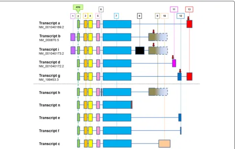
Previously described gene arrangements for HTR4 are summarized in Figure 4. Transcripts a, b, i, d and g are currently reported by NCBI (December 2012); transcripts h, n, e, f and c have been described in the literature [10]. These data illustrate the complex splicing that exists at this gene locus, including multiple functionally relevant
C-Figure 2Immunohistochemistry of 5-HT4R in COPD lung tissue.Weak 5-HT4R staining was found in the nuclei and cytoplasm of pneumoctyes in
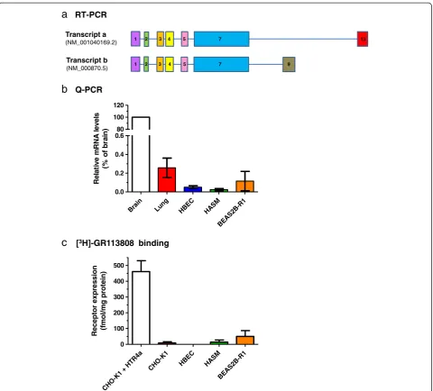
[image:5.595.58.540.400.697.2]terminal variants, leading to alterations in cyclic AMP signalling responses [10]. Analysis by RT-PCR indicated expression of mRNA encodingHTR4in total brain tissue, but failed to detect expression in HASM, undifferentiated HBEC, PBMC or total lung tissue (data not shown). Targeted sequencing of PCR products derived from brain cDNA confirmed the presence of both variants a (acces-sion NM_001040169.2) and b (acces(acces-sion NM_000870.5), distinguished by the presence of exons 9 and 13 respect-ively. Additionally, exon 1 was present in variant a, al-though this contradicts data currently reported by NCBI (Figure 5a). Q-PCR data indicated very low levels of mean HTR4mRNA expression in airway cells and tissues when compared to total brain tissue: total lung (0.26%), undiffer-entiated HBEC (0.05%), HASM (0.02%) and BEAS2B-R1 (0.16%) (Figure 5b).
In addition, radioligand binding (using the highly 5-HT
4R-selective ligand [3H]-GR113808 [24]) failed to detect
any significant 5-HT4R expression in undifferentiated
HBEC (none detected; n = 3), HASM (14.1 ± 13.2 fmol/ mg protein; n = 4) or BEAS2B-R1 (50.1 ± 36.6 fmol/mg protein; n = 3) cells (Figure 5c), in good agreement with the Q-PCR data in these cell types. In contrast, a substan-tial 5-HT4R population could be detected in CHO-K1 cells
transiently transfected with pcDNA3-HTR4a (460.7 ± 69.3 fmol/mg protein; n = 4), but not in un-transfected CHO-K1 cells (8.3 ± 7.8 fmol/mg protein; n = 4). These data suggest that there is limited 5-HT4R expression in
cul-tured structural cells of the adult lung, in agreement with the low levels of IHC staining for 5-HT4R in adult lung
tissue.
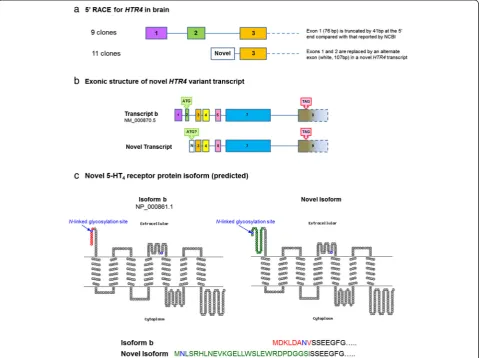
[image:6.595.52.540.92.233.2]5′ RACE showed variation of HTR4 transcripts in cDNA derived from total brain tissue only; it was not pos-sible to amplify HTR4 transcripts from total lung tissue, HASM, HBEC (undifferentiated and differentiated) and PBMC due to the extremely low abundance. Approxi-mately half of the analyzed clones derived from brain cDNA showed the presence of exons 1–3, although exon 1 was truncated by 41bp at the 5′end compared to the se-quence reported by NCBI (Figure 6a). The remaining clones possessed a novel exon of 107bp, in place of exons 1 and 2. Two consecutive RT-PCR procedures in cDNA derived from total brain,i.e.using the amplicon from the first as a template in a second, and confirmation by se-quencing showed that the exonic structure of the novel exon-containing variant was comparable to transcript b, since sequence alignment was of good quality for con-secutive exons 3, 4, 5, 7 and 9 (Figure 6b). However, consecutive RT-PCR procedures failed to detect this novel transcript in total lung tissue or differentiated HBEC (data not shown). Using BLAST, we found that the sequence of the novel exon aligns with a region on the same genomic contig as the HTR4 gene (accession NG_029052.1). Using the same translational stop codon as that in transcript b, the novel transcript variant has an in-frame translational start codon (ATG) within this novel exon. The predicted protein structure of the novel transcript has an extendedN-terminus (Figure 6c). Fur-thermore, the N-linked glycosylation site in the N -ter-minal region of transcript b is replaced in the novel transcript by an alternate site further upstream.
Figure 3Immunohistochemistry of 5-HT4R in fetal lung tissue.Weak staining is apparent in early Embryonic stages which increases in intensity with age leading to strong staining in the Pseudoglandular and moderate staining in the Canalicular stages.a-lrepresent age ordered samples of 19 days(a,b), 21 days(c,d), 23 days(e,f), 10 weeks(g,h), 12 weeks(i,j), 17 weeks(k)and 19 weeks(l). All isotype controls were negative (data not shown). X40 magnification.
Table 1 Fetal lung gene array data forHTR4expression during Pseudoglandular and Canalicular stages of lung development
Probe ID AveExpr t P.value Adj.p.val Beta-coefficient
216939_s_at 3.2999 0.9672 0.3393 0.5156 0.0009
207577_at 3.6980 3.3242 0.0019 0.0121 0.0024
207578_s_at 7.0472 0.3855 0.7020 0.8160 0.0008
AveExpr, average expression between all samples;t, t-statistic describing differential expression;P.Value, Unadjusted p value;adj.P.Val, Adjusted p value controlling for false discovery rate; Beta-coefficient= log-odds ratio
[image:6.595.57.291.636.691.2]Analysis of HTR4 gene regulation using the ENCODE dataset
Annotation of the region containing the HTR4 gene (shown in Figure 7) illustrates the regulatory elements contained within this region, as predicted by the ENCODE database. There are no obvious CpG islands in this region, however substantial DNase I hypersensitivity clusters, transcription factor binding sites (identified by ChIP-seq) and H3K27Ac marks (at least in HUVEC and K562 erythroleukemia cells) are identified within the 3′end of the HTR4 gene. These regulatory features coincide with the region containing the variants in linkage disequlibrium (LD) (r2>0.80) with the sentinel SNPs (rs3995090 and rs6889822) identified in the SpiroMeta lung function GWAS [6], as indicated in Figure 7. Less pronounced his-tone modification peaks, in addition to clusters of tran-scription factor binding and DNase I hypersensitivity sites are found towards the 5′end of theHTR4gene. Using the HaploReg database focussed to one of the sentinel SNPs (rs3995090) identified in the SpiroMeta lung function GWAS [6], we found that this SNP was in LD (r2>0.80) with an additional 28 SNPs in the 1000 Genomes dataset, which included the second sentinel SNP rs6889822 (r2>0.96) (Additional file 1: Table S1). We identified a wide range of alterations associated with these SNPs in: i)
enhancer histone marks, ii) DNase I hypersensitivity sites, iii) proteins bound and iv) regulatory motifs. Of note,
three of the SNPs (rs7733088, rs4705259 and
5:147836450) in LD (r2>0.80) with sentinel SNP rs3995090 significantly alterFoxp1 binding motifs within theHTR4gene (Additional file 1: Table S1). The Fox fam-ily of transcription factors are key regulators of lung development.
Discussion
The translation of GWAS findings to human biology re-mains a challenge. In the current study we have begun to interpret our recent finding that SNPs spanning the HTR4 gene are associated with FEV1 in the general
population [6]. To this end we have extensively expres-sion profiled the 5-HT4R protein and HTR4 mRNA in
human lung tissue and cells. We identified that 5-HT4R
protein is only weakly expressed in the adult human lung, irrespective of the presence of COPD. In agree-ment, we identified very low levels of HTR4mRNA and were unable to detect a significant 5-HT4R population
in cultured airway structural cells, including bronchial epithelial and airway smooth muscle cells. Interestingly, when we investigated 5-HT4R in human fetal lung tissue
[image:7.595.60.539.88.391.2]appeared to be differentially regulated through lung de-velopment. In agreement, gene expression array data also showed differential HTR4 mRNA lung expression levels across gestational stages. These data are novel and potentially suggest a role forHTR4in lung development. We also identified a novel N-terminal splice variant in brain with potential implications for 5-HT4R function.
Fi-nally we investigated the functional relevance of key genome-wide significant SNPs and identified multiple
potential functional mechanisms e.g. through the alter-ation of developmental transcription factor binding motifs.
The very low level ofHTR4mRNA detected in lung in the present study is consistent with a number of previous studies. For instance, Bach et al. [14] used RT-PCR to screen a variety of tissues for the expression of HTR4a andHTR4band successfully detected both in a number of tissues (e.g. brain and colon). However, they only showed
b
Q-PCRa
RT-PCRTranscript a (NM_001040169.2)
Transcript b (NM_000870.5)
1 2 3 4 5 7
1 2 3 4 5 7 9
13
R
e
la
ti
ve m
R
N
A
l
evel
s
(%
of
br
a
in)
Brain Lung HBEC HASM
BE AS
2B-R 1 0.0
0.2 0.4 0.6 80 100 120
c
[3H]-GR113808 bindingRe
cepto
r e
x
pre
ssion
(f
mol
/mg prot
ein)
CH O-K
1 + HTR4
a
CH O-K
1
HB EC
HA SM
BE AS2
B-R 1
[image:8.595.59.540.87.521.2]0 100 200 300 400 500
Figure 5Expression profiling ofHTR4in airway cells.RT-PCR and sequencing confirmed expression ofHTR4transcripts a (NM_001040169.2) and b (NM_000870.5) in total brain tissue(a). Sequence encoding exon 1 was found in both transcripts, which contradicts information provided by NCBI for transcript a, however transcripts a and b were differentiated by the presence of exon 13 or 9 respectively. Q-PCR analysis(b)indicated highest expression in total brain tissue, but≤0.26% relative to this in total lung tissue, HBEC, HASM and the BEAS2B-R1 cell line. Error bars indicate SEM (n≥3). Radioligand binding experiments(c), using a saturating concentration of [3H]-GR113808 (approx. 1 nM), defined 5-HT4R expression in CHO-K1 cells
transiently transfected with pcDNA3-HTR4a, un-transfected CHO-K1 cells, HBEC, HASM and BEAS2B-R1 cells. Error bars indicate SEM (n≥3). While the pcDNA3-HTR4a-transfected CHO-K1 cells expressed a substantial 5-HT4R population (approx. 460 fmol/mg protein), no other cell type expressed a
“barely detectable” levels of HTR4b (and were unable to detect HTR4a at all) in the lung. Medhurst et al. [33]
employed a Q-PCR approach using a pan HTR4 PCR
assay and detected only very low levels ofHTR4in lung. In contrast, Brattelid et al. [15] were able to detect both HTR4a and HTR4b in lung by RT-PCR, while Q-PCR identifiedHTR4a, HTR4band HTR4gin the same tissue. Specifically in airway smooth muscle cells, a gene micro-array study identifiedHTR4gas the only significantHTR4 species [17]. Together with our findings in control and COPD subjects, the consensus from the literature would suggest thatHTR4is expressed in adult lung, albeit at very low levels.
The majority of the literature is limited to whole lung HTR4expression so, in addition to human lung tissue, we chose to focus on two specific, functionally and clinically relevant primary airway cell types (cultured human bron-chial epithelial cells (HBEC) and airway smooth muscle cells (HASM)), as well as the human bronchial epithelial
cell line BEAS2B-R1, to investigate whether HTR4 was enriched within a specific lung cell type. We observed similar (or even lower) levels ofHTR4mRNA in these in-dividual cell types as in whole lung. We also failed to de-tect a significant level of expression at the protein level (using [3H]-GR113808 radioligand binding) in these cells, despite successfully detecting robust 5-HT4R expression
in pcDNA3-HTR4a-transfected CHO-K1 cells. Since [3H]-GR113808 has also previously been used to measure relatively low levels of endogenous 5-HT4R expression
(60–223 fmol/mg protein) in human brain regions [34], our findings suggest that there is little enrichment of 5-HT4R in bronchial epithelial and airway smooth muscle
cells. While the radioligand and IHC approaches to deter-mine 5-HT4R protein levels in airway structural cells are
in broad agreement, we cannot exclude that the lack of 5-HT4R protein expression observed in cultured cells is due
[image:9.595.59.540.90.448.2]BEAS2B and A549 airway epithelial cell lines and in pri-mary type II alveolar pneumocytes by RT-PCR, providing evidence for expression ofHTR4in airway epithelial cells (albeit cell lines). Our immunohistochemical data indicate the strongest 5-HT4R protein expression in alveolar
pneumocytes, with only weak and variable expression in bronchial epithelial cells. Taken together, these findings suggest that the main site of 5-HT4R expression in adult
lung may be in alveolar pneumocytes.
In contrast to the adult human lung analyses, we ob-served clear, robust staining for 5-HT4R in the fetal lung
tissue using immunohistochemistry. The predominantly nuclear localization of 5-HT4R in lung tissue suggests that
the protein could have distinct roles during development, possibly beyond the scope of a classical plasma membrane-localized GPCR. Interestingly, there is a growing body of evidence for the nuclear localization and function of endogenous GPCRs in a variety of systems (see [35] for review). Notwithstanding the predominantly nuclear localization, our qualitative data identified a differential expression of 5-HT4R with gestational age, with the
in-tensity of staining being particularly prominent in the Pseudoglandular stage of lung development. In agree-ment, using more quantitative mRNA data, differential expression through development was observed, with
elevated HTR4 expression across the Pseudoglandular (7–16 weeks) and Canalicular (17–26 weeks) stages. Al-though both immunohistochemical protein and expres-sion array mRNA data indicate that there is differential HTR4 gene expression during lung development, the timing of the increase in expression differs between mRNA (higher expression during the Canalicular stage) and protein (peak expression in the Pseudoglandular stage). For the gene expression microarray analysis, 26 samples in the Pseudoglandular stage (specifically 53–110 days post-conception) and 12 samples in the Canalicular stage (specifically 113–154 days post-conception) contrib-uted to these analyses, whereas tissue used for immunohis-tochemistry included 4 samples at the Pseudoglandular stage (70–94 days post-conception) and only 2 samples at the Canalicular stage (specifically 119 and 133 days post-conception). It is, therefore, possible that the differences in sample sizes and/or sample cohorts used in the two ana-lyses could underlie this discrepancy.
Nonetheless, these lung-specific data are novel and suggestHTR4 is likely to be of relevance to lung devel-opment. Of interest, differential 5-HT4R protein
expres-sion during human hypothalamus development has been described, with the appearance of 5-HT4R being
[image:10.595.57.538.89.325.2]associ-ated with the later stages (31–32 weeks) of development Sentinel SNPs (and SNPs r2>0.80) associated with lung function
Figure 7Regulatory motifs and lung function-associated single nucleotide polymorphisms (SNPs) within theHTR4gene.
[36]. Similarly a role for 5-HT4R and other serotonin
re-ceptors has also been shown in the prefrontal cortex during post-natal development, again with elevated 5-HT4R expression reported during the later stages [37].
Taken together these data suggest a role forHTR4in the development of multiple human tissues.
The low level of HTR4 mRNA detected in adult lung precluded a thorough investigation of the splice variant expression profile in human airways. However, there is ex-tensive evidence for functionally relevant HTR4 splicing, in particular at theC-terminus [10]. In brain, we were able to successfully perform 5′RACE, which indicated signifi-cant expression of a previously un-reportedHTR4 splice variant, arising from the replacement of exons 1 and 2 by a novel exon. The resulting variant is predicted to possess an extendedN-terminus (14 amino acids longer than the other 5-HT4R isoforms) and a distinctN-glycosylation site
(at position 2 in the novel variant, as opposed to position 7 in all other reported human 5-HT4R variants).
Interest-ingly, although our findings represent the first example of variation at theN-terminus of the human 5-HT4R, several
transcripts encoding differentN-termini have been identi-fied in mouse brain [38]. Sequence predictions based upon these novel transcripts indicated changes in N-terminal length and in some cases altered phosphorylation, acetyl-ation and glycosylacetyl-ation sites [38], highlighting parallels with our own findings in human brain.N-terminal struc-ture and in particular the N-glycosylation of GPCRs may influence a range of receptor functions, including the traf-ficking of receptors to the cell surface, ligand binding, re-ceptor activation and down-regulation [39].
SNPs withinHTR4have been associated with lung func-tion [5,6] and COPD/airway obstrucfunc-tion [8,9] in several populations. Recently,HTR4SNPs have also been associ-ated with asthma in a Korean population, with the most significant associations localized to introns within HTR4 that were previously identified in the SpiroMeta/CHARGE lung function meta-analyses [5,6,40]. However, large scale analyses of asthma utilizing 10,365 asthma and 16,110 un-affected controls as part of the GABRIEL study did not identify an association forHTR4SNPs [41].
The functional relevance of associated HTR4 SNPs re-mains to be determined, however the potential localization of the key SNPs (i.e. rs3995090 and rs6889822) from the SpiroMeta lung function meta-analyses to intron 6 of the gene (variant a) provided an initial focus for the current study. The ENCODE database identified a number of fea-tures within this locus (histone modifications, DNase I hypersensitivity clusters and transcription factor binding sites) consistent with a regulatory region. Interestingly, according to the HaploReg database, three of the SNPs (rs7733088, rs4705259 and 5:147836450) in LD (r2>0.80) with the sentinel SNP (rs3995090) identified in the SpiroMeta lung function GWAS significantly alter Foxp1
binding motifs withinHTR4. The Fox family of transcrip-tion factors are key regulators of lung development. In particular, the sub-family of Foxp1/2/4 is highly expressed in the developing airway epithelium, as well as in the adult lung [42]. Foxp1 and Foxp2 co-operatively regulate distal lung epithelial development [43], while Foxp1 and Foxp4 together modulate lung secretory epithelial cell fate during development and regeneration, by restricting the goblet cell differentiation program [44]. Given the presence of a Foxp1 binding motif within the region ofHTR4associated with lung function, it is tempting to speculate that Foxp1 binding in this locus might be important in regulating HTR4during lung development.
Influences on airway growth during development can have long-term physiological effects within the lung [45]. Our data suggest thatHTR4 is differentially expressed in the developing lung. When taken together with the finding that SNPs in LD with those associated with lung function alter transcription factor binding sites for factors known to be involved in lung development, our results suggest thatHTR4expression may be important for lung develop-ment. In agreement with this interpretation, it is import-ant to note that the association betweenHTR4SNPs and lung function was apparent in children included in GWAS meta-analyses of lung function,e.g.in the ALSPAC (Avon Longitudinal Study of Parents and Children) and in the Raine Studies [6,7].
It is also important to note that a role for 5-HT4R
in post-natal lung related functions has been described including: the modulation of cytokine release in human alveolar type II cells [16], influencing the effect of 5-HT on cholinergic contraction in human bronchial strips [46] and bronchopulmonary C-fibre mediated cough and dyspnea [47]. Similarly, 5-HT4R is expressed in the
pre-Bötzinger complex in the CNS, and has been dem-onstrated to regulate spontaneous respiratory activity [48].
Conclusions
the first steps towards the translation of population-based genetic association into respiratory biology.
Additional file
Additional file 1: Table S1.Predicted regulatory effects of keyHTR4 SNPs.
Abbreviations
5HT:5-hydroxytryptamine; 5-HT4R: 5-HT receptor type 4; COPD: Chronic
obstructive pulmonary disease; ENCODE: Encyclopedia of DNA elements consortium; FEV1: Forced expiratory volume in 1 s; FVC: Forced vital capacity;
GPCR: G protein-coupled receptor; GWAS: Genome-wide association study; HASM: Human airway smooth muscle; HBEC: Human bronchial epithelial cells;HTR4: Gene encoding 5-HT receptor type 4; IHC: Immunohistochemistry; LD: Linkage disequilibrium; PCR: Polymerase chain reaction; Q-PCR: Quantitative PCR; RACE: Rapid amplification of cDNA ends; RT-PCR: Reverse transcription PCR; SNP: Single nucleotide polymorphism.
Competing interests
The authors have no competing interests to declare.
Authors’contributions
IS, IPH and AM conceived and designed the study. EH/CN/IS drafted the manuscript. EH, CES, CKB, AH and CS performed the RT-PCR, while CG and CS completed the Q-PCR. AH, CKB and CS performed the RACE studies. SM completed the IHC. CN performed the radioligand binding experiments. CN and AH performed the bioinformatic analysis. EM collaborated on the Affymetrix data analyses. All authors contributed to the final draft of the manuscript.
Acknowledgements
We would like to thank Professor James Lowe for help with the use of human tissues under the HTA and in assistance with immunohistochemical optimization and interpretation. The human embryonic and fetal material was provided by the Joint MRC/Wellcome Trust (grant # 099175/Z/12/Z) Human Developmental Biology Resource (www.hdbr.org). EM has received funding from The Swedish Research Council, The Swedish Heart-Lung Foundation and Stockholm County Council (ALF).
This study was funded by grants from the Medical Research Council, UK (G1000861) and Pfizer Inc.
Author details 1
Division of Respiratory Medicine, University of Nottingham, Queen’s Medical Centre, Nottingham NG7 2UH, UK.2Institute of Environmental Medicine, Karolinska Institutet and Sachs’Children’s Hospital, Stockholm, Sweden. 3Precision Medicine Unit, Pfizer Global Research and Development,
Cambridge, UK.
Received: 13 June 2013 Accepted: 23 July 2013 Published: 26 July 2013
References
1. Berger M, Gray JA, Roth BL:The expanded biology of serotonin.Annu Rev Med2009,60:355–366.
2. Kroeze WK, Kristiansen K, Roth BL:Molecular biology of serotonin
receptors structure and function at the molecular level.Curr Top Med
Chem2002,2:507–528.
3. Hoyer D, Hannon JP, Martin GR:Molecular, pharmacological and
functional diversity of 5-HT receptors.Pharmacol Biochem Behav2002,
71:533–554.
4. Milligan G:G protein-coupled receptor hetero-dimerization: contribution
to pharmacology and function.Br J Pharmacol2009,158:5–14.
5. Hancock DB, Eijgelsheim M, Wilk JB, Gharib SA, Loehr LR, Marciante KD, Franceschini N, van Durme YM, Chen TH, Barr RG,et al:Meta-analyses of genome-wide association studies identify multiple loci associated with
pulmonary function.Nat Genet2010,42:45–52.
6. Repapi E, Sayers I, Wain LV, Burton PR, Johnson T, Obeidat M, Zhao JH, Ramasamy A, Zhai G, Vitart V,et al:Genome-wide association study identifies
five loci associated with lung function.Nat Genet2010,42:36–44.
7. Soler Artigas M, Loth DW, Wain LV, Gharib SA, Obeidat M, Tang W, Zhai G, Zhao JH, Smith AV, Huffman JE,et al:Genome-wide association and large-scale follow up identifies 16 new loci influencing lung function.
Nat Genet2011,43:1082–1090.
8. Soler Artigas M, Wain LV, Repapi E, Obeidat M, Sayers I, Burton PR, Johnson T, Zhao JH, Albrecht E, Dominiczak AF,et al:Effect of five genetic variants associated with lung function on the risk of chronic obstructive lung
disease, and their joint effects on lung function.Am J Respir Crit Care Med
2011,184:786–795.
9. Wilk JB, Shrine NR, Loehr LR, Zhao JH, Manichaikul A, Lopez LM, Smith AV, Heckbert SR, Smolonska J, Tang W,et al:Genome-wide association studies identify CHRNA5/3 and HTR4 in the development of airflow obstruction.
Am J Respir Crit Care Med2012,186:622–632.
10. Coupar IM, Desmond PV, Irving HR:Human 5-HT(4) and 5-HT(7) receptor
splice variants: are they important?Curr Neuropharmacol2007,5:224–231.
11. Bender E, Pindon A, van Oers I, Zhang YB, Gommeren W, Verhasselt P, Jurzak M, Leysen J, Luyten W:Structure of the human serotonin 5-HT4
receptor gene and cloning of a novel 5-HT4 splice variant.J Neurochem
2000,74:478–489.
12. Bockaert J, Claeysen S, Compan V, Dumuis A:5-HT(4) receptors: history,
molecular pharmacology and brain functions.Neuropharmacology2008,
55:922–931.
13. Hegde SS, Eglen RM:Peripheral 5-HT4 receptors.FASEB J1996,10:1398–1407. 14. Bach T, Syversveen T, Kvingedal AM, Krobert KA, Brattelid T, Kaumann AJ,
Levy FO:5HT4(a) and 5-HT4(b) receptors have nearly identical pharmacology and are both expressed in human atrium and ventricle.
Naunyn Schmiedebergs Arch Pharmacol2001,363:146–160.
15. Brattelid T, Kvingedal AM, Krobert KA, Andressen KW, Bach T, Hystad ME, Kaumann AJ, Levy FO:Cloning, pharmacological characterisation and tissue distribution of a novel 5-HT4 receptor splice variant, 5-HT4(i).
Naunyn Schmiedebergs Arch Pharmacol2004,369:616–628.
16. Bayer H, Muller T, Myrtek D, Sorichter S, Ziegenhagen M, Norgauer J, Zissel G, Idzko M:Serotoninergic receptors on human airway epithelial cells.
Am J Respir Cell Mol Biol2007,36:85–93.
17. Einstein R, Jordan H, Zhou W, Brenner M, Moses EG, Liggett SB:Alternative splicing of the G protein-coupled receptor superfamily in human airway
smooth muscle diversifies the complement of receptors.Proc Natl Acad
Sci USA2008,105:5230–5235.
18. Liu B, Peel SE, Fox J, Hall IP:Reverse mode Na+/Ca2+ exchange mediated by STIM1 contributes to Ca2+ influx in airway smooth muscle following
agonist stimulation.Respir Res2010,11:168.
19. Stewart CE, Nijmeh HS, Brightling CE, Sayers I:uPAR regulates bronchial epithelial repair in vitro and is elevated in asthmatic epithelium.
Thorax2012,67:477–487.
20. Reddel RR, Ke Y, Kaighn ME, Malan-Shibley L, Lechner JF, Rhim JS, Harris CC:
Human bronchial epithelial cells neoplastically transformed by v-Ki-ras: altered response to inducers of terminal squamous differentiation.
Oncogene Res1988,3:401–408.
21. Livak KJ, Schmittgen TD:Analysis of relative gene expression data using
real-time quantitative PCR and the 2(−Delta Delta C(T)) Method.
Methods2001,25:402–408.
22. Melen E, Kho AT, Sharma S, Gaedigk R, Leeder JS, Mariani TJ, Carey VJ, Weiss ST, Tantisira KG:Expression analysis of asthma candidate genes during
human and murine lung development.Respir Res2011,12:86.
23. Kho AT, Bhattacharya S, Tantisira KG, Carey VJ, Gaedigk R, Leeder JS, Kohane IS, Weiss ST, Mariani TJ:Transcriptomic analysis of human lung
development.Am J Respir Crit Care Med2010,181:54–63.
24. Ponimaskin E, Dumuis A, Gaven F, Barthet G, Heine M, Glebov K, Richter DW, Oppermann M:Palmitoylation of the 5-hydroxytryptamine4a receptor regulates receptor phosphorylation, desensitization, and
beta-arrestin-mediated endocytosis.Mol Pharmacol2005,67:1434–1443.
25. Krogh A, Larsson B, von Heijne G, Sonnhammer EL:Predicting transmembrane protein topology with a hidden Markov model:
application to complete genomes.J Mol Biol2001,305:567–580.
26. Rosenbloom KR, Dreszer TR, Long JC, Malladi VS, Sloan CA, Raney BJ, Cline MS, Karolchik D, Barber GP, Clawson H,et al:ENCODE whole-genome data
in the UCSC Genome Browser: update 2012.Nucleic Acids Res2012,
40:D912–D917.
27. Kent WJ, Sugnet CW, Furey TS, Roskin KM, Pringle TH, Zahler AM, Haussler D:The human genome browser at UCSC.Genome Res2002,
28. Meyer LR, Zweig AS, Hinrichs AS, Karolchik D, Kuhn RM, Wong M, Sloan CA, Rosenbloom KR, Roe G, Rhead B,et al:The UCSC Genome Browser database:
extensions and updates 2013.Nucleic Acids Res2013,41:D64–D69.
29. Ward LD, Kellis M:HaploReg: a resource for exploring chromatin states, conservation, and regulatory motif alterations within sets of genetically
linked variants.Nucleic Acids Res2012,40:D930–D934.
30. Blondel O, Vandecasteele G, Gastineau M, Leclerc S, Dahmoune Y, Langlois M, Fischmeister R:Molecular and functional characterization of a 5-HT4
receptor cloned from human atrium.FEBS Lett1997,412:465–474.
31. Claeysen S, Sebben M, Becamel C, Bockaert J, Dumuis A:Novel brain-specific 5-HT4 receptor splice variants show marked constitutive activity: role of the
C-terminal intracellular domain.Mol Pharmacol1999,55:910–920.
32. Vilaro MT, Domenech T, Palacios JM, Mengod G:Cloning and characterization
of a novel human 5–HT4 receptor variant that lacks the alternatively spliced
carboxy terminal exon. RT-PCR distribution in human brain and periphery of
multiple 5–HT4 receptor variants.Neuropharmacology2002,42:60–73.
33. Medhurst AD, Lezoualc’h F, Fischmeister R, Middlemiss DN, Sanger GJ:
Quantitative mRNA analysis of five C-terminal splice variants of the human 5-HT4 receptor in the central nervous system by TaqMan real
time RT-PCR.Brain Res Mol Brain Res2001,90:125–134.
34. Bonaventure P, Hall H, Gommeren W, Cras P, Langlois X, Jurzak M, Leysen
JE:Mapping of serotonin 5-HT(4) receptor mRNA and ligand binding
sites in the post-mortem human brain.Synapse2000,36:35–46.
35. Tadevosyan A, Vaniotis G, Allen BG, Hebert TE, Nattel S:G protein-coupled receptor signalling in the cardiac nuclear membrane: evidence and
possible roles in physiological and pathophysiological function.J Physiol
2012,590:1313–1330.
36. Wai MS, Lorke DE, Kwong WH, Zhang L, Yew DT:Profiles of serotonin
receptors in the developing human thalamus.Psychiatry Res2011,
185:238–242.
37. Lambe EK, Fillman SG, Webster MJ, Shannon Weickert C:Serotonin receptor expression in human prefrontal cortex: balancing excitation
and inhibition across postnatal development.PLoS One2011,6:e22799.
38. Azim S, Banday AR, Tabish M:Identification of alternatively spliced
multiple transcripts of 5-hydroxytryptamine receptor in mouse.Brain Res
Bull2012,87:250–258.
39. Unal H, Karnik SS:Domain coupling in GPCRs: the engine for induced
conformational changes.Trends Pharmacol Sci2012,33:79–88.
40. Kim TH, An SH, Cha JY, Shin EK, Lee JY, Yoon SH, Lee YM, Uh ST, Park SW, Park JS, et al:Association of 5-hydroxytryptamine (serotonin) receptor 4 (5-HTR4)
gene polymorphisms with asthma.Respirology2011,16:630–638.
41. Moffatt MF, Gut IG, Demenais F, Strachan DP, Bouzigon E, Heath S, von Mutius E, Farrall M, Lathrop M, Cookson WO:A large-scale, consortium-based
genomewide association study of asthma.N Engl J Med2010,363:1211–1221.
42. Lu MM, Li S, Yang H, Morrisey EE:Foxp4: a novel member of the Foxp subfamily of winged-helix genes co-expressed with Foxp1 and Foxp2
in pulmonary and gut tissues.Mech Dev2002,119(Suppl 1):S197–S202.
43. Shu W, Lu MM, Zhang Y, Tucker PW, Zhou D, Morrisey EE:Foxp2 and Foxp1 cooperatively regulate lung and esophagus development.
Development2007,134:1991–2000.
44. Li S, Wang Y, Zhang Y, Lu MM, DeMayo FJ, Dekker JD, Tucker PW, Morrisey
EE:Foxp1/4 control epithelial cell fate during lung development and
regeneration through regulation of anterior gradient 2.
Development2012,139:2500–2509.
45. Stick S:Pediatric origins of adult lung disease. 1. The contribution of airway
development to paediatric and adult lung disease.Thorax2000,55:587–594.
46. Dupont LJ, Pype JL, Demedts MG, De Leyn P, Deneffe G, Verleden GM:The effects of 5-HT on cholinergic contraction in human airways in vitro.
Eur Respir J1999,14:642–649.
47. Potenzieri C, Meeker S, Undem BJ:Activation of mouse
bronchopulmonary C-fibres by serotonin and allergen-ovalbumin
challenge.J Physiol2012,590:5449–5459.
48. Manzke T, Guenther U, Ponimaskin EG, Haller M, Dutschmann M, Schwarzacher S, Richter DW:5-HT4(a) receptors avert opioid-induced breathing
depression without loss of analgesia.Science2003,301:226–229.
doi:10.1186/1465-9921-14-77
Cite this article as:Hodgeet al.:HTR4 gene structure and altered expression in the developing lung.Respiratory Research201314:77.
Submit your next manuscript to BioMed Central and take full advantage of:
• Convenient online submission
• Thorough peer review
• No space constraints or color figure charges
• Immediate publication on acceptance
• Inclusion in PubMed, CAS, Scopus and Google Scholar
• Research which is freely available for redistribution