ISSN Online: 2165-7416 ISSN Print: 2165-7408
DOI: 10.4236/ojoph.2018.82011 May 8, 2018 75 Open Journal of Ophthalmology
Haemogram Parameters in the Development of
Retinopathy of Prematurity
Muberra Akdogan
1, Didem Arda Demirag
2, Ipek Guney Varal
3, Sadık Gorkem Cevik
1,
Yasemin Ustundag
41Department of Ophthalmology, University of Health Sciences (SBU) Bursa Yüksek İhtisas Training and Research Hospital,
Bursa, Turkey
2The Ministry of Health, Bursa Zubeyde Hanım Maternity Hospital, Bursa, Turkey
3Newborn Clinic, University of Health Sciences, Bursa Yuksek Ihtisas Training and Research Hospital, Bursa, Turkey 4Deparment of Clinical Biochemistry, University of Health Sciences, Bursa Yuksek Ihtisas Training and Research Hospital,
Bursa, Turkey
Abstract
Purpose: Retinopathy of prematurity (ROP), an abnormal proliferation of re-tinal vessels in premature infants with low birth weight, develops due to many factors. This study investigated a possible correlation between haematological parameters and ROP development. Method: This study included 189 infants without ROP and 128 with ROP. All were born at 35 weeks’ gestation or earli-er, had a CBC drawn within 72 hours of birth, and had haemogram data for the first month. Haemoglobin (Hb), haematocrit (Hct), mean corpuscular volume (MCV), mean corpuscular haemoglobin (MCH), mean corpuscular haemoglobin concentration (MCHC), red cell distribution width (RDW), platelet counts (PLT), mean platelet volume (MPV) and platelet distribution width (PDW) values were obtained from hospital data and retrospectively analysed. Results: The mean gestational age was 31 weeks and 29 weeks for the control and ROP groups, respectively; the mean birth weights were 1757 g and 1332 g, respectively. The ROP group’s birth Hb, MCV and RDW were significantly lower than the control group (p < 0.001), whereas PLT and MPV were significantly higher (p < 0.001). The ROP group’s Hb and PLT in the first month were significantly lower than the control group (p < 0.001), whe-reas RDW, MPV and PDW were significantly higher (p < 0.001). Conclusion: Premature infants with ROP had lower Hb and increased platelet, MPV and PDW in early postnatal life than infants without ROP. In the first month, when ROP develops, low PLT, PDW and Hb and high MPV could be indica-tors for ROP diagnosis, follow-up and treatment.
Keywords
Retinopathy of Prematurity (ROP), Platelet, Erythrocyte How to cite this paper: Akdogan, M.,
Demirag, D.A., Varal, I.G., Cevik, S.G. and Ustundag, Y. (2018) Haemogram Parame-ters in the Development of Retinopathy of Prematurity. Open Journal of Ophthal-mology, 8, 75-83.
https://doi.org/10.4236/ojoph.2018.82011
Received: March 23, 2018 Accepted: May 5, 2018 Published: May 8, 2018
Copyright © 2018 by authors and Scientific Research Publishing Inc. This work is licensed under the Creative Commons Attribution International License (CC BY 4.0).
DOI: 10.4236/ojoph.2018.82011 76 Open Journal of Ophthalmolog
1. Introduction
Retinopathy of prematurity (ROP) is a disease caused by abnormal proliferation of retinal vessels in infants with low birth weight and preterm birth [1][2]. De-spite the significant improvement in the conditions of neonatal intensive care and the advancements in screening and treatment, ROP still emerges as a cause of the main ophthalmologic problem in the later lives of premature infants. Ad-vanced stage ROP may be associated with vision loss ranging from severe vision loss to full blindness. In the USA alone, 14,000 - 16,000 infants have different stages of ROP, and more than 50,000 children worldwide are blind due to ROP
[3][4]. ROP is the most important global cause of visual impairment and blind-ness in premature infants [5].
Retinopathy of prematurity is a two-phase disease consisting of phase 1 (in utero 22 - 30 weeks, hyperoxic phase-angiogenesis phase) and phase 2 (in utero 31 - 45 weeks, hypoxic phase-vasculogenesis phase). Phase 1 infants, who are normally in a hypoxic environment in the mother’s womb, are exposed to high concentrations of oxygen in the atmosphere due to premature birth. In addition, this is the phase that causes the inhibition of retinal vascularisation in the im-mature retina resulting from a reduction in the release of vascular endothelial growth factor (VEGF), while receiving O2 therapy due to the immature lungs. The release of Ꙍ3 polyunsaturated fatty acids, erythropoietin, insulin-like growth factor-binding protein is also reduced. In Phase 2, a proliferation of usually abnormal, and rarely normal, vessels appear due to the release of hypox-ia-related VEGF resulting from inhibition of retinal vascularisation.
Phase 1 is characterised by a reduction in insulin-like growth factor (IGF-1) in premature infants. Hypoxia arising due to undeveloped vessels is followed by the release of VEGF, which results in an increase in IGF-1 level. Decreases occur in levels of haemoglobin (Hb), the protein that carries oxygen from the lungs to the tissues in the circulation, and in the haematocrit (Hct), for various reasons in infants with ROP, and these decreases may be an additional enhancing factor for VEGF secretion by increasing the retinal hypoxia in premature infants. The sulting cytokines are responsible for a destructive new vascularisation in the re-tina (Phase II ROP) [3]. Angiogenesis regulated by local release of proangiotic or angiogenic factors is a further critical step in the development pathological ROP
[6]. According to recent findings, platelets are also involved in the development of angiogenesis and retinochoroidal vascular diseases [7][8].
DOI: 10.4236/ojoph.2018.82011 77 Open Journal of Ophthalmolog haematological parameters (Hb and MPV, PDW and platelet counts) and the development of ROP by comparing the values in the first month, known as the period of ROP development, in premature infants who have developed and have not developed ROP.
2. Materials and Methods
We retrospectively analysed the CBC drawn within <72 hours of age and the first month haemogram results of premature infants born at 35 weeks of gesta-tion and earlier with an average weight of 1550 grams or less and who did not have ROP or who developed ROP at different stages. The infants attended our ROP diagnosis and treatment centre between February 2016 and February 2017 and consisted of infants who were followed up in our hospital intensive care unit and who were referred to our centre for the diagnosis and treatment of ROP from outer centres. In total, 317 of the 786 infants—those that had a CBC drawn within 72 hours of birth, and had haemogram data for the first month. are in-cluded in the study. Infants that had undergone blood transfusion, had sepsis or were diagnosed with necrotising enterocolitis were excluded from the study. Additional exclusion criteria included gestational age greater than 35 weeks, birth weight greater than 2850 g, and family history of refraction errors.
The study group consisted of infants with ROP and was evaluated according to the severity and prevalence of vascular proliferation following the instructions of the International Classification of Retinopathy of Prematurity (ICROP) 2005
[11]. The infants were divided into 4 groups: Group I (47 infants) with stage 0 - 1 disease, Group II (39 infants) with stage 1 - 2 disease, Group III (18 infants) with stage 3 disease and above and Group IV (24 infants) with no diffuse find-ings but showing a rapid progression from stage 1 to stage 5 and having Aggres-sive Posterior Retinopathy of Prematurity (APROP) (Type II or rush-type ROP which has the following characteristic: 1) more posterior location; 2) rapid pro-gression rather than through the classic stages 1 - 5; and 3) poor prognosis de-spite early treatment) defined as a form usually seen in infants born at 28 weeks of gestation and earlier [12]. Only one infant in Group III was stage 4A and no infant had stage 4A or 5 premature retinopathy. All examinations were carried out by the same physician after administering 2.5% phenylephrine (Mydfrin, Alcon, USA) and 0.5% tropicamide (tropamide, BilimIlac, Turkey) eye drops 3 times at 10 minute intervals to dilate the pupil. Fundal views were recorded with an Archimed Medical video archiving system (Pronova, Ankara, Turkey).
Routine haemogram test results were obtained with LH780 Analysers (Beck-man Coulter Inc., Fullerton, CA) and retrieved from the hospital data system.
Statistics
DOI: 10.4236/ojoph.2018.82011 78 Open Journal of Ophthalmolog Descriptive statistics were given as frequency and percentages for qualitative da-ta, as mean ± standard deviation for normally distributed quantitative dada-ta, and medians (minimum-maximum). The Mann-Whitney U test or Student’s t-test was used to compare the variables. Correlation tests were used to correlate the data sets with each other. The Spearman test was used for correlation. A P value of less than 0.05 was considered statistically significant.
3. Results
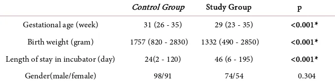
[image:4.595.208.538.627.710.2]The control group consisted of 189 premature babies with a mean gestational age of 31 (26 - 35) weeks and a birth weight of 1757 (820 - 2830) grams. The study group included 128 babies with a mean gestational age of 29 (23 - 35) weeks and a birth weight of 1332 (490 - 2850) grams and with ROP at different stages.
Table 1 lists the gestational week, birth weights and lengths of stay in incuba-tors for the control and study group infants.
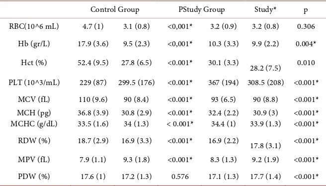
Measurements included red blood cell count (RBC; 106 mL), haemoglobin (HB; gr/L), haematocrit (HCT; %), platelet count (PLT; 103/mL), mean corpus-cular volume (MCV; fL), mean corpuscorpus-cular haemoglobin (MCH; pg), mean corpuscular haemoglobin concentration (MCHC; g/dL), red cell distribution width (RDW; %), mean platelet volume (MPV; fL) and platelet distribution width (PDW; %).
During birth, the values of RBC, Hb, MCV, MCH, RDW were significantly lower (p < 0.001) and the values of MCHC, PLT, MPV were significantly higher in the infants who developed ROP than in the infants who did not develop ROP. Likewise, the values of Hb, MCV, MCH, MCH, PLT were significantly lower but the values of RDW, MPV, PDW were significantly higher in haemogram results of infants born with ROP in the first month after birth (p < 0.001) (Table 2).
Comparison of the subgroups of the study group with respect to MPV values revealed differences between the control group infants and the group I infants (p = 0.234), as well as the group II and group IV, although the differences were not statistically significant (p = 0.415). However, the difference between the control group and group IV was statistically significant (p < 0.001). A correlation test showed a weak correlation between the ROP stage and the gestational week (r = −441; p < 0.001) and the birth weight (r = −449; p < 0.001) of the study group and the control group infants for the haemogram data in the first month. Again,
Table 1. Clinical characteristics of the control and study group infants.
Control Group Study Group p
Gestational age (week) 31 (26 - 35) 29 (23 - 35) <0.001*
Birth weight (gram) 1757 (820 - 2830) 1332 (490 - 2850) <0.001*
Length of stay in incubator (day) 24(2 - 120) 46 (6 - 195) <0.001*
Gender(male/female) 98/91 74/54 0.304
DOI: 10.4236/ojoph.2018.82011 79 Open Journal of Ophthalmolog
Table 2. Statistical comparison of hemogram data of the control and study group infants
in the first month.
Control Group PStudy Group Study* p
RBC(10^6 mL) 4.7 (1) 3.1 (0.8) <0,001* 3.2 (0.9) 3.2 (0.8) 0.306
Hb (gr/L) 17.9 (3.6) 9.5 (2.3) <0,001* 10.3 (3.3) 9.9 (2.2) 0.004*
Hct (%) 52.4 (9.5) 27.8 (6.5) <0.001* 30.1 (3.3) 28.2 (7.5) 0.010
PLT (10^3/mL) 229 (87) 299.5 (176) <0.001* 367 (194) 308.5 (208) <0.001*
MCV (fL) 110 (9.6) 90 (8.4) <0.001* 93 (6.5) 90 (8.8) <0.001*
MCH (pg)
MCHC (g/dL) 36.8 (3.9) 33.5 (1.6) 30.8 (2.9) 34 (1.3) < 0.001* <0.001* 32.4 (2.2) 34.4 (1) 33.9 (1.3) 30.9 (3) <0.001* <0.001* RDW (%) 18.7 (2.9) 16.9 (3.3) <0.001* 16.9 (2.2) 17.8 (3.1) <0.001*
MPV (fL) 7.9 (1.1) 9.3 (1.8) <0.001* 8.3 (1.3) 9.2 (1.9) <0.001*
PDW (%) 17.6 (1) 17.2 (1.3) 0.576 17.1 (1.3) 17.7 (1.4) <0.001*
Hemogram values of the control and study group in the first month. *p < 0.005. RBC (10^6 mL); red blood cell, Hb(gr/L); hemoglobin, HCT(%); hematocrit, PLT(10^3/mL); platelet, MCV(fL); mean corpuscular volume, MCH(pg); mean corpuscular hemoglobin, MCHC (g/dL); mean corpuscular hemoglobin concen-tration, RDW(%); red cell distribution width, MPV(fL); mean platelet volume, PDW(%); platelet distribu-tion width.
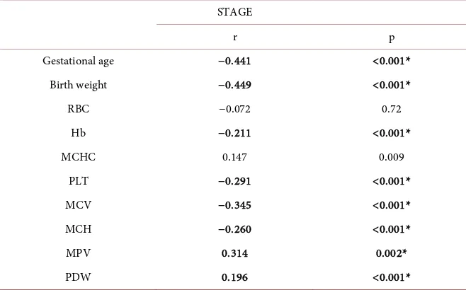
a weak correlation was evident between the ROP stage and RBC (r =-0.72; p = 0.2), HCT (r = −183; p < 0.001), PLT (r = −291; p < 0.001), MCV( r = −345; p < 0.001, MCH (r = −260; p < 0.001), MCHC (r = −147; p = 0.009), RDW (r = 174; p = 0002), MPV (r = 314; p < 0.001), PDW (r = 196; p < 0.001) and HB (r= −211; p < 0.001) (Table 3).
Modelling of birth Hb, PLT and MPV data following logistic regression analy-sis revealed a significant OR for Hb of 0.378 (p < 0.001, an OR for PLT of 1.004 that was not significant (p = 0.2) and an OR for MPV of 2.595 (p = 0.001) that was significant (Table 4). The modelling of Hb, PLT and MPV in the first month revealed a significant OR for Hb of 0.781 (p < 0.001), for PLT of 0.997 (p = 0.004) and for MPV of 1.421 (p = 0.002) (Table 5).
4. Discussion
In our study, we detected a marked decrease in levels of Hb, the oxygen carrier molecule, in infants with ROP. Oxygen transport is important during the devel-opment of ROP, and low levels of haemoglobin in infants with ROP can be a contributing factor to VEGF secretion from the hypoxic retina [8][13][14].
DOI: 10.4236/ojoph.2018.82011 80 Open Journal of Ophthalmolog
Table 3. Correlation between the stage of ROP development and hemogram parameters
and gestational age-birth weight of infants.
STAGE
r p
Gestational age −0.441 <0.001*
Birth weight −0.449 <0.001*
RBC −0.072 0.72
Hb −0.211 <0.001*
MCHC 0.147 0.009
PLT −0.291 <0.001*
MCV −0.345 <0.001*
MCH −0.260 <0.001*
MPV 0.314 0.002*
PDW 0.196 <0.001*
[image:6.595.208.540.374.445.2]P < 0.005. RBC (10^6 mL); red blood cell, Hb (gr/L); hemoglobin, MCHC (g/dL); mean corpuscular he-moglobin concentration, PLT (10^3/mL); platelet, MCV (fL); mean corpuscular volume, MCH (pg); mean corpuscular hemoglobin, MPV(fL); mean platelet volume, PDW(%); platelet distribution width.
Table 4. Multi-factor logistic regression analysis according to birth hemogram.
OR (95% confidence interval) p
Hb 0.378 (0.284 - 0.503) <0.001*
PLT 1.004 (0.998 - 1.010) 0.2
MPV 5.595 (1.495 - 1.504) 0.001*
P < 0.005*. Hb(gr/L); hemoglobin, PLT(10^3/mL); platelet, MPV(fL); mean platelet volume.
Table 5. Multi-factor logistic regression analysis according to hemogram in the first
month.
OR (95% confidence interval) p
Hb 0.781 (0.693 - 0.880) <0.001*
PLT 0.997 (0.995 - 0.999) 0.004*
MPV 1.421 (1.137 - 1.777) 0.002*
*p < 0.005. Hb(gr/L); hemoglobin, PLT(10^3/mL); platelet, MPV(fL); mean platelet volume.
[image:6.595.206.541.504.573.2]DOI: 10.4236/ojoph.2018.82011 81 Open Journal of Ophthalmolog a role in the abnormal vasculogenesis (phase 2) or abnormal angiogenesis (phase 1) underlying the pathoanatomy of severe ROP. In aggressive APROP, atypical and rapidly developing haemorrhages lead to the occurrence of APROP+ disease and the formations of flattened new blood vessels [11][16] [17]. Thrombocyte growth factors (including VEGF-A) also play a role in the transport of the thrombospondin, cytokines and chemokines known as the stromal cell-derived factor. When the platelet count is low, platelets cannot perform this role [18].
Conversely, the MPV level was higher in both birth and first-month haemo-gram values of patients with ROP. MPV, another indicator of platelet function, is an important predictive factor for cardiovascular diseases in adults. It also has an important involvement in diseases such as diabetes, obesity, rheumatologic diseases, psoriasis and FMF [17] [18] [19]. Although only a small number of studies have focused on thrombocyte and platelet disorders in the development of ROP, transporter and cleaner characteristics have been demonstrated for these proteins (both pro- and anti-angiogenetic) that regulate angiogenesis. Both proangiogenetic and antiangiogenic agents are stored in different subpopula-tions of α-granules in platelets and megakaryocytes, and when a signal is re-ceived, these factors are gradually released. The possible roles of active platelets in the pathogenesis of ROP can be explained in this way [18].
The correlation between MPV and the likelihood of ROP development has been investigated previously by Cekmez et al. [20], who found no difference be-tween a control group and infants with ROP. However, in that study, the blood samples were obtained from the infants within the first 2 hours immediately af-ter birth, whereas the critical period for the development of ROP is postmen-strual 31 - 34 weeks. In fact, our data indicate significantly higher values for MPV in the haemogram data obtained in the first month in infants with and without ROP, and especially in group IV infants with ROP. This demonstrates a role for active thrombocytes in the development of ROP.
In our study, the PDW increase was significantly higher in the first month in the haemogram data of infants with and without ROP (p < 0.001). One of the platelet function indicators is the platelet distribution range (PDW) and related increases in the heterogeneity of the platelet volume distribution. In adult di-abetic retinopathy patients, PDW values were significantly higher in patients with T2 DM than in controls [19]. A study conducted by Sadiq et al. in 2015 [8]
also demonstrated the importance of platelet-derived growth factor (PDGF) in the angiogenesis cascade of retinochoroidal vascular diseases, such as diabetic retinopathy, retinal vein occlusion, age-related macular degeneration, patholog-ical myopia and ROP. To the best of our knowledge, the present study is the first to examine the role of PDW in patients with ROP.
DOI: 10.4236/ojoph.2018.82011 82 Open Journal of Ophthalmolog
References
[1] Hah, P.K., Prabhu, V., Karandikar, S.S., Ranjan, R., Narendran, V. and Kalpana, N. (2016) Retinopathy of Prematurity: Past, Present and Future. World Journal of Clinical Pediatrics, 5, 35-46. https://doi.org/10.5409/wjcp.v5.i1.35
[2] Chew, E.Y. (1999) Major Clinical Trials of Vitreoretinal Diseases. In: Regillo, C.D., Brown, G.C. and Flyn, H.W., Eds., Vitreoretinal Disease: The Essentials, Theme Medical Publisher Inc, New York, 667-677.
[3] Pudraza, W., Pudraza, H., Jezierska, K., Szwed, J., Modrejewska, M., Rudnicki, J., Kordek, A. and Domek, H. (2011) The Role of Hemoglobin Variant Replacement in Retinopathy of Prematurity. The Indian Journal of Pediatrics, 78, 1498-1502.
https://doi.org/10.1007/s12098-011-0460-7
[4] Rothman, A.L., Mangalesh, S., Chen, X. and Toth, C.A. (2016) Optical Coherence Tomography of the Preterm Eye: From Retinopathy of Prematurity to Brain Devel-opment. Eye and Brain, 8, 123-133.
[5] Gilbert, C. (2008) Retinopathy of Prematurity: A Global Perspective of the Epidem-ics, Population of Babies at Risk and Implications for Control. Early Human De-velopment, 84, 77-82. https://doi.org/10.1016/j.earlhumdev.2007.11.009
[6] Rivera, J.C., Sapieha, P., Joyal, J.S., Dulhamel, F., Shao, Z., Sitaras, N., Picard, E., Hardy, P., Lachapella, P. and Chemtob, S. (2010) Understanding Retinopathy of Prematurity: Update on Pathogenesis. Neonatology, 100, 343-353.
https://doi.org/10.1159/000330174
[7] Patzelt, J. and Langer, H.F. (2012) Platelets in Angiogenesis. Current Vascular Pharmacology, 10, 570-577. https://doi.org/10.2174/157016112801784648
[8] Sadiq, M.A., Hanout, M,. Sarwar, S., Hassan, M., Do, D.V., Nquyen, Q.D. and Se-pah, Y.J. (2015) Platelet Derived Growth Factor Inhibitors: A Potential Therapeutic Approach for Ocular Neovas Cularization. European Journal of Ophthalmology, 29, 287-291. https://doi.org/10.1159/000438953
[9] Chu, S.G., Becher, R.C., Berger, P.B., Bhatt, D.L., Eikelboom, J.W., Konkle, B., Mohler, E.R., Reilly, M.P. and Berger, J.S. (2010) Mean Platelet Volume as a Pre-dictor of Cardiovascular Risk: A Systematic Review and Meta-Analysis. Journal of Thrombosis and Haemostasis, 8, 148-156.
https://doi.org/10.1111/j.1538-7836.2009.03584.x
[10] Gionia, S., Piazze, J., Anceschi, M.M., Cerekia, A., Alberini, A., Giancotti, A., Larci-prete, G. and Cosmi, E.V. (2007) Mean Platelet Volume: Association with Adverse Neonatal Outcome. Platelets, 18, 284-288.
https://doi.org/10.1080/09537100601078448
[11] International Committee for the Classification of Retinopathy of Prematurity (2005) The International Classification of Retinopathy of Prematurity Revisited. Archives of Ophthalmology, 123, 991-999.
[12] Ahn, S.J., Kim, J.H., Kim, S.J. and Yu, Y.S. (2013) Capillary-Free Vascularized Reti-na in Patients with Aggressive Posterior Retinopathy of Prematurity and Late Re-tinal Capillary Formation. Korean Journal of Ophthalmology, 27, 109-115.
[13] Lubetzky, R., Stolovitch, C. and Dolberg, S. (2005) Nucleated Red Cells in Preterm Infants with Retinopathy of Prematurity. Pediatrics, 116, 619-622.
https://doi.org/10.1542/peds.2005-0915
[14] Kandasamy, Y., Kumar, P. and Hartley, L. (2014) The Effect of Erythropoietinon the Severity of Retinopathy of Prematurity. Eye (Lond), 28, 814-818.
DOI: 10.4236/ojoph.2018.82011 83 Open Journal of Ophthalmolog
Subconjunctival Haemorrhages Unmasking İmmune Thrombocytopenic Purpura during Retinopathy of Prematurity Screening. BMJ Case Reports.
https://doi.org/10.1136/bcr-2017-221444
[16] Vinekar, A., Hegde, K., Gilbert, C., Braganza, S., et al. (2010) Do Platelets Have a Role in the Pathogenesis of Aggressive Posterior Retinopathy of Prematurity. Reti-na, 30, 520-523.https://doi.org/10.1097/IAE.0b013e3181cafc30
[17] İlhan, D., Özbabalık, D., Gülcan, E., Özdemir, O. and Gülbağ, S.Z. (2010) Evalua-tion of Platelet ActivaEvalua-tion, CoagulaEvalua-tion, and Fibrinolytic ActivaEvalua-tion in Patients with Symptomatic Lacunar Stroke. Neurologist, 16, 188-191.
https://doi.org/10.1097/NRL.0b013e318198d8bc
[18] Tao, Y., Dong, Y., Lu, C.W. and Li, Q. (2015) Relationship between Mean Platelet Volume and Retinopathy of Prematurity. Graefe’s Archive for Clinical and Experi-mental Ophthalmology, 253, 2972-2978.
[19] Akdogan, M., Budak, Y.U. and Huysal, K. (2016) The Association of Hematologic Inflammatory Markers with Atherogenic Index in Type 2 Diabetic Retinopathy Pa-tients. Clinical Ophthalmology, 10, 1797-1801.
https://doi.org/10.2147/OPTH.S110749