z
RESEARCH ARTICLE
VISFATIN UP-REGULATED SVCAM-1 AND ASSOCIATED WITH THE CHRONIC KIDNEY DISEASE
RELATED TO METABOLIC SYNDROME
*1,2
Enas N. Morgan and
1Abeer A. Khalefa
1
Physiology Department-Faculty of Medicine-Zagazig University-Egypt
2
PhD, Assist. Professor, College of Medical Rehabilitation, Qassim University, Saudi Arabia
ARTICLE INFO ABSTRACT
Objective: The present study aimed to clarify the role of visfatin in the pathogenesis of Chronic Kidney Disease (CKD) complicated Metabolic Syndrome (MS).
Materials and methods: 30 adult albino rats were included. Rats were divided into three equal groups: group I: served as control; group II: rats received high fat diet (HFD); group III: rats received visfatin injection. The serum levels of visfatin, sVCAM-1, insulin and fasting Serum glucose (FSG) levels were investigated for all groups. Moreover, proteinurea, mean arterial blood pressure (MABP) and histopathological examination were detected.
Results: Results showed a development of major systemic alterations similar to human MS, including obesity, hyperglycemia, hyperinsulinemia, and hypertension in rats on a HFD. These systemic changes were accompanied by renal pathophysiological alterations including, high creatinin levels, proteinurea, and atrophy of the glomeruli and sclerosis of the glomerular capillaries. Injection of visfatin was associated with lower levels of FSG, insulin, and HOMA-IR in comparison with both control and HFD-fed groups and high levels of creatinin, proteinurea, and sVCAM-1 in comparison with control. Moreover, mild glomerular atrophy and sclerosis was detected. Visfatin was positively correlated with proteinurea, and sVCAM-1 in both HFD-fed group and visfatin injected group.
Conclusion: Visfatin plays an important role in the development of renal injury associated with MS. As it increased the sVCAM-1 level that is considered as a biomarker for endothelial dysfunction and it also caused glomerular sclerosis.
Copyright © Enas N. Morgan and Abeer A. Khalefa, This is an open access article distributed under the Creative Commons Attribution License, which permits unrestricted use, distribution, and reproduction in any medium, provided the original work is properly cited.
INTRODUCTION
Metabolic syndrome has been reported to be associated with chronic
kidney disease (CKD), but the mechanisms remain unclear(Deji et al.
2009). Diabetic nephropathy ranks top amongst the causes of end stage renal disease in patients older than 40 years old. It is accounting for 44% of new cases of chronic kidney disease in United States, (diabetes fact sheet, 2007) 20% of cases of end stage renal disease in
England(Anse et al., 2007) and it is the leading known cause of
chronic renal failure in Pakistan (Rizvi & Manzoor 2002).Moreover,
the prevalence of microalbuminurea was 10.6% among the relatives of patients with CKD in Egypt and this prevelance is increased with
age, diabetes and obesity (Gouda et al., 2011).Ejerblad et al. (2006)
was throwing light on the association of adiposity with CKD. This
emerging connection has led to the generation of hypothesesabout
the possible role of adipokines in the pathogenesis of CKD. Visfatin is one of newly emerging adipokines. It is also known as B-cell colony-enhancing factor (PBEF) and Nicotinamide phosphoribosyl
transferase (NAMPT), it was first identified by Fukuhara et al. in
2005. It has much greater expression in visceral fat and thus named as
Visfatin (Root, 2005). Fukuhara et al. (2005) identified an insulin
mimetic action for visfatin as it binds to insulin receptor at a site
different from insulin binding site and increases glucose uptake. It
also acts as a Nicotinamide phosphoribosyl transferase and is thus
involved in production of reactive oxygen species (ROS) (Revollo et
al., 2007).Many recent reports have provided evidence that visfatin is
a novel marker of inflammation and its levels are elevated in a number of acute and chronic inflammatory diseases including sepsis, acute lung injury, rheumatoid arthritis, inflammatory bowel disease
*Corresponding author:Enas N. Morgan, Physiology Department-Faculty of
Medicine-Zagazig University-Egypt
(Garcia & Moreno Vinasco 2006, Brentano et al., 2007). Moreover,
visfatin expression is increased in foam cell macrophages that have a role in plaque destabilization within unstable atheroscletic regions
(Dahl et al., 2007). Axelson et al. in 2007 for the first time reported
an increased serum level of visfatin in CKD and later on, several other studies reproduced similar relationship between visfatin and
CKD (Yilmaz et al., 2008a, Nüsken et al., 2009).Plasma visfatin
concentrations are related to endothelial dysfunction in type-2
diabetic patients and patients with CKD (Malyszko et al., 2009,
Yilmaz et al., 2008a). As mentioned before, visfatin levels are
elevated in a number of acute and chronic inflammatory diseases (Garcia & Moreno Vinasco 2006). Collectively, these results suggest a new physiological role of visfatin in organ injury. The fibrotic
buildup observed by Song et al. (2008)and possibility of reactive
oxygen species via its activity as a Nicotinamide phosphoribosyl transferase (Nampt) strongly supports the concept that visfatin could be one of the cytokines responsible for renal damage in diabetic nephropathy.
Kang et al. (2010) indicated an increase in the visfatin synthesis from
renal cells under high glucose conditions that possibly contributes to increased glucose influx into cells through GLUT1, so visfatin may cause acceleration of the diabetic nephropathy through increasing the
metabolic alterations. Furthermore, Axelsson et al. (2009) also
indicated that visfatin was associated with sVCAM-1(Soluble Vascular Adhesion Molecule-1) which is a biomarker of endothelial damage in chronic kidney disease. In addition an association was observed between proteinuria and visfatin level, and proteinuria is considered as an important predictor of endothelial dysfunction in
early diabetic nephropathy (Matthews et al., 1985).It was not clear
whether the early increase in plasma visfatin levels indiabetic rats
ISSN: 0975-833X
Available online at http://www.journalcra.com
International Journal of Current Research
Vol. 5, Issue, 08, pp.2305-2310, August,2013
INTERNATIONAL JOURNAL
OF CURRENT RESEARCH
Article History:
Received 24th May, 2013
Received in revised form 09th June, 2013
Accepted 26th July, 2013
Published online 23rd August, 2013
Key words:
Visfatin, Obesity, High fat diet, Proteinurea,
had a causative role in tissue injury as a pro-inflammatorymolecule
or its elevation was a compensatory response to decrease insulin
resistance. In the current study, we investigated the association between visfatin serum levels and renal damage complicating high fat diet (HFD) induced MS in rats through measuring the sVCAM-1 (biomarker of endothelial damage) and proteinuria (the best indicator of endothelial damage). Moreover, its association with serum levels of glucose, and insulin, HOMA-IR and mean arterial blood pressure (MABP) was detected. The direct effect of visfatin injection on the serum level of Svcam-1, proteinuria, serum levels of glucose and insulin, HOMA-IR and MABP was investigated in healthy rats. Hisopathological examination of the renal tissue for all groups was performed.
MATERIAL AND METHODS
The current study was carried on 30 male adult albino rats (body weight, 184-205gm). Rats were housed under hygienic conditions, in the animal house of the faculty of medicine Zagazig University at 21°C–24°C in a 12 hr/12 hr light/dark cycle (Lesourd & Mazari 1999). They were given free access to water and food. Rats were divided into three equal groups (n=10). Group I: that was served as a control group; rats were fed standard rat chow that consisted of 25.8% protein, 62.8% carbohydrates and 11, 4% fat (total12.6 kJ/g), (Ahren
& Scheurink 1998). Group II: rats were given HFD with 60% kcal%
fat that consists of 16.45 proteins, 25.6% carbohydrate and 55.0% fat (total 23.4kJ/g) in the form of cotton seed oil added to the laboratory
chow (Ahren & Scheurink 1998, Cha et al., 2000) for 12 weeks and
Group III: rats fed standard rat chow and received visfatin in a dose of
240ρmol intravenously as a single bolus injection daily for 12 weeks
(Naz et al., 2011).For all groups, body weight was recorded per
week, and at the end of the study period. At the end of the
experimental period (the end of 12th week) rats were placed in
separate metabolism cages and food was removed during urine collection. Water was available ad libitum at all times. Urine samples from all surviving animals were collected overnight (approximately
16 hrs) in containers placed on dry ice for proteinurea analysis.After
overnight fasting, at 8:00 a.m, blood pressure was measured for all groups. Then the serum sample collected, serum was obtained by allowing the blood samples to clot at room temperature before centrifuging at approximately 3000 rpm for 15 minutes. The serum
was stored at -20°Cto examine the visfatin levels, sVCAM-1, insulin
levels, fasting serum glucose (FSG) level.One Kidney was excised from each animal and was prepared to histopathology.
The histopathological examination
The kidney tissues were fixed in Bouin’s fluid for 48 h. Later, they were dehydrated in graded levels of ethanol, cleared in xylene, and
embedded in paraffin wax for sectioning. The 5-μm thick sections
were cut, mounted on glass slides, and stained with hematoxylin and eosin for light microscopic analysis.
Measuring the blood pressures (MABP) for rats
After ketamine hydrochloride (45mg/kg) intraperitoneal injection, the animals were implanted with cannulae (PE50) inserted into the left common carotid artery and the right jugular vein. The arterial cannula was connected to a pressure transducer fitted with F.C. 137 coupler connected to MD4-Oscillograph (Bioscience, Washington) (Martin
et al., 2005).
Biochemical Analysis
1-Determination of Serum visfatin levels
Serum Visfatin was measured using RayBio_Visfatin Enzyme Immunoassay Kit Protocol Catalog No: EIA-VIS-1 LOT No.: 601344. The performance characteristics of this assay were Intra-Assay: CV<10% Inter-Intra-Assay: CV<15%. The minimum detectable concentration of Visfatin is 379 pg/ml or 6.82pM.
2-Determination of serum glucose levels: According to Trinder
(1969) using glucose enzymatic (GOD-PAP)-liquizyme Kits (Biotechnology, Egypt).
3-Determination of serum insulin level: By a solid phase enzyme
amplified sensitivity immunoassay according to Starr et al. (1978)
using KAP1251-INS-EASIA (Enzyme Amplified Sensitivity
Immunoassay) Kits (BioSource Europe S.A., Belgium).
4-HOMA-IR: it was assessed by homeostasis model assessment
(where HOMA= (fasting insulin (μU/ml) x fasting plasma glucose
(mg/dl) /405 (Matthews et al., 1985).
5-Determination of sVCAM-1serum levels: Serum sVCAM-1 was
measured using Quantikine sVCAM-1 Immunoassay Kit, Catalog No. MVC00. Microplate reader capable of measuring absorbance at 450nm, with the correction wavelength set at 540 nm or 570 nm.
6-Determination of proteinurea: Protein concentration in urine was
determined by Microlab 300: Vital Scientific, Germany. According to the manufacturer's protocol. Proteinuria is measured by the principle of turbidimetry by using 5% trichloroacetic acid. All the biochemical and hormonal parameter were measured in the Department of Medical Biochemistry, the proteinurea, and creatinin were measured in the Department of Clinical Pathology, while the histopathological examination was done in the Pathology Department, Faculty of Medicine, Zagazig University, Egypt.
Statistical Analysis
The data obtained in the present study were expressed as meanSD
for quantitative variables and statistically analyzed by using SPSS
program (version 17 for windows) (SPSS Inc. Chicago, IL, USA).
Analysis of variance (ANOVA)was used to compare the results of all
examined cases in all studied groups within group comparisons. Correlations were performed by Pearson 2 tailed test (Kirkwood
1989).
RESULTS
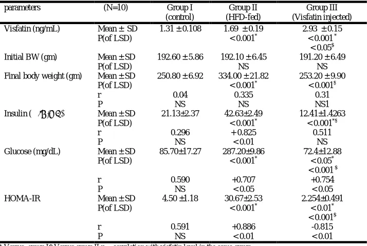
Table 1shows the serum visfatin levels (ng/mL), initial body weight
“BW” (gm), final BW and some glucose metabolic parameters
(glucose”mg/dL”, insulin”IU/mL” and calculated HOMA-IR) in the
While glucose and HOMA-IR correlated negatively with visfatin levels (r=-0.754; P<0.05 and -0.641; P<0.05 respectively) insulin did not correlated with visfatin levels. In control group no significant correlations was detected between visfatin levels and insulin, glucose or HOMA-IR.
Table 2shows the serum Creatinine (mg/dL), sVCAM, Proteinuria,
and Mean arterial blood pressure in the three studied groups. In HFD-fed group, it was found that the mean value of serum Creatinine, serum sVCAM and Proteinuria levels (3.15±0.62, 7.65±0.93 and 1.20±0.13 respectively) were significantly higher than that of control group (0.70±0.07, 3.48±0.43 and 0.70±0.08 respectively, P<0.001) and that of visfatin injected group (1.39±0.12, 5.89±0.72 and 0.92±0.09 respectively, P<0.001). In addition these parameters in visfatin injected group were significantly lower than that of HFD-fed group (P<0.001) and significantly higher than that of control group (P<0.001). There were significant positive correlations between visfatin levels and Creatinine (mg/dL), Proteinuria and sVCAM (r=+0.870; P<0.01, +0.889, P<0.01 and r=+0.851, P<0.01 respectively) in HFD-fed group, and there was no significant correlation detected in control group. In visfatin injected group, there
was a significant positive correlation between serum visfatin levels and sVCAM (r=+0.965, P<0.001). Moreover, there were positive correlation between sVCAM levels and Proteinuria levels in HFD-fed group and visfatin injected group (r=+0.674, P<0.05 and r=+0.843; P<0.01 respectively). As regard Mean arterial blood pressure, the mean value of HFD-fed group (123.37±12.70) was significantly higher than that of control group (90.2 ± 6.49, P <0.01) and that of Visfatin injected group (101.40±7.03 p<0.001). Moreover, visfatin injected group it was significantly higher than that of control group (P<0.05). The correlation between serum visfatin levels and Mean
arterial blood pressure was significant only in HFD-fed group (r = +0.900 P<0.01). The histopathological examination of the
[image:3.595.120.477.86.326.2]Kidney’s tissue showed normal renal tissue formed of normal glemeruli and tubules separated by scanty connective tissue stroma in the control group (Fig. 1). While that for the HFD- received group showed completely atrophic glemeruli with total hyalinization and sclerosis of the glermular capillaries. Moreover, the renal tubules showed cloudy swelling (Fig. 2). The visfatin injected group showed some atrophic glomeruli with mild sclerosis of the glomerular capillaries (Fig. 3).
Table 1. Serum visfatin levels (ng/mL), initial body weight “BW” (gm), final BW and some glucose metabolic parameters (glucose”mg/dL”, insulin”IU/mL” and calculated HOMA-IR) in the three studied groups
parameters (N=10) Group I
(control)
Group II (HFD-fed)
Group III (Visfatin injected)
Visfatin (ng/mL) Mean ± SD 1.31 ± 0.108 1.69 ± 0.19 2.93 ± 0.15
P(of LSD) < 0.001* < 0.001 *
< 0.05$
Initial BW (gm) Mean ± SD 192.60 ± 5.86 192.10 ± 6.45 191.20 ± 6.49
P(of LSD) NS NS
Final body weight (gm) Mean ± SD 250.80 ± 6.92 334.00 ± 21.82 253.20 ± 9.90
P(of LSD) < 0.001* < 0.001$
r P
0.04 NS
0.335 NS
0.31 NS1
Insulin ( IU/mL) Mean ± SD 21.13±2.37 42.63±2.49 12.41±1.4263
P(of LSD) < 0.001* < 0.001*$
r P
0.296 NS
+ 0.825 < 0.01
0.511 NS
Glucose (mg/dL) Mean ± SD 85.70±17.27 287.20±9.86 72.4±12.88
P(of LSD) < 0.001* < 0.05*
< 0.001 $
r P
0.590 NS
+0.707 < 0.05
+0.754 < 0.05
HOMA-IR Mean ± SD 4.50 ±1.18 30.67±2.53 2.254±0.491
P(of LSD) < 0.001* < 0.01*
< 0.001$
r P
0.591 NS
+0.886 < 0.01
-0.815 < 0.01 * Versus group I$ Versus group II r = correlation with visfatin level in the same group
Table 2. Markers for renal function and MABP in the three studied groups
parameters N=10 Group I
(control)
Group II (HFD-fed)
Group III (Visfatin injected) Creatinine (mg/dL) Mean ± SD 0.70 ± 0.07 3.15 ± 0.62 1.39 ± 0.12
P(of LSD) < 0.001* < 0.001*$
r P
0.277 NS
0.870 < 0.01
0.276 NS
Proteinuria Mean ± SD 0.70 ± 0.08 1.20 ± 0.13 0.92 ± 0.09
P(of LSD) > 0.001* < 0.001*$
r P
0.401 NS
0.889 < 0.01
0.276 NS
sVCAM Mean ± SD 3.48 ± 0.43 7.65± 0.93 5.89±0.72
P(of LSD) < 0.001* < 0.001*$
r P
0.177 NS
+ 0.851 < 0.01
+0.965 < 0.001 r @
P
0.357 NS
0.674 < 0.05
0.843 < 0.01 Mean arterial blood
pressure
Mean ± SD 90.2 ± 6.49 123.37±12.70 101.40±7.03
P(of LSD) < 0.001* < 0.05*
< 0.001 $
r P
0.356 NS
0.900 < 0.01
0.314 NS
* Versus group I$ Versus group II r = correlation with visfatin level in the same group r @ = correlation with proteinuria level in the same group
[image:3.595.135.466.353.530.2]Fig. 1. Photomicrogtaph of normal renal tissue formed of normal glemeruli and tubules separated by scanty connective tissue stroma
[image:4.595.50.279.243.390.2](H&E X100)
Fig. 2. Photomicrograph of a case of severe diabetic nephropathy showing completely atrophic glemeruli (↑) with total hyalinization and sclerosis of the glermular capillaries – The renal tubules showed cloudy swelling ()
(H &E X 400)
Fig. 3: Photomicrogtaph of a case of mild diabetic nephropathy showing normal sized glemeruli (↑) and atrophic one ( ) showing mild sclerosis of
the glemerular capillaries (H& E X400)
DISCUSSION
The results of the current study approved a development of major systemic alterations similar to human metabolic syndrome; including obesity, hyperglycemia, hyperinsulinemia, and hypertension in rats on a HFD for 12 weeks. Furthermore, these systemic changes were accompanied by renal pathophysiological alterations including, proteinurea, elevated creatinine level, and atrophy of the glomeruli with total hyalinization and sclerosis of the glomerular capillaries. This renal pathophysiological alterations may caused by Systemic
lipid overload “lipotoxicity,” which can induce systemic
inflammation and oxidative stress (Van Gaal et al., 2006, Weinberg
2006) and/or the local alteration of lipid metabolism in the kidney
(Jiang et al., 2005, Kume, 2007). Moreover, obesity which is an
important character of metabolic syndrome is considered as an important source for several factors called adipokines or
adipocytokines (Kershaw & Flier, 2004).Many adipokines have a
role in the inflammatory process accompanied with obesity (Bulcao
et al., 2006). Visfatin is a novel adipokine that is secreted by visceral
and subcutaneous fat with insulin mimetic effect (Fukuhara et al.,
2005). This insulin mimic effect was confirmed in the current study, as injection of visfatin caused decrease in the fasting serum glucose levels, insulin levels and HOMA-IR. These results are also congruent
with the results of Naz et al. (2011) who showed that the
administration of exogenous visfatin in obese and diabetic groups of mice resulted in significant reduction in their fasting blood glucose levels but it remained higher than the glucose levels of normoglycaemic mice. However this insulin minic effect cannot be detected in the HFD-fed group as it showed high levels of visfatin, fasting serum glucose, insulin and HOMA-RI. These results are in line with the results of many other publications in which high fat/high
sucrose diets have been used to induce obesity (Boyd et al., 1990,
Yaspelkis et al., 2004). In spite of the higher levels of visfatin
detected in the HFD-received group, the levels of serum glucose and insulin were high. This may be caused by the development of visfatin resistance during obesity similar to that of insulin resistance as
visfatin uses the same receptors of insulin (Hammarstedt et al., 2006).
Moreover, in obesity, various adipocytokines have been released from the adipocytes that may neutralize the effect of visfatin and have
resulted in visfatin resistance and hyperglycaemia (Naz et al., 2011,
Arner 2005). Many researches indicated relation between visfatin and inflammation as its levels were elevated in a number of acute and chronic inflammatory diseases including sepsis, acute lung injury, rheumatoid arthritis, inflammatory bowel disease (Garcia & Moreno
Vinasco 2006, Brentano et al., 2007). Also visfatin acts as a
Nicotinamide phosphoribosyl transferase and is thus involved in
production of reactive oxygen species (Revollo et al., 2007). These
findings directed us to propose that visfatin may be released from the adipose tissue as a compensatory mechanism for glucose intolerance however due to its role in inflammation and oxidative reaction; it may cause many systemic worse effects.
In 2007, Axelsson et al. reported for the first time an increased serum
level of visfatin in CKD and later on, several other studies demonstrated similar relationship between visfatin and CKD (Yilmaz
et al., 2008, Nüsken et al., 2009). In the current study we detected significant positive correlations related visfatin levels with creatinine, proteinuria and sVCAM-1 in the HFD-fed group, these results suggested a role for visfatin in CKD that complicating the metabolic syndrome. And these results were in line with the results of other researches that indicated an association of visfatin with sVCAM-1
(Axelsson et al., 2007) and proteinurea (Yilmaz et al., 2008).
sVCAM-1 is considered as a biomarker of endothelial damage in
chronic kidney disease (Axelsson et al., 2007) and proteinurea is
considered as an important predictor of endothelial dysfunction in
early diabetic nephropathy (Matthews et al., 1985). In addition our
results detected a positive correlation between the sVCAM-1 and the proteinurea in both HFD-fed and visfatin injected groups. Moreover, injection of visfatin in healthy rats caused increase in the levels of proteinurea, creatinin and sVCAM-1, also positive correlation was detected between visfatin levels and sVCAM-1 in this group in addition to mild atrophy and sclerosis in the renal glomeruli. These results strongly support the concept that visfatin has a direct role in the pathogenesis of CKD associated with metabolic syndrome. This
association was also confirmed by Mahmood et al. (2010) who
indicated an association between visfatin and CKD secondary to diabetic nephropathy and other risk factors and they also considered visfatin as a new marker for endothelial dysfunction related to CKD. In other study, the plasma visfatin concentration was found to correlate with hsCRP and resistin levels, this result makes it to be considered as a potential link with inflammation and as inflammation and hyperlipidemia are risk factors of atherosclerosis, visfatin may play some role in linking lipid dysregulation and inflammation to
[image:4.595.48.281.445.578.2]our study as high level of visfatin in HFD-fed group and also visfatin injection were accompanied by sclerosis of the glomerular capillaries
and increased MABP. Kim et al. (2008) investigated the effect of
visfatin on vascular inflammation, and they concluded that visfatin induced leukocyte adhesion to endothelial cells by induction of the cell adhesion molecules (CAMs), intracellular adhesion molecule-1 (ICAM-1) and VCAM-1 and it also stimulated ROS generation in endothelial cells through its NADPH dependent ROS production. These observations, support the concept that visfatin may be able to
accelerate vascular diseases. At the molecular level Song et al. (2008)
indicated that treatment of cultured mesangial cells with recombinant visfatin caused activation of protein kinase a and b, and dramatic increase in the synthesis of profibrotic molecules including TGF-ß (Tranasforming growth factor beta), Plasminogen activation inhibiting factor-1 (PAI-1) and type-1 collagen which are considered as contributors in pathogenesis of diabetic nephropathy.
Conclusion
This study suggests that visfatin may play an important role in the development of renal injury associated with metabolic syndrome. As it increased the sVCAM-1 level that is considered as a biomarker for endothelial dysfunction and it also caused glomerular sclerosis. Visfatin may also play its role in vascular inflammation; through its NADPH dependent ROS production, so the exact mechanism is needed to be elucidated. We would like to address this issue in our future studies.
Acknowledgment
The author is grateful to faculty of Medicine- Zagazig University for providing the necessary facilities and also would like to acknowledge all the contributors in the Departments of Clinical Pathology, Pathology and Medical Biochemistry for providing greet help in the analytic part of the research.
REFERENCES
Ahren, B. and Scheurink, A.J.W. 1998. Marked hyperleptinemia after high fat diet associated with severe glucose intolerance in mice. Eur. J. Endocrinol., 139: 461–467.
Ansell, D., Feehally, J., Feest, T.G., Tomson, C., Williams, A.J., and Warwick, G. 2007. The Renal Association UK Renal Registry. The Tenth Annual Report December.
Arner, P. 2005. Insulin resistance in type 2 diabetes-role of adipokines. Curr Mol Med., 5:333–339.
Axelsson, J., Witasp, A., Carrero, J.J., Qureshi, A.R., Suliman, M.E., Heimbürger, O., Bárány, P., Lindholm, B., Alvestrand, A., Schalling, M., Nordfors, L. and Stenvinkel, P. 2007. Circulating levels of visfatin/pre-B-cell colony-enhancing factor 1 in relation to genotype, GFR, body composition, and survival in patients with CKD. Am J Kidney Dis, 49: 237–44.
Boyd, J., Contreras, I., Kern, M., Tapscott, E., Downes, D., Frisell, W. and Dohm, G. 1990. Effect of a high-fat-sucrose diet on in vivo insulin receptor kinase activation. Am J Physiol.,259: E111–116.
Brentano, F., Schorr, O., Ospelt, C., Stanczyk, J., Gay, R.E., Gay, S. and Kyburz, D. 2007. Pre-B cell colony-enhancing factor/visfatin, a new marker of inflammation in rheumatoid arthritis with proinflammatory and matrix-degrading activities. Arthritis Rheum; 56: 2829-39.
Bulcao, C., Ferreira, S.R., Giuffrida, F.M., and Ribeiro-Filho, F.F. 2006. The new adipose tissue and adipocytokines. Curr Diabetes Rev., 2(1):19–28.
Centers for Disease Control and Prevention. National diabetes fact sheet: general information and national estimates on diabetes in United States, 2007.
Cha, M.C., Chou, J. and Boozer, C.N. 2000. High-fat diet feeding reduces the diurinal variation of plasma leptin concentration in rats. Metab., 48:503–507.
Dahl TB, Yndestad A, Skjelland M, Øie E, Dahl A, Michelsen A, Damås J K, Tunheim S H, Ueland T, Smith C, Bendz B, Tonstad S, Gullestad L, Frøland S, Krohg-Sørensen K, Russell D, Aukrust P, Halvorsen B. Increased expression of visfatin in macrophages of human unstable carotid and coronary atherosclerosis: possible role in inflammation and plaque destabilization. Circulation 2007; 115: 972–980.
Deji, N., Kume, S., Araki, S., Soumura, M., Sugimoto, T., Isshiki, K., Chin-Kanasaki, M., Sakaguchi, M., Koya, D., Haneda, M., Kashiwagi, A. and Uzu, T. 2009. Structural and functional changes in the kidneys of high-fat diet-induced obese mice. Am J Physiol Renal Physiol., 296: F118–F126.
Ejerblad, E., Fored, C.M., Lindblad, P., Fryzek, J., McLaughlin, J.K., and Nyrén, O. 2006. Obesity and risk for chronic renal failure. J Am Soc Nephrol., 17: 1695–702.
Fukuhara, A., Matsuda, M., Nishizawa, M., Segawa, K., Tanaka, M., Kishimoto. K., Matsuki, Y., Murakami, M., Ichisaka, T., Murakami. H., Watanabe, E., Takagi, T., Akiyoshi, M., Ohtsubo, T., Kihara, S., Yamashita, S., Makishima, M., Funahashi, T., Yamanaka, S., Hiramatsu, R., Matsuzawa, Y., Shimomura, I. 2005. Visfatin: a protein secreted by visceral fat that mimics the effects of insulin. Science, 307: 426–30.
Garcia, J.G., and Moreno Vinasco, L. 2006. Genomic insights into acute inflammatory lung injury. Am J Physiol Lung Cell Mol Physiol., 291:L1113-7.
Gouda, Z., Mashaal, G., Bello, A.K., Attar, A.E., Kemmery, T.E., .El Reweny, A., El Nahas M.Egypt information, Prevention and treatment of chronic kidney disease (EGIPT-CKD) programme: prevalence and risk factors of microalbuminurea among the relatives of patients with CKD in Egypt. Saudi J Kidney Dis Transpl 2011; 22 (5):1055-1063.
Hammarstedt, A., Pihlajamaki, J., Rotter Sopasakis, V., Gogg S., Jansson P.A., Laakso, M., and Smith, U. 2006. Visfatin is an adipokine, but it is not regulated by thiazolidinediones. J Clin Endocrinol Metab., 91:1181–1184.
Jiang, T., Wang, Z., Proctor, G., Moskowitz, S., Liebman, S.E., Rogers, T., Lucia, M.S., Li, J., and Levi, M. 2005. Diet-induced obesity in C57BL/6J mice causes increased renal lipid accumulation and glomerulosclerosis via a sterol regulatory element-binding protein-1c-dependent pathway. J Biol Chem., 280: 32317–32325.
Kang, Y.S., Song, H.K., Lee, M.H., Ko, G.J., Han, J.Y., Han, S.Y, Han, K.H., Kim, H.K., and Cha, D.R. 2010. Visfatin is upregulated in type-2 diabetic rats and targets renal cells. Kidney Int., 78:170–81.
Kershaw, E. and Flier, J.S. 2004. Adipose tissue as an endocrine organ. J Clin Endocrinol Metab, 89: 2548–56.
Kim, S.R., Bae, Y.H., Bae, S.K., Choi, K.S., Yoon, K.H., Koo, T.H., Jang, H.O., Yun, I., Kim, K.W., Kwon, Y.G., Yoo, M.A. and Bae, M.K. 2008. Visfatin enhances ICAM-1 and VCAM-1 expression through ROS-dependent NF-kappaB activation in endothelial cells. Biochim Biophys Acta., 1783(5):886–95.
Kirkwood, B.1989. Essentials of medical statistics. Black well Scientific Publication, Oxford, London, 151.
Kume, S., Uzu, T., Araki, S., Sugimoto, T., Isshiki, K., Chin-Kanasaki, M., Sakaguchi, M., Kubota, N., Terauchi, Y., Kadowaki, T., Haneda, M., Kashiwagi, A. and Koya, D. 2007. Role of altered renal lipid metabolism in the development of renal
injury induced by a high-fat diet. J Am Soc Nephrol.,18: 2715–2723.
Lesourd, B. and Mazari, L.1999. Nutrition and immunity in the elderly. Proceeding of Nutrition Society.,58 (3):94–200.
Mahmood, N., Junejo, A.M., Jamal, Q. and Awan, R. 2010. Association of visfatin with chronic kidney disease in a cohort of patients with and without diabetes. J Pak Med Assoc., 60 (11), 922–926
Malyszko, J., Malyszko, J.S., and Mysliwiec, M. 2009. Visfatin, a new adipocytokine, is predominantly related to inflammation/
endothelial damage in kidney allograft recipients. Transplant Proc., 41: 150–3.
Martin, D.S., Biltoft, S., Redetzke, R., and Vogel, E. 2005. Castration reduces blood pressure and autonomic venous tone in male spontaneously hypertensive rats. Journal of Hypertension, 23(12), 2229–2236.
Matthews, D.R., Hosker, J.P., and Rudenski, A.S., Naylor, B.A., Treacher, D.F., and Turner, R.C. 1985. Homeostasis model assessment: insulin resistance and beta-cell function from fasting plasmaglucose and insulin concentrations in man. Diabetologia., 28:412–419.
Mu, J., Feng, B., Ye, Z., Yuan, F., Zeng, W., Luo, Z. and Qi, W. 2011. Visfatin is related to lipid dysregulation, endothelial dysfunction and atherosclerosis in patients with chronic kidney disease. J Nephrol., 24(2):177–84
Naz, R., Hameed, W., Hussain, M.M., and Aslam, M. 2011. Glucose lowering effect of visfatin in obese and insulin dependent diabetes mellitus. Pak J Physiol.,7(1):4-7
Nüsken, K.D., Petrasch, M., Rauh, M., Stöhr, W., Nüsken, E., Schneider, H., and D¨otsch, J. 2009. Active visfatin is elevated in serum of maintenance haemodialysis patients and correlates inversely with circulating HDL cholesterol. Nephrol Dial Transplant, 24: 2832–8.
Revollo, J.R., Grimm, A.A., and Imai, S. 2007. The regulation of
nicotinamide adenine dinucleotide biosynthesis by
Nampt/PBEF/visfatin in mammals. Curr Opin Gastroenterol., 23: 164-70.
Rizvi, S.A. and Manzoor, K. 2002. Causes of Chronic Renal Failure in Pakistan: A Single Large Center Experience. Saudi Journal of Kidney Diseases and Transplantation, 13:376–9.
Root, A.W. 2005. Visfatin a new visceral fat adipokine. Growth, Genetics and Hormones, 21:21-4.
Song, H.K., Lee, M.H., Kim, B.K., Park, Y.G., Ko, G.J., Kang, Y.S., Han, J.Y., Han, S.Y., Han, K.H., Kim, H.K., and Cha, D.R. 2008. Visfatin: a new player in mesangial cell physiology and diabetic nephropathy. Am J Physiol Renal Physiol., 295: 1485–94. Starr, J.I., Mako, M.E., and Juhn, D.1978. Rubenstein AH.
Measurement of serum pro-insulin–like material: cross reactivity of porcine and human proinsulin. J. Lab. Clin. Med., 1978; 91:691–692.
Trinder, P.1969. Enzymatic determination of glucose. An. Clin. Bioch., 6:24–27.
Van Gaal, L.F., Mertens, I.L., and De Block, C.E. 2006. Mechanisms linking obesity with cardiovascular disease. Nature, 444:875–880. Weinberg, J.M. 2006. Lipotoxicity. Kidney Int., 70: 1560–1566. Yaspelkis, B., Singh, M., Krisan, A., Collins, D., Kwong, C.,
Bernard, J. and Crain, A. 2004. Chronic leptin treatment enhances insulin-stimulated glucose disposal in skeletal muscle of high-fat fed rodents. Life Sci., 74: 1801–1816.
Yilmaz, M.I., Saglam, M., Carrero, J.J., Qureshi, A.R., Caglar, K., Eyileten, T., Sonmez, A., Cakir, E., Yenicesu, M., Lindholm, B., Stenvinkel, P., Axelsson, J. 2008. Serum visfatin concentration and endothelial dysfunction in chronic kidney disease. Nephrol Dial Transplant, 23: 959–65.