ISSN Online: 2158-2750 ISSN Print: 2158-2742
Effect of Black Tea Polyphenol on Cell-ECM
Interaction and MMP
Nabanita Bhattacharyya
1, Subhajit Mondal
1, Shuvojit Moulik
1, Sathi Paul
1,
Shreoshi Bhattacharrya
1, Alok Kumar Hazra
1, Md. Nasim Ali
2, Anjan Adhikari
3,
Amitava Chatterjee
1*1Ramakrishna Mission Vivekananda University, IRDM, Kolkata, India 2B.C.K.V, Kalyani, India
3R.G. Kar Medical College, Kolkata, India
Abstract
Tea is one of the most widely consumed beverages in the world. Black tea, ob-tained from the leaves of Camellia sinensis is the preferred beverage in India and in most western countries. Epidemiological studies on black tea and can-cer are limited. However, preliminary studies indicate a positive correlation between black tea consumption and a lower incidence of breast and ovarian cancer. In the present communication, we wanted to see the effect of black tea extract and the polyphenol theaflavin on cell-ECM interaction, MMP activity etc. to strengthen the anti-cancer effect of black tea.
Keywords
Black Tea, Polyphenol, MMP, ECM, Cancer
1. Introduction
Tea is one of the most widely consumed beverages in the world. Black tea, ob-tained from the leaves of Camellia sinensis is the preferred beverage in India and in most western countries [1] [2]. Theaflavins are anti-oxidant polyphenols formed via co-oxidation of pairs of epimerized catechins during fermentation of tea leaves in black tea and account for 2% - 6% of dry weight. Theaflavins are characterized by benzotropolone ring structure and contribute to the unique taste of black tea. Principal theaflavins include theaflavin (TF-1), theaflavin-3-gallate (TF-2A), theaflavin-3’-gallate (TF-2B), and theaflavin-3, 3’-digallate (TF-3) [1] [3]. Theaflavins are the major antioxidant constituents of black tea, inhibiting free radical generation and pro-oxidative enzyme activities and chelating metal ions to prevent lipid peroxidation. TF-3 has also been shown to possess a higher How to cite this paper: Bhattacharyya, N.,
Mondal, S., Moulik, S., Paul, S., Bhat-tacharrya, S., Hazra, A.K., Ali, Md.N., Adhikari, A. and Chatterjee, A. (2017) Effect of Black Tea Polyphenol on Cell- ECM Interaction and MMP. American Journal of Plant Sciences, 8, 856-866. https://doi.org/10.4236/ajps.2017.84058
Received: February 2, 2017 Accepted: March 26, 2017 Published: March 30, 2017
Copyright © 2017 by authors and Scientific Research Publishing Inc. This work is licensed under the Creative Commons Attribution International License (CC BY 4.0).
antioxidative activity than catechins, including epigallocatechin gallate (EGCG) [1] [2] [4].
Epidemiological studies on black tea and cancer are limited. However, pre-liminary studies indicate a positive correlation between black tea consumption and a lower incidence of breast and ovarian cancer [5] [6]. A number of studies have indicated that black tea polyphenols possess anti-inflammatory and anti- tumorigenic properties, can inhibit proliferation and induce apoptosis in various tumours, including skin and colon cancers [6] [7].
Tumour invasion and formation of secondary tumours require coordinated matrix degradation and modulation of cell-cell and cell matrix attachment. Ma-trix metalloproteinases (MMPs), a family of zinc dependant endopeptidases, de-grade various extra cellular matrix (ECM) components, creating pathways through which tumour cells can invade [8] [9]. MMPs are synthesised as inactive pro-MMPs; activation requires proteolytic removal of the prodomain. MMP-2 (gelatinase A), a 72 kDa type IV collagenase, is activated at the tumour cell sur-face by an “activation complex” composed of membrane type-1 matrix metal-loproteinase (MT1-MMP), tissue inhibitor of metalmetal-loproteinase-2 (TIMP-2) and pro-MMP-2 [8] [9] [10] [11]. MMP activity is regulated in vivo by cellular activators and inhibitors e.g. extra-cellular matrix metalloproteinase inducer (EMMPRIN) which upregulates MMP activity and tissue inhibitors of matrix metalloproteinases (TIMPs) which inhibit their activity [8] [9].
Elevated expression and activity of MMPs (e.g. MMP-2) strongly correlate with increased metastatic potential, tumour spread and poor prognosis in a number of cancers including melanomas, breast, lung, thyroid, oral, stomach and colon carcinomas [8] [9] [12] [13] [14]. MMPs play multiple roles in promot-ing tumour progression by increaspromot-ing cancer cell growth, migration, metastasis and angiogenesis. Numerous studies indicate that highly invasive cells become less aggressive when MMP expression or activity is reduced, making MMPs promising targets for compounds with anti-metastatic and anti-tumorigenic properties. Black tea polyphenols, including theaflavin and its derivatives, down regulate MMP and urokinase expression in human stomach and colon carcinomas and inhibit MMP-2 and MMP-9 activity in mouse Lewis lung carcinoma cells, thus inhibiting tumour invasion and metastasis [15] [16].
autophos-phorylation in mouse epidermal cell line JB6Cl41 and may exert its chemopre-ventive effects through the downregulation of EGFR [21]. Integrins are a family of transmembrane heterodimeric proteins which mediate cell adhesion to a vari-ety of ECM and plasma proteins (e.g. fibronectin, fibrinogen). Integrin receptors play important roles in cell anchoring and are critically involved in multiple sig-nal transduction cascades [22] [23]. Theaflavin and its derivatives have been re-ported to reduce adhesion of MO4 cells to laminin and adhesion of A375 mela-noma cells fibronectin, vitronectin and laminin via integrin receptors [17] [24]. Cytoskeletal components like actin filaments and microtubules play an impor-tant role in regulation of cell motility and migration [22]. Microtubule targeting agents have a broad spectrum of activity against both haematological malignan-cies and solid tumours [25]. Theaflavins have been reported to act as micro-tubule depolymerizers and disrupt the micromicro-tubule network, leading to by al-teration of cellular morphology and apoptosis in human cervical cancer HeLa cells [26].
FAK acts as a transducer of integrin-growth factor stimulated signals to downstream targets like extracellular signal related kinase (ERK), phospho-inositol-3-kinase (PI-3K), c-Jun kinase (Jnk) and other mitogen activated pro-tein kinase (MAPK) cascades [22] [27]. Signalling through FAK, ERK, PI-3K and other MAPK pathways can increase MMP expression and activity and also influence cytoskeletal organization and migration. FAK is overexpressed in in-vasive human carcinomas and activation of FAK plays an important role in cell migration [22] [27] [28]. Nuclear transcription factor κβ (NF-κB), a dimeric transcription factor, is retained in the cytosol in the form of complexes with in-hibitory proteins and upon activation, translocates to the nucleus. Signalling through NF-κB plays an important role in inflammation, invasion, angiogenesis and regulation of MMPs. Many malignant tumours display constitutive NF-κB activation that allows malignant cells to escape apoptosis and resist the action of chemotherapeutic agents [29].
damaged or infected cells which pose a threat to the organism’s integrity. p53, the product of tumour suppressor gene TP53, has been implicated in control of cell proliferation and tumour progression, as well as in the maintenance of ge-nome integrity in response to DNA-damaging events [35]. Treatment with black tea polyphenols has been shown to induce apoptosis in prostate cancer cells by induction of p53 and down-regulation of NF-κB activity [36]. Theaflavins have been reported to induce apoptosis in mammary epithelial carcinomas through inhibition of PI-3K-Akt-pBad survival pathway [37]. Theaflavin and extracts from black tea have been shown to induce apopotosis in human leukemic cell lines HL-60 and K-562 [40].
In the present communication we wanted to see the effect of black tea extract and the polyphenol theaflavin on cell-ECM interaction, MMP activity etc. to strengthen the anti-cancer effect of black tea.
2. Materials and Methods
2.1. Materials
Black Tea (Darjeeling variety), B16F10 cells, gelatin and other biochemicals from Sigma, USA, different antibodies are from Santa Cruz, USA
2.2. Tea Extract
10 gms of Tea were added to a sterile tube adding 100 ml of distilled water and were percolated for 1 hr at 90˚C. The suspension was run at 10,000 rpm for 10 mins at room temp and the clear supernatant was saved and used as black tea extract.
2.3. Cell Adhesion Assay
B16F10 cells were grown in DMEM medium (10% FCS). 300,000 cells were col-lected, washed and grown in DMEM (without FCS) in presence of 20/40/80 ul of tea extract/ml or 20/40/80 μg of theaflavin/ml for 24 and 48 hrs. Cells were col-lected and cell adhesion assay was performed in a fibronectin coated (1 μg/ml) 96 well plate (in trplicate). Controls were run side by side [11] [17].
2.4. Immunoblot
B16 cells were treated with tea extract and or Theaflavin for 48 hrs in DMEM. The cells were collected, extracted and the protein concentration was measured. 100 μg of proteins were then run on SDS-PAGE and transferred onto nitrocellu-lose membrane. The immune blot was developed with respective primary anti-bodies (Nf-κB, ERK1/2, PI-3K) followed by 2nd antibody coupled to alkaline
phosphatase. The colour was developed with NBT/BCIP [11] [17].
2.5. Zymography
done with human salivary MMPs incubating it with black tea extract and theaflavin separately (in vitro) for 30 mins at 37˚C [11] [17]. We also assayed salivary MMP in vivo. The volunteer was requested to donate 0.5 ml of normal saliva and then after collection the volunteer was requested to drink 200 ml of black tea liquor without milk and sugar. After 30 mins the saliva of the same volunteer was collected. The control and the experimental saliva (100 μg pro-teins) were subjected to zymography.
2.6. Immunocytochemistry
B16F10 cells were grown on clean glass slides. The cells were then washed and incubated with 3% BSA blocking buffer for 1 hr at 37˚C followed by incubation with alfa5 monoclonal antibody for 1 and half hour. The cells were then washed ×3 with PBS and then incubated with 2nd antibody coupled to FITC for 1 hr at
37˚C. The cells were washed x 5 with PBS and observed under fluorescence mi-croscope [11] [17].
3. Results
3.1. Cell Adhesion Assay
Cell adhesion assay (Figure 1(a), Figure 1(b)) clearly shows that treatment of B16F10 cells with black tea extract (20, 40, 80 μl/ml) or theaflavin (20, 40, 80 μg/ml) in DMEM medium (without FBS) for 24 and 48 hrs affect the adhesion of tumor cells to ECM protein fibronectin drastically along with time and concen-trations.
3.2. Zymography
Zymogram shows clearly the 72 and 64 kD pro and activated MMP-2 (Figure 2(a), Figure 2(b)). After 24 and 48 hrs of treatment in case of black tea extract the activated 68 kD band was absent within 48 hrs but in case of theaflavin treated cells both the pro and activated bands of MMP-2 were absent within 48 hrs of treatment. Figure 2(c) shows the control salivary MMP (C) and black tea extract (1) and theaflavin (2) treated salivary MMP activity (in vitro) and tea ex-tracts in vivo (3). Figure 2(d) shows Control (C), FibronectB16F10 cells in-teraction (1), alfa5 antibody treated B16F10 cells (2).
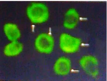
3.3. Immunocytochemistry
To see the presence of alfa5beta1 integrin on B16F10 cells the immunocyto-chemistry (Figure 3) clearly demonstrate the presence of alfa5beta1 on the sur-face of the B16F10 cells.
3.4. Effect of Black Tea Extract on ERK/PI-3K/NF-
κ
B
Figure 1. Cell Adhesion Assay: Cell adhesion assay of B16F10 cells treated with black tea extract for 24 hrs & 48 hrs ((a) Control and 20/40/80 μl/ml) and with theaflavin for 24 & 48 hrs; ((b) Control and 20/40/80 μg/ml) was performed following the method described in methods & materials.
Figure 2. Zymography: Zymography of cell culture medium (FBS free DMEM) of black tea extract ((a) Control—C, 24 hrs—1 and 48 hrs—2) and theaflavin ((b), Control—C, 24 hrs—1, 48 hrs—2) treated B16F10 cells was done following the method described in method section. (c) Effect of black tea extract & theaflavin (C—Control, 1—25 μl tea ex-tract, 2—50 μl tea exex-tract, 3—theaflavin 20 μg/ml) on salivary MMP following the method described in method section. (d) C—Control, 1—Fn-Cell interaction overnight, 2—Fn-cells (reacted with alfa 5 monoclonal antibody) interaction
Figure 3. Immunofluorescence: B16F10 cells were grown on slide. Blocked with 3% BSA, reacted with alfa5 monoclonal antibody followed by FITC coupled 2nd antibody. The fluorescence was observed under fluorescence microscope ×40. The arrow indicates the fluorescence on cell surface.
[image:6.595.280.468.537.678.2]Figure 4. Immunoblot: B16F10 cells were treated with theaflavin at 40 μg/ml conc. The control and treated cell extract (100 μg of protein) was run to develop immunoblot using antibodies against ERK 1/2 (a), PI-3K (b) and NF-κB (c) using method described in method section.
4. Discussion
MMPs especially MMP-2 is sitting in a pivotal position of cancer biology and should be looked at seriously. Role of MMP 2 and 9 has been studied and re-ported in development of cancer by numerous researchers. Our experimental observations are in agreement with the possibility that black tea (Darjeeling va-riety) extract and the polyphenol theaflavin have anti-cancer potential. Whole world is looking for synthetic or organic compound(s) playing role as efficient inhibitor of MMP-2 and or MMP-9. Here in a cup of black tea we have such compound (black tea polyphenols, theaflavin) which may have some long lasting medicinal effect on tissue microenvironment in general causing prevention of onset of the disease like cancer.
Acknowledgements
We acknowledge Vice Chancellor, Ramakrishna Mission Vivekananda Univer-sity, Natonal Tea Research Foundation for funding the project. Code No: NTRF 171/2014 started on June 6, 2014.
Conflict of Interest
None.References
[1] Wu, Y.Y., Li, W., Xu, Y., Jin, E.H. and Tu, Y.Y. (2011) Evaluation of the Antioxi-dant Effects of Four Main Theaflavin Derivatives through Chemiluminescence and DNA Damage Analyses. Journal of Zhejiang University Science B, 12, 744. https://doi.org/10.1631/jzus.B1100041
[2] Yang, C.S., Wang, X., Lu, G. and Picinich, S.C. (2009) Cancer Prevention by Tea: Animal Studies, Molecular Mechanisms and Human Relevance. Nature Reviews Cancer, 9, 429-439. https://doi.org/10.1038/nrc2641
[3] Balentine, D.A., Wiseman, S.A. and Bouwens, L.C. (1997) The Chemistry of Tea Flavonoids. Critical Reviews in Food Science & Nutrition, 37, 693-704.
https://doi.org/10.1080/10408399709527797
[4] Miller, N.J., Castelluccio, C., Tijburg, L. and Rice-Evans, C. (1996) The Antioxidant Properties of Theaflavins and Their Gallate Esters—Radical Scavengers or Metal Chelators? FEBS Letters, 392, 40-44. https://doi.org/10.1016/0014-5793(96)00780-6 [5] Sun, C.L., Yuan, J.M., Koh, W.P. and Mimi, C.Y. (2006) Green Tea, Black Tea and
Breast Cancer Risk: A Meta-Analysis of Epidemiological Studies. Carcinogenesis, 27, 1310-1315. https://doi.org/10.1093/carcin/bgi276
[6] George, J., Singh, M., Srivastava, A.K., Bhui, K., Roy, P., Chaturvedi, P.K. and Shukla, Y. (2011) Resveratrol and Black Tea Polyphenol Combination Synergisti-cally Suppress Mouse Skin Tumors Growth by Inhibition of Activated MAPKs and p53. PLoS ONE, 6, e23395. https://doi.org/10.1371/journal.pone.0023395
[7] Roy, P., Nigam, N., George, J., Srivastava, S. and Shukla, Y. (2009) Induction of Apoptosis by Tea Polyphenols Mediated through Mitochondrial Cell Death Path-way in Mouse Skin Tumors. Cancer Biology & Therapy, 8, 1281-1287.
https://doi.org/10.4161/cbt.8.13.8728
https://doi.org/10.1038/nrc745
[9] Nagase, H., Visse, R. and Murphy, G. (2006) Structure and Function of Matrix Met-alloproteinases and TIMPs. Cardiovascular Research, 69, 562-573.
https://doi.org/10.1016/j.cardiores.2005.12.002
[10] Strongin, A.Y., Collier, I., Bannikov, G., Marmer, B.L., Grant, G.A. and Goldberg, G.I. (1995) Mechanism of Cell Surface Activation of 72-kDa Type IV Collagenase Isolation of the Activated Form of the Membrane Metalloprotease. Journal of Bio-logical Chemistry, 270, 5331-5338. https://doi.org/10.1074/jbc.270.10.5331
[11] Banerji, A., Chakrabarti, J., Mitra, A. and Chatterjee, A. (2005) Cell Mem-brane-Associated MT1-MMP-Dependent Activation of pro-MMP-2 in A375 Mela-noma Cells. Journal of Environmental Pathology, Toxicology and Oncology, 24, 3-18. https://doi.org/10.1615/JEnvPathToxOncol.v24.i1.20
[12] Kamiya, N., Kishimoto, T., Suzuki, H., Sekita, N., Nagai, Y., Oosumi, N., Kito, H., Tochigi, N., Shinbo, M., Nemori, R. and Ichikawa, T. (2003) Increased in Situ Ge-latinolytic Activity in Renal Cell Tumor Tissues Correlates with Tumor Size, Grade and Vessel Invasion. International Journal of Cancer, 106, 480-485.
https://doi.org/10.1002/ijc.11272
[13] Tester, A.M., Waltham, M., Oh, S.J., Bae, S.N., Bills, M.M., Walker, E.C., Kern, F.G., Stetler-Stevenson, W.G., Lippman, M.E. and Thompson, E.W. (2004) Pro-Matrix Metalloproteinase-2 Transfection Increases Orthotopic Primary Growth and Ex-perimental Metastasis of MDA-MB-231 Human Breast Cancer Cells in Nude Mice.
Cancer Research, 64, 652-658. https://doi.org/10.1158/0008-5472.CAN-0384-2 [14] Yoon, S.O., Park, S.J., Yun, C.H. and Chung, A.S. (2003) Roles of Matrix
Metallo-proteinases in Tumor Metastasis and Angiogenesis. Journal of Biochemistry and Molecular Biology, 36, 128-137.
[15] Ann Beltz, L., Kay Bayer, D., Lynn Moss, A. and Mitchell Simet, I. (2006) Mecha-nisms of Cancer Prevention by Green and Black Tea Polyphenols. Anti-Cancer Agents in Medicinal Chemistry (Formerly Current Medicinal Chemistry-Anti- Cancer Agents), 6, 389-406.
[16] Sazuka, M., Imazawa, H., Shoji, Y., Mita, T., Hara, Y. and Isemura, M. (1997) Inhi-bition of Collagenases from Mouse Lung Carcinoma Cells by Green Tea Catechins and Black Tea Theaflavins. Bioscience, Biotechnology, and Biochemistry, 61, 1504- 1506. https://doi.org/10.1271/bbb.61.1504
[17] Sil, H., Sen, T., Moulik, S. and Chatterjee, A. (2010) Black Tea Polyphenol (Theafla-vin) Downregulates MMP-2 in Human Melanoma Cell Line A375 by Involving Multiple Regulatory Molecules. Journal of Environmental Pathology, Toxicology and Oncology, 29, 55-68.
https://doi.org/10.1615/JEnvironPatholToxicolOncol.v29.i1.80
[18] Herbst, R.S. (2004) Review of Epidermal Growth Factor Receptor Biology. Interna-tional Journal of Radiation Oncology*Biology*Physics, 59, S21-S26.
https://doi.org/10.1016/j.ijrobp.2003.11.041
[19] Olayioye, M.A., Neve, R.M., Lane, H.A. and Hynes, N.E. (2000) The ErbB Signaling Network: Receptor Heterodimerization in Development and Cancer. The EMBO Journal, 19, 3159-3167. https://doi.org/10.1093/emboj/19.13.3159
[20] Seshacharyulu, P., Ponnusamy, M.P., Haridas, D., Jain, M., Ganti, A.K. and Batra, S.K. (2012) Targeting the EGFR Signaling Pathway in Cancer Therapy. Expert Opinion on Therapeutic Targets, 16, 15-31.
https://doi.org/10.1517/14728222.2011.648617
Re-ceptor Downregulation. Molecular carcinogenesis, 45, 204-212. https://doi.org/10.1002/mc.20174
[22] Juliano, R.L. (2002) Signal Transduction by Cell Adhesion Receptors and the Cy-toskeleton: Functions of Integrins, Cadherins, Selectins, and Immunoglobulin-Su- perfamily Members. Annual Review of Pharmacology and Toxicology, 42, 283-323. https://doi.org/10.1146/annurev.pharmtox.42.090401.151133
[23] Jin, H. and Varner, J. (2004) Integrins: Roles in Cancer Development and as Treat-ment Targets. British Journal of Cancer, 90, 561-565.
https://doi.org/10.1038/sj.bjc.6601576
[24] Liotta, L.A., Nageswara Rao, C. and Wewer, U.M. (1986) Biochemical Interactions of Tumor Cells with the Basement Membrane. Annual Review of Biochemistry, 55, 1037-1057. https://doi.org/10.1146/annurev.bi.55.070186.005133
[25] Pasquier, E., Andre, N. and Braguer, D. (2007) Targeting Microtubules to inhIbit Angiogenesis and Disrupt Tumour Vasculature: Implications for Cancer Treatment.
Current Cancer Drug Targets, 7, 566-581. https://doi.org/10.2174/156800907781662266
[26] Chakrabarty, S., Das, A., Bhattacharya, A. and Chakrabarti, G. (2011) Theaflavins Depolymerize Microtubule Network through Tubulin Binding and Cause Apoptosis of Cervical Carcinoma HeLa Cells. Journal of Agricultural and Food Chemistry, 59, 2040-2048. https://doi.org/10.1021/jf104231b
[27] Sieg, D.J., Hauck, C.R., Ilic, D., Klingbeil, C.K., Schaefer, E., Damsky, C.H. and Schlaepfer, D.D. (2000) FAK Integrates Growth-Factor and Integrin Signals to Promote Cell Migration. Nature Cell Biology, 2, 249-256.
https://doi.org/10.1038/35010517
[28] Sieg, D.J., Hauck, C.R. and Schlaepfer, D.D. (1999) Required Role of Focal Adhesion Kinase (FAK) for Integrin-Stimulated Cell Migration. Journal of Cell Science, 112, 2677-2691.
[29] Weber, W.M., Hunsaker, L.A., Roybal, C.N., Bobrovnikova-Marjon, E.V., Abcou-wer, S.F., Royer, R.E., Deck, L.M. and Vander Jagt, D.L. (2006) Activation of NFκB Is Inhibited by Curcumin and Related Enones. Bioorganic & Medicinal Chemistry, 14, 2450-2461.
[30] Gosslau, A., Jao, E., Li, D., Huang, M.T., Ho, C.T., Evans, D., Rawson, N.E. and Chen, K.Y. (2011) Effects of the Black Tea Polyphenol Theaflavin-2 on Apoptotic and Inflammatory Pathways in Vitro and in Vivo. Molecular Nutrition & Food Re-search, 55, 198-208. https://doi.org/10.1002/mnfr.201000165
[31] Kim, S. and Joo, Y.E. (2011) Theaflavin Inhibits LPS-Induced IL-6, MCP-1, and ICAM-1 Expression in Bone Marrow-Derived Macrophages through the Blockade of NF-κB and MAPK Signaling Pathways. Chonnam Medical Journal, 47, 104-110. https://doi.org/10.4068/cmj.2011.47.2.104
[32] Khan, N., Afaq, F. and Mukhtar, H. (2008) Cancer Chemoprevention through Die-tary Antioxidants: Progress and Promise. Antioxidants & Redox Signaling, 10, 475- 510. https://doi.org/10.1089/ars.2007.1740
[33] Ukil, A., Maity, S. and Das, P.K. (2006) Protection from Experimental Colitis by Theaflavin-3,3’-Digallate Correlates with Inhibition of IKK and NF-κB Activation.
British journal of Pharmacology, 149, 121-131. https://doi.org/10.1038/sj.bjp.0706847
[35] Agarwal, M.L., Taylor, W.R., Chernov, M.V., Chernova, O.B. and Stark, G.R. (1998) The p53 Network. Journal of Biological Chemistry, 273, 1-4.
https://doi.org/10.1074/jbc.273.1.1
[36] Kalra, N., Seth, K., Prasad, S., Singh, M., Pant, A.B. and Shukla, Y. (2007) Theaflavins Induced Apoptosis of LNCaP Cells Is Mediated through Induction of p53, Down-Regulation of NF-Kappa B and Mitogen-Activated Protein Kinases Pathways. Life Sciences, 80, 2137-2146.
[37] Lahiry, L., Saha, B., Chakraborty, J., Bhattacharyya, S., Chattopadhyay, S., Banerjee, S., Choudhuri, T., Mandal, D., Bhattacharyya, A., Sa, G. and Das, T. (2008) Contri-bution of p53-Mediated Bax Transactivation in Theaflavin-Induced Mammary Epithelial Carcinoma Cell Apoptosis. Apoptosis, 13, 771-781.
https://doi.org/10.1007/s10495-008-0213-x
[38] Kundu, T., Dey, S., Roy, M., Siddiqi, M. and Bhattacharya, R.K. (2005) Induction of Apoptosis in Human Leukemia Cells by Black Tea and Its Polyphenol Theaflavin.
Cancer Letters, 230, 111-121. https://doi.org/10.1016/j.canlet.2004.12.035
[39] Pal, S., Kumar Ganguly, K., Moulik, S. and Chatterjee, A. (2012) Modulation of MMPs by Cell Surface Integrin Receptor α5β1. Anti-Cancer Agents in Medicinal Chemistry (Formerly Current Medicinal Chemistry-Anti-Cancer Agents), 12, 726- 732. https://doi.org/10.2174/187152012802650183
[40] Murugan, R.S., Vinothini, G., Hara, Y. and Nagini, S. (2009) Black Tea Polyphenols Target Matrix Metalloproteinases, RECK, Proangiogenic Molecules and Histone Deacetylase in a Rat Hepatocarcinogenesis Model. Anticancer Research, 29, 2301- 2305.
[41] Hou, Z., Lambert, J.D., Chin, K.V. and Yang, C.S. (2004) Effects of Tea Polyphenols on Signal Transduction Pathways Related to Cancer Chemoprevention. Mutation Research/Fundamental and Molecular Mechanisms of Mutagenesis, 555, 3-19.
Submit or recommend next manuscript to SCIRP and we will provide best service for you:
Accepting pre-submission inquiries through Email, Facebook, LinkedIn, Twitter, etc. A wide selection of journals (inclusive of 9 subjects, more than 200 journals)
Providing 24-hour high-quality service User-friendly online submission system Fair and swift peer-review system
Efficient typesetting and proofreading procedure
Display of the result of downloads and visits, as well as the number of cited articles Maximum dissemination of your research work