RESEARCH ARTICLE
A COMPARATIVE EVALUATION BETWEEN SNORERS AND NON SNORERS USING
CEPHALOMETRIC ANALYSIS OF SOFT TISSUES
1
Nilotpol Kashyap,
2,*Brij Kumar,
3Soumendu Bikash Maiti,
4Nilesh Bhanawat,
5Pallavi Pawar and
6Rashi Dubey
1Department of Pedodontics and Preventive Dentistry, Rungta College of Dental Sciences and Research,
Bhilai, Chhattisgarh, India
2Department of Pedodontics and Preventive Dentistry, Rungta College of Dental Sciences and Research,
Bhilai, Chhattisgarh, India
3Department of Oral Medicine and Radiology, Pacific Dental College and Research Center,
Udaipur, Rajasthan, India
4Oral Medicine and Radiology to Oral and Maxillofacial Surgery, Pacific Dental College and Research Center 5Department of Pedodontics and Preventive Dentistry, Rungta College of Dental Sciences and Research,
Bhilai, Chhattisgarh, India
6Department of Pedodontics and Preventive Dentistry, Rungta College of Dental Sciences and Research,
Bhilai, Chhattisgarh, India
ARTICLE INFO ABSTRACT
Introduction: Sleep-disordered breathing (SDB) is a collective clinical term encompassing primary
snoring, upper airway resistance syndrome (UARS), and obstructive sleep apnea (OSA). These syndromes currently are regarded to fall along a spectrum of severity concerning the same pathophysiological condition.
Material and method: The study was conducted on 60 participants with 18 males and 12 female
participants in study group and 16 males and 14 female participants among control group. Cephalogram and panoramic radiographs of each study individual were made using X MIND PANO D+ X ray unit and results were analyzed using Digora software. Four parameters (length, width of soft palate, distance to pharynx from maximum width and width of tongue) related to soft tissue were analyzed.
Results: Observations reveal that the length and width of the soft palate was more among snorers in
comparison to non snorers and was statistically significant. The posterior superior pharyngeal space was found to be less among snorers as compared to non snorers and was found to be statistically highly significant it was also found that the maximum width of tongue was slightly more among snorers as compared to non snorers and was found to be statistically non significant.
Conclusion: This method of detecting snoreres from non snorers may help in deriving definite
parameters to identify the risk and/or arriving at the diagnosis of snoring and its management, helping snorers and the sufferers to lead a better life and have a good sleep.
Copyright © 2018, Nilotpol Kashyap et al. This is an open access article distributed under the Creative Commons Attribution License, which permits unrestricted use, distribution, and reproduction in any medium, provided the original work is properly cited.
INTRODUCTION
In times bygone, snoring was regarded as a symbol of a great man and large snoring sound was considered to reflect deep sleep.
*Corresponding author: Brij Kumar
Department of Pedodontics and Preventive Dentistry, Rungta College of Dental Sciences and Research,
Bhilai, Chhattisgarh, India
As a matter of fact, is associated with two problems
The first problem is that loud snoring produces noise during night when it should be naturally quiet. The person himself is usually unaware of his snoring, but his snoring disturbs the sleep of others. The second problem is that loud every night snoring is suspected to be associated with sleep apnea syndrome (SAS). In other words, snoring is an inevitable symptom of sleep apnea syndrome (SAS).
ISSN: 0975-833X
International Journal of Current Research
Vol. 10, Issue, 04, pp.67691-67695, April, 2018
INTERNATIONAL JOURNAL OF CURRENT RESEARCH
Article History:
Received 09th January, 2018
Received in revised form 21st February, 2018
Accepted 23rd March, 2018
Published online 30th April, 2018
Citation: Nilotpol Kashyap, Brij Kumar, Soumendu Bikash Maiti, Nilesh Bhanawat, Pallavi Pawar and Rashi Dubey, 2018. “A comparative evaluation
between snorers and non snorers using cephalometric analysis of soft tissues”, International Journal of Current Research, 10, (04), 67691-67695.
Key words:
(Tadao, 2003) Snorers can also be divided into occasional snorers and habitual snorers (who snore always every night) (Ramanathan and Revathi, 2005). Snoring is part of the spectrum of sleep disordered breathing (SDB), from obstructive sleep apnoea/ hypopnoea syndrome (OSAHS) at one end to simple snoring at the other. The entire spectrum is characterized by changes in the physical conformation, structural properties and neuromuscular function of the pharynx. (Paul and Janet, 2008). Habitual snoring has been associated with arterial hypertension and angina pectoris. Snoring has also been suggested as a risk factor for brain infarction. Heavy snoring is almost always present in obstructive sleep apnea, and obstructive sleep apnea syndrome has many harmful effects on the cardiovascular system.5 Soft
palate is a soft tissue flap-like structure hinged to the back of the hard palate with a free posterior margin. Although it is considered as a part of roof of oral cavity, it is also related to pharynx. It is a type of ‘flutter valve’ that swings up (elevate) to close the pharyngeal isthmus (Drake et al., 2005). It can swing down (depress) to close the oropharyngeal isthmus and seal off the oral cavity from oropharynx. Snoring is the vibration involving soft palate and the following sound, while obstructive sleep apnea (OSA) is partial or complete obstruction of airway usually also at soft palate level. Lateral cephalometry is a simple, well-standardized, inexpensive and readily accessible method of screening associated with low radiation exposure. This method has commonly been used from early fifties in order to assess the growth of dentofacial skeleton, nevertheless it has been used in patients with Sleep Disordered Breathing (SDB) since 1983 (Eugene and Renaud, 2010; Hudgel, 1988). Hence the present study was designed to evaluate the structural anatomy of the pharynx and tongue of the study volunteers comprising known snorers and non-snorers using lateral cephalogram and panoramic radiographs.
Aim and Objective: To evaluate soft tissues anatomy of the
pharynx among habitual snorers and non snorers using lateral cephalogram and panoramic radiograph.
MATERIALS AND METHODS
This cross sectional study was conducted in the patients visiting the out patients department of the institution. Patients with known history of regular snoring with history of snoring revealed by the patient based on questionnaire were included in the study. Patients with the following criteria were excluded from the study. Individuals who could not give a written consent for exposure to X- rays, or those in whom X-ray exposure would be more riskier, Patients with history of injury or environmental factors known to cause craniofacial deformity, Patients undergoing or previously undergone orthodontic/ orthognathic surgery, Presence of gross facial asymmetry, History of surgical intervention involving craniofacial skeleton and soft tissue surgery of head and neck region, History and / or clinical features suggestive of endocrine disturbances, neuromuscular disorder, hereditary, nutritional or developmental or any prolong illness affecting oropharynx and Patients with completely edentulous arch. After understanding aims and objectives and pros and cons of the study a total of 80 individuals volunteered to participate in study. Among these only 60 individuals were selected for completing the study based on inclusion and exclusion criteria. These 60 individuals were grouped into snorers and non snorers based on their answers for the study questionnaire related to snoring.
Among the study group- 18 were males and 12 were females and among control group- 16 were males and 14 were females. All the study participants were distributed equally into 3 age groups- 20-30 years, 31-40 years, 41-50 years.
Standardization of Radiographs
Cephalogram and panoramic radiographs of each study individual were made using X MIND PANO D + X ray unit. The study participants were instructed to stand maintaining the natural head position for lateral cephalograms (Figure 1).
Figure 1. X Mind Pano D+ Xray Unit
Exposure Parameters
All the exposures were made at 75kvp, 10mAs using scanography technique. A second radiograph was made to produce to optimum images of soft tissues and hard tissues with necessary correction in exposure parameters. Radiographs with optimum contrast and less noice were considered for study. Necessary radiation protection was adopted while taking radiographs.
Method of Analyzing the Radiograph
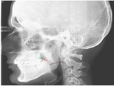
[image:2.595.349.518.180.316.2]Present study was planned to evaluate the soft tissue anatomy influencing the pharyngeal space among the study participants. All images were stored digitally and the image quality was optimized separately for soft tissue and hard tissue landmarks using the inbuilt software (Digora for windows). Soft tissues analyzed in the present study were soft palate, anterior and posterior wall of pharynx and tongue. Parameters related to soft palate analyzed using lateral cephalogram, were, it’s length, width, it’s distance to anterior and posterior wall of pharynx and to the tongue. Soft Tissue Landmarks On Lateral Cephalogram (Figure 2 & 3)
Figure 2. Black line represents length of soft palate, green line represents width of soft palate at junction, blue line represents maximum width of soft palate, yellow line represents distance to
Figure 3. Green line represents distance between tongue at soft palate at junction, red line represents distance between tongue and soft palate at maximum width and pink line represents distance between tongue and soft palate at tip and blue line
represents distance between tongue and pharynx
Length of Soft Palate: The length of the soft palate
was measured from junction of hard and soft palate till the distal most point (POINT P) using multiple points for measuring the curvature.
Maximum Width of Soft Palate (W): Maximum
width of soft palate was measured between two points; point on the soft palate facing superior pharyngeal surface and point on the soft palate towards the oral cavity.
Distance of Pharynx from Maximum Width of Soft Palate
Width of the Tongue
RESULTS
Present study was planned to evaluate the soft tissue status influencing the pharyngeal space among the study participants, to evaluate the difference in anatomy of these structures among known snorers and non snorers. After understanding aims and objectives and pros and cons of the study a total of 280 individuals volunteered to participate in study. Among these only 60 individuals were selected for completing the study based on inclusion and exclusion criteria. These 60 individuals were grouped into snorers and non snorers based on their answers for the study questionnaire related to snoring. Among the study group- 18 were males and 12 were females and among control group- 16 were males and 14 were females Among study group population, the minimum and maximum known history of snoring was 5 and 20 years respectively. It was also observed that overall physical appearance of study group population was suggestive of obese built as compared to control group, but for 8 individuals who were non-obese, whereas 5 individuals in control group were having physical appearance suggestive of obesity.
Among the study group minimum age was 20, maximum age was 50 and they were equally distributed into 3 age groups i.e. 20-30 years, 31-40 years, and 41-50 years. Similarly among the study group minimum age was 20, maximum age was 50 and they were equally distributed into 3 age groups i.e. 20-30 years, 31-40 years, and 41-50 years. Cephalogram and panoramic radiographs of each study individual were made using X MIND PANO D + x ray unit. The study participants were instructed to stand maintaining the natural head position for lateral cephalograms. All the exposures were made at 75 kvp, 10 mAs using scanography technique. A second radiograph was made to produce to optimum images of soft tissues and hard tissues with necessary correction in exposure parameters. All images were stored digitally and the image quality was optimized separately for soft tissue and hard tissue landmarks using the inbuilt software (Digora for Windows). Soft tissues analyzed in the present study were soft palate, anterior and posterior wall of pharynx and tongue. Following observations were made in soft tissues parameters among snorers and non snorers.
Soft Tissue Analysis
Length of the Soft Palate
The length of the soft palate was measured from junction of hard and soft palate till the distal most point using multiple points for measuring the curvature. Minimum length measured in the entire study population was 24.42 mm and maximum length was 53.84 mm, with a mean value of 39.11 mm. Among snorers, the minimum and maximum length of the soft palate was 33.17 mm and 53.84 mm respectively with a mean value of 43.64 mm and it was found to be statistically significant (p < .001), whereas among non snorers minimum and maximum length was 24.42 mm and 44.56 mm respectively with a mean value of 34.58 mm (Table 1, Graph 1). The above observations reveal that the length of the soft palate is more (mean= +9.06 mm) among snorers in comparison to non snorers.
[image:3.595.89.511.693.781.2]Graph 1. Graph Showing Maximum, Minimum and Mean Values of Soft Palate Length, Among Snorers and Non Snorers
Table 1. Showing Mean Values, Standard Deviation and P-Values of Soft Tissue Landmarks
Parameter Group N Mean Std Deviation t-value p-value
Length of soft palate Snorer
Non snorer
30 30
43.64 34.58
4.78 4.003
7.966 .000
Maximum width of soft palate Snorer
Non snorer
30 30
12.0450 9.9720
1.13 1.21
6.84 .000
Distance to pharynx from max width Snorer Non snorer
30 30
11.75 15.68
1.73 1.81
-8.58 .000
Maximum width of tongue Snorer
Non snorer
30 30
228.18 226.31
6.71 10.91
Width of the Soft Palate
Minimum width of the soft palate among the entire study population was 8.03 mm and maximum was 15.36 mm, with a mean value of 11mm. Among snorers minimum and maximum width was 10.46 mm and 15.36 mm respectively with a mean of 12.04 mm whereas among non snorers minimum and maximum width was 8.03 mm and 12.47 mm respectively with a mean value of 9.97 mm (Table 1, Graph 2). The above observations revealed that the maximum width of soft palate was more (mean = + 2.07 mm) among snorers as compared to non snorers. The above values were subjected to paired ‘t’ test and was found to be statistically highly significant (p < .001) (Table 1).
Distance of Pharynx from Maximum Width of Soft Palate
Minimum distance of pharynx from maximum width of soft palate among the entire study population was 8.76 mm and maximum distance was 19.09 mm with a mean value of 13.72 mm. The distance of pharynx from maximum width of soft palate was compared with the maximum width of the soft palate to analyze their co-relationship and was found that out of 60 individuals, the distance to pharynx was in direct co-relationship with maximum width of 43 individuals. In rest of 17 individuals, the maximum width of soft palate was varying without any co-relationship. Among snorers minimum and maximum distance to pharynx from maximum width was 8.76 mm and 16.83 mm respectively with a mean of 11.75 mm whereas among non snorers minimum and maximum distance was 12.32 mm and 19.09 mm respectively with a mean value of 15.68 mm (Table 1, Graph 3). The above observations revealed that the posterior superior pharyngeal space to be less (-3.93 mm) among snorers as compared to non snorers. The above values were subjected to paired ‘t’ test and was found to be statistically highly significant (p < .001) (Table 1).
Graph 2. Graph Showing Maximum, Minimum And Mean Values Of Width Of Soft Palate, Among Snorers and Non Snorers
Graph 3. Graph Showing maximum, Minimum And Mean Values Of Distance Of Pharynx From Soft Palate Among Snorers and
Width of the Tongue
Minimum width of the tongue, among entire study population was 207.41 mm and maximum width was 244.51 mm with a mean value of 227.24 mm. Among snorers minimum and maximum value was 215.37 mm and 240.84 mm respectively with a mean value of 228.18 mm whereas among non snorers minimum and maximum values were 207.41 mm and 244.51 mm respectively with a mean value of 226.31 mm (Table 1, Graph4). The above observations revealed that the maximum width of tongue to be slightly more (mean =+ 1.87 mm) among snorers as compared to non snorers. The above values were subjected to paired ‘t’ test and was found to be statistically non significant (p < .05) (Table 1).
Graph 4. Bar Graph Showing width of Tongue among Snorers and Non Snorers
DISCUSSION
The present study was about evaluation of the pharyngeal anatomy and the structures influencing the same among snorers and non snorers. Sleep-disordered breathing (SDB) is a collective clinical term encompassing primary snoring, upper airway resistance syndrome (UARS), and obstructive sleep apnea (OSA). These syndromes currently are regarded to fall along a spectrum of severity concerning the same pathophysiological condition (Hudgel and Hendricks, 1988). The pathogenesis of snoring is vibrating tissues accompanied by increased collapsibility and incomplete pharyngeal obstruction or narrowing of the pharyngeal airway which is same for obstructive sleep apnea syndrome which has been related to increased upper airway collapsibility and reduction of upper airway size, alterations in craniofacial structure and enlargement of surrounding soft tissue structures (i.e., tongue and lateral pharyngeal walls (Moses, 2008). Habitual snoring is harmful as it heralds the development of OSAHS. It may progress over the course of years. Owing to obstructed breathing there may be episodes of oxygen desaturation in sleep which is harmful. Sleep disorders are being increasingly recognized in clinical practice as they can result in organic disorders. For example, obstructive sleep apnoea is a risk factor for cardiovascular disorders as well as cognitive disorders (Ramanathan and Revathi, 2005; Andreou et al., ?). Habitual snoring has been associated with arterial hypertension and angina pectoris. Snoring has also been suggested as a risk factor for brain infarction (Eugene and Renaud, 2010). In the
decibels), and severe (>60 decibels) and severity of OSA was graded as no OSA (AHI < 5), mild (AHI 5 to 15), moderate (AHI 15 to 30), severe (AHI 30 to 50), and very severe OSA (AHI > 50). They concluded that the intensity of snoring increases as OSA becomes more severe (Maimon and Hanly, 2010). In the present study length and width of the soft palate was found to be significantly more among snorers (p- value= 000.000). Intensity of the snoring had a direct co-relationship with length of the soft palate. It was also observed the height of the study participants had direct co-relationship with length of the soft palate however it was statistically more significant among snorers whereas the width of the soft palate had direct co-relation with obesity of the study participants, that in turn was significantly more among the snorers. Distance to pharynx was significantly less among snorers in the region of oropharynx and hypopharynx. Among snorers a direct co-relation was found between length of the soft palate and its distance to pharynx from its tip. In the present study it was also observed that length of the soft palate was in direct co-relationship with loudness of snoring. This could be due to longer the soft palate greater could be the resonance of sound of snore. Further in the present study length of the soft palate also influenced the distance between the tip of soft palate and posterior wall of pharynx which was less among snorers (n = 22).
Distance between tongue and posterior pharyngeal wall was measured at the level of tip of soft palate which was found to be less among snorers (mean = - 4.94 mm). Further in the present study length of the soft palate also influenced the distance between the tip of soft palate and posterior wall of pharynx which was less among snorers (n = 22). Studies done by Maltais et al, Kurt et al, Miyao et al confirmed similar findings (Maltais et al., 1991; Kurt et al., 2011; Miyao et al., 2000). The fact is further confirmed in the studies conducted by Fujita et al, Kamani et al where after surgical reduction of length of soft palate, reduction in snoring has been reported. A surgical relief of snoring has been reported when stiffening of soft palate was done with help of laser by Ellis et al (Fujita et al., 1981). Distance between tongue and posterior pharyngeal wall was measured at the level of tip of soft palate which was found to be less among snorers (mean = - 4.94 mm). In the present study it was also observed that length of the soft palate was in direct co-relationship with loudness of snoring. This could be due to longer the soft palate greater could be the resonance of sound of snore
CONCLUSION
Observation of the present study indicate the advantage of using simple and affordable images of lateral cephalogram and panoramic radiograph in identifying the anatomical differences of pharynx and size of the tongue among snorers as compared
to non snorers of the study population. Analysis of the same in large sample involving different population may help in deriving definite parameters to identify the risk and/or arriving at the diagnosis of snoring and its management, helping snorers and the sufferers to lead a better life and have a good sleep.
REFERENCES
Andreou, G., Vlachos, F. and Makanikas, K. Neurosognitive Disorder. Neurocognitive Deficits in Patients with Obstructive Sleep Apnea Syndrome (OSAS). In Tech. Greece.
Drake, LR., Wayne, V. and Mitchell, MW. 2005. Gray’s Anatomy For Students. China: Elsevier Inc;
Eugene, L. and Renaud, M. 2010. Snoring-Causes, Diagnosis and Treatment. New York: Nova Science Publisher; Fujita, S., Conway, W. and Zorick, F. 1981. Surgical
correction ofanatomic abnormalities in obstructive sleep apnea syndrome: Uvulopalatopharyngoplasty. Otolaryngol
Head Neck Surg., 89 : 923-34.
Hudgel, DW. and Hendricks, C. 1988. Palate and hypopharynx--sites of inspiratory narrowing of the upper airway during sleep. 138 (6): 1542-1547.
Kurt G, Sisman C, Akin E, Akcam T. Cephalometric measurements of pharyngeal airway in snoring and non snoring patients. European journal of dentistry, 2011; 5: 84-88.
Maimon, N. and Hanly, PJ. 2010. Does Snoring Intensity Correlate With The Severity of Obstructive Sleep Apnea?. Ijsm. Journal of clinical sleep medicine, 6(5): 475-478. Maltais, F., Carrier, G. and Cormier, Y. 1991. Cephalometric
measurements in snorers, non snorers and in patients with sleep apnea. Thorax, 46: 419-423.
Miyao, E., Miyao, M., Ohtat Okawa, M., Inafuku, S., Nakayama, M. and Goto, S. 2000. Differential diagnosis of obstructive sleep apnea patients and snorers using lateral cephalograms. Psychiatry and clinical neurosciences, 54: 659-654.
Moses, A. 2008. Protocol for Primary Treatment of Snoring by Dentists. Sleep diagnosis and therapy. 3(6): 21-22.
Palomaki, H., Marrku, P., Seppo, J. and Marrku, K. 1989. Snoring as a risk factor for sleep-related brain infarction. Stroke. 20 (10).
Paul, C. and Janet, W. 2008. The management of simple snoring. Sleep medicine reviews. 8: 433-441.
Ramanathan, I. and Revathi, I. 2005. Snoring-harmful effects.www.apiindia.org
Tadao, N. 2003. Treatment of snoring. JMAJ., 46(3): 133-138.