Original Article
Abnormal baseline brain activity in patients with
cancer-induced bone pain: a resting-state
functional magnetic resonance imaging study
Qian Liao1,Rong Hu1,2, Dong Huang1, XuebinYan1, Gangwen Guo1, Qin Liao1,2, Xin Li1
1Pain Management, Third Xiangya Hospital, Pain Research Institution of Central South University, Central South
University, Changsha, Hunan Province, China; 2Anesthesiology, Third Xiangya Hospital, Central South University,
Changsha, Hunan Province, China
Received October 12, 2015; Accepted June 8, 2016; Epub July 15, 2016; Published July 30, 2016
Abstract:Objective: To investigate the alternations of brain activities in patients with cancer-induced bone pain at resting state. Methods: The present study uses resting-state functional magnetic resonance imaging (fMRI) to ex-plore functional brain activities in 12 lung cancer patients with cancer-induced bone pain, 12 lung cancer patients without cancer-induced bone pain, and 12 healthy controls. All of the lung cancer patients were in stage M1 at be-ginning of the study. The amplitude of low-frequency fluctuations (ALFF) was analyzed on the whole brain in all sub -jects. Results: There was significantly ALFF in the anterior cingulated cortex (ACC) and the medial prefrontal cortex (mPFC) in patients with cancer-induced bone pain in comparison with patients without cancer-induced bone pain. In addition, when compared with healthy controls, the patients with cancer-induced bone pain and patients without cancer-induced bone pain both showed altered activities, mainly in the posterior cingulate cortex (PCC). There was a significantly positive correlation between the visual analogue scale (VAS) and ALFF in the mPFC in patients with cancer-induced bone pain. Conclusion: Patients with cancer-induced bone pain displayed abnormal variations in baseline brain activity in some brain regions related to prefrontal-limbic network based on ALFF. All abnormalities in the brain regions may be viewed as a key marker and meanwhile could add insight into the judgment of the thera-peutic effect of pain in patients with cancer-induced bone pain in future.
Keywords: Resting-state, amplitude of low-frequency fluctuations, cancer-induced bone pain, functional magnetic resonance imaging
Introduction
For people with bone metastasis, the ongoing
pain is the most common symptom and can be excruciating. 30-40% of patients present the symptom of pain during early stages of the dis-ease, and the number rises to 70-90% in advanced stages of the disease [1]. Patients with pain resulting from bone metastasis are highly maladaptive. Some studies have report-ed that severe pain of patients can result in anxiety and/or depression, which ultimately alters physiological processes [2-4]. The under-lying brain mechanisms of pain in patients with cancer-induced bone pain are largely unknown.
A neuroimaging approach will be utilized to gain
a further understanding on this subject. During the last few decades, the pathophysio-logical mechanisms of pain have been
measured through changes in blood flow what
is referred as blood-oxygen-evel-dependent signal [16]. As already reported, relying on method of resting state analysis a number of brain networks have been discovered. These resting state networks consist of the human brain’s intrinsic functional networks [16, 17]. Detecting abnormal brain activities may be considered a marker to reveal the progress state of diseases. The brain’s intrinsic
function-al architecture has been widely visufunction-alized and
measured through the resting-state fMRI in patients with several different medical
disor-ders, for instance depression [18], Alzheimer [19], and Schizophrenia [20]. However, until
now, rarely studies had identified the brain
activities of resting-state networks in patients with cancer-induced bone pain.
In our study, we utilizedALFF to study the alter -nations of brain activities in patients with can-cer-induced bone pain at resting state. These abnormalities may serve as a key marker and could add insight into the therapeutic effect of pain in patients with cancer-induced bone pain
in future. We hypothesized that an abnormal ALFF is found at resting state in certain brain
areas in patients with cancer-induced bone pain in comparison to patients without cancer-induced bone pain.
Materials and methods
Central South University (CSU) Institutional Review Board committee approved all the human fMRI experiment in the study. All partici-pants gave informed consent according to guidelines set by MRI Center of Central South University.
Participants
A total of 36 participants, including 24 cancer patients and 12 healthy subjects, participated in this study. Cancer patients had been clini-cally diagnosed with stage M1 lung cancer. The informed written consent was taken. Twelve right-handed patients with cancer-induced bone pain, twelve right-handed patients with-out cancer-induced bone pain, and twelve right-handed healthy subjects (control) from the Third Xiangya Hospital participated in this study. Exclusion criteria included tumor metas-tasis into the brain, other chronic pain condi-tions, history of head injury, head trauma, epi-lepsy and clinical depression, which excluded potential participants via the use of the Beck’s
sics agents were instructed to stop taking these at least 7 days before the start of experiment. Before the functional scan, rate the
spontane-ous fluctuations of bone metastasis pain, by
evaluating spontaneous fluctuations in the
intensity of cancer patients’ ongoing pain with
a visual analogue scale (VAS) that ranged from 0 to 10, with zero representing no pain and 10
representing maximum pain. Subjects selected words that most closely described painful stim-uli using Short McGill Pain Questionnaires.
Imaging protocol
A 1.5-T (Siemens; Tokyo, Japan) MRI scanner at the Third Xiangya Hospital in Hunan, China, was used to obtain images, and an Echo planar imaging (EPI) sequence was used to collect
functional images. For every participant, 210
images were obtained using following imaging parameters: echo time = 40 ms; repetition time
= 2000 ms; slice = 26; slice gap = 1 mm; Flip Angle = 90°; voxel size = 4 × 4 × 5 mm3; matrix
size = 64 × 64. A high-resolution, three-dimen -sional T1-weighted structural image was also
scanned with following parameters: Flip Angle
=15°; echo time = 2.92 ms; repetition time =
1900 ms; voxel size= 1 × 1 × 1 mm3; slice =
176; matrix size = 240 × 240. During 30 minute scanning, all participants were asked to keep their eyes closed, remain awake, and rest with-out thinking abwith-out anything in particular.
Data processing and analysis
SPM8 (http://www.fil.ion.ucl.ac.uk14k),
Stati-stical Parametric Mapping was used for carry-ing out imagcarry-ing preprocesscarry-ing. We discarded the beginning 10 volumes to eliminate any
non-equilibrium effects of magnetization. Then, the
resting 200 time points were corrected for acquisition delay between slices and realigned
to the first image for head motion correction.
No participants were excluded due to head motions exceeding 2 mm or 2° of rotation in any direction during fMRI scanning [21]. The
functional images were spatially normalized to
the standard SPM8 Montreal Neurological Institute (MNI) template, re-sampled to 3 mm cubic voxels, and smoothed by a Gaussian ker-nel with the full width set at a half maximum of 8 mm.
ALFF calculation
REST software (http://resting-fmri.sourceforge.
cessing. Primarily, the linear trend must be
removed and every voxel was band-pass fil
-tered (0.01~0.08 Hz) to remove the effects of
low-frequency drift and high-frequency noise. The interference of head motion was then elim-inated using linear regression, but white matter
and cerebral cerebrospinal fluid (CSF) were not regressed out. The procedure for the ALFF cal
-culation is as follows: 1) The filtered time series
was transformed to a frequency domain with a
fast Fourier transform; 2) After obtaining power
spectrum, square root was calculated at each frequency of the power spectrum and then
across 0.01 Hz to 0.08 Hz each voxel the aver -aged square root was aver-aged to calculate the
ALFF; 3) For standardization purposes, the
mean was subtracted and divided by global standard deviation to convert the resulting
ALFF into z-scores [22].
Statistical analysis
For the comparison among groups, a two-sam
-ple t-test was performed to examine ALFF dif
-ferences amongst: (1) cancer patients with bone metastasis pain, (2) cancer patients with-out bone metastasis pain patients, (3) healthy
volunteers. Significant clusters (cluster size ≥
432 mm3, 16 voxels) of activation for the two sample t- tests were determined by using a (P <
0.05) FDR corrected threshold. Using the Monte
Carlo simulations, we determined this correc-tion on REST software.
Correlation analysis
An analysis of partial correlation between ALFF values and visual analogue scale (VAS) out -comes was performed to give a test in
correla-tion of ALFF abnormality and intensity of pain in
patients with bone metastasis pain. It showed
the most significant differences between the
bone metastasis pain group and the non-bone metastasis pain group. Anatomical Automatic
[image:3.612.94.522.74.354.2]Labeling atlas toolbox [23] defined the intersec -tion of the corresponding region, which means each of the clusters and the within group two sample t-test map allowing for threshold set at P = 0.05.
Results
Generic parameters
Based on clinical and demographic characteris-tics of all subjiects, gender and age (t = 1.297,
P = 0.186; t = 1.712, P = 0.240) had not signifi
-cant differences among three groups. For VAS
(t = 9.268, P < 0.001), scores of patients with
cancer-induced bone pain were significantly
higher than those of cancer patients without cancer-induced bone pain.
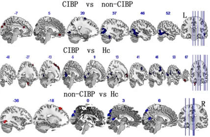
Brain activity for spontaneous pain
Patients with cancer-induced bone pain (CIBP) versus patients without cancer-induced bone pain (non-CIBP): Compared with CIBP, non-CIBP
showed a decreased ALFF in right middle occip -ital gyrus, the right middle temporal gyrus, right superior frontal gyrus and right lingual gyrus.
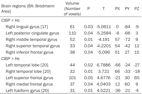
be significantly lower in CIBP than in Hc in infe -rior frontal gyrus, right middle temporal gyrus, the left posterior cingulate gyrus, right lingual gyrus and right superior temporal gyrus.
Patients without cancer-induced bone pain (non-CIBP) versus healthy controls (Hc): As shown in Figure 1 and Table 3, the group of (Hc)
demonstrated a decreased ALFF in the right
precuneus, the left cuneus and right posterior cingulate gyrus in comparison with healthy vol-unteers. Conversely, left occipital lobe and the left superior frontal gyrus demonstrated an
increased ALFF in non-CIBP.
Statistical correlations between VAS and ALFF values
[image:4.612.91.520.85.227.2]The statistical correlations between VAS and ALFF values in brain regions, including mPFC
Table 1. Showing altered ALFF of brain regions between CIBP and non-CIBP
Brain regions [BA: Brodmann Area] (Number of voxels)Volume P T PX PY PZ CIBP < non-CIBP
Right middle temporal gyrus/right middle occipital gyrus 400 0.04 -6.1739 42 -63 12
Right lingual gyrus [17] 23 0.01 -3.5875 18 -87 0
Right superior frontal gyrus [6] 17 0.01 -4.8251 24 -3 69
CIBP > non-CIBP
Right medial frontal gyrus 15 0.01 4.0087 3 42 -18
Anterior cingulate gyrus [24] 16 0.02 4.9619 -6 24 21
Left cuneus [19] 20 0.03 4.0773 -6 -90 27
Left precuneus 17 0.01 3.961 -9 -81 42
Table 2. Showing altered ALFF of brain regions between CIBP and
Hc
Brain regions [BA: Brodmann Area]
Volume (Number
of voxels) P T PX PY PZ CIBP < Hc
Right lingual gyrus [17] 61 0.03 -5.0611 0 -84 -9 Left posterior cingulate gyrus 110 0.04 -5.2584 -6 -66 3 Right middle temporal gyrus 52 0.01 -4.191 57 -72 9 Right superior temporal gyrus 33 0.04 -4.2201 54 -42 12 Right inferior frontal gyrus 38 0.04 -5.096 51 27 15 CIBP > Hc
Left temporal lobe [20] 44 0.02 6.7886 -66 -24 -27 Right temporal lobe [20] 32 0.01 3.721 66 -33 -18 Left superior frontal gyrus 101 0.05 4.6776 -21 30 60 Right medial frontal gyrus 37 0.04 4.0403 12 60 9 Left fusiform gyrus [20] 31 0.01 4.5221 -36 -21 -4
right medial frontal gyrus, left cuneus, anterior cingulated gyrus and precuneus. In Fi- gure 1 and Table 1, specific
contents including clusters and Montreal Neurological Institute (MNI) coordinates are shown.
Patients with cancer-induced bone pain (CIBP) versus hea- lthy controls (Hc): In Figure 1
and Table 2, the group differ-ences between CIBP and Hc
are shown. The ALFF in CIBP
was higher than in Hc in the left superior frontal gyrus, temporal lobe, right medial frontal gyrus and fusiform
[image:4.612.90.382.271.465.2](CIBP verse. non-CIBP) were examined. A
sig-nificant statistical correlation was found only between VAS and ALFF values in mPFC (r =
0.771, P = 0.003).
Discussion
The goal of this study was to investigate the alterations in resting-state brain activities in patients with bone metastasis pain using fMRI,
and we found an increased ALFF the right medi -al front-al gyrus, left cuneus, anterior cingulated gyrus and precuneus in patients with cancer-induced bone pain when compared with patients without cancer-induced bone pain.
Inversely, a decreased ALFF (CIBP < non-CIBP)
was observed in right middle occipital gyrus, the right middle temporal gyrus, right superior frontal gyrus and right lingual gyrus. When compared with healthy controls, the patients with cancer-induced bone pain and patients without cancer-induced bone pain both dis-played changed activities, mainly in the poste-rior cingulate gyrus. In addition to this, there
was a significant positive correlation between the VAS and ALFF in the mPFC in patients with
cancer-induced bone pain.
The frontal cortex is one of the most prominent areas associated with abnormal brain activi-ties in chronic pain. Multiple factors can
con-tribute to sustained mPFC activity after decre -ment of the nociceptive elicitor [24]. Previous
studies have suggested that the mPFC may play a specific role in mediating the attenuation
of pain perception via cognitive control
mecha-nisms and that the mPFC is involved in response conflict, detection of unfavorable outcomes
and emotions [24, 25]. Therefore, cognitive and mental disorders may arise from abnormal
correlation between VAS and ALFF values w
ith-in mPFC. In lith-ine with our results, hyperactivity of mPFC in association with pain has been
determined by previous studies [26-29], which may be considered as a critical trait marker for chronic pain. Prior study has already
demon-strated an increase in the firing activities of mPFC neurons during the delay period in
delayed-choice task [30], which suggests that working memory processing may rely heavily on
increased activitie of mPFC in the experiment.
This is in accordance with suggestion that
mPFC instability may play a crucial role in
pain-induced memory dysfunction. Cross-sectional
fMRI studies indicate that mPFC, encoding fluc -tuations of ongoing pain in multitudinous chron-ic pain syndromes, is prior to take part in the brain’s emotion and limbic circuitry [31]. Particularly, in chronic back pain, spontaneous
pain primarily activates the mPFC [24]. Frank
Seifert et al. [32] found that during
hyperalge-sia, activity increase was found in mPFC and posterior cingulate gyrus (PCC). Activity in mPFC
correlates inversely with the individual’s extent of central hyperalgesia and predicts the in-dividual’s pharmacological antihyperalgesic treatment response. With the help of these results, it can be speculated that hyperactivity
in mPFC may be a principal element of the
development and progression of cancer-induced bone pain.
Pain perception is also modulated through
important cerebral influences on the limbic sys -tem. ACC is an important limbic system area for pain-related perception. In this study, we have shown that activity is increased within the ACC in patients with cancer-induced bone pain. Previous research has demonstrated that the ACC results of acute pain in healthy
partici-Table 3. Showing altered ALFF of brain regions between non-CIBP
pain and Hc
Brain regions [BA: Brodmann Area]
Volume (Number
of voxels) P T PX PY PZ Non-CIBP < Hc
Right posterior cingulate gyrus 19 0.02 -4.7369 6 -63 12 Right precuneus 32 0.01 -6.4373 3 -78 48
Left cuneus 26 0.01 -4.8602 0 -87 30
Non-CIBP > Hc
Left superior frontal gyrus 26 0.02 5.1674 -18 21 60 Left occipital lobe 15 0.03 4.7132 -36 -72 -6
activity in the mPFC. This
abnormal activity may partly contribute to cognitive de- cline and a lack of interest or pleasure in patients with chronic pain. Using resting-state fMRI, the current research perceived an in-
creased ALFF in the mPFC
[image:5.612.91.386.96.224.2]pants versus participants with chronic pain are
significantly different [33]. Involvement of the
ACC, one of the most commonly studied regions regarding acute pain and chronic pain, is highly prominent in pathological pain. Acute nocicep-tive responses in patients with lesions of the
ACC were significantly increased [34], and stud -ies based on human beings manifest that ACC activity is involved in social exclusion due to pain, the empathy of pain, chronic migraine and
hypothesized pain [35, 36]. A meta-analysis on
the structure and function of the ACC indicated that ACC activations found in neuroimaging
studies of pain predominantly reflect affective
pain processing [37]. The suppression of affec-tive responses to a negaaffec-tive stimulus was asso-ciated with the activity of the ACC. It is true that the ACC, which plays a crucial role in emotion generation and regulation, is connected with
the mPFC, the amygdale, and the hippocam -pus. Thus, the increased activity in ACC in patients with cancer-induced bone pain may
give rise to an imbalance of mPFC-activated influence on activities leading to affective pain.
Our data, however, cannot declare that the observed increase of activity levels in the ACC is due to the variational relevance between ACC
and mPFC. On the basis of previous studies, we
believe that this conceive should be examined in the following studies.
The active brain regions, including the mPFC and the ACC, found to have an increased ALFF
in patients with cancer-induced bone pain are both a part of prefrontal-limbic network that plays an important role in affective pain pro-cessing. It is likely that interactions between intrinsic resting state networks and networks of brain areas that receive nociceptive input become dysfunctional in pain states. Existence of patients with pain exhibiting abnormal varia-tions in brain regions of prefrontal-limbic net-work has been demonstrated in previous brain imaging studies. In this study, we found the existence of patients with cancer-induced bone pain having aberrant neural activities in pre-frontal-limbic network, bringing about a poten-tial insight to the neurobiological mechanisms of pain in patients with cancer-induced bone pain.
In opposition to the increased activities in brain regions located at prefrontal-limbic network,
this study detected a decreased ALFF in the
posterior cingulate cortex (PCC) when com-pared with Hc in both patients with cancer-induced bone pain and patients without can-cer-induced bone pain. In a meta-analysis,
Nielsen FA et al. showed that instances of
memory activation are predominantly found in the caudal part of the PCC, whereas pain is found in the rostral part of the PCC [38]. In a previous study, PCC was linked to stimuli intended to elicit a feeling of disgust rather
than a feeling of pain [39]. Significant PCC acti -vation was detected during the painful stimula-tions of patients with chronic pain. In recent studies, Han S. Duke et al. used resting-state fMRI approaches and found that patients with chronic musculoskeletal pain were provided with different pattern of functional brain activi-ties. Notably, these patients displayed greater functional connectivity of PCC to the left supe-rior temporal gyrus, left cerebellum and left insula [40]. In addition, J Keltner et al. found
that HIV-associated neuropathic pain is associ -ated with smaller ventral PCC [41]. In our study, during spontaneous painful stimulations to patients with cancer-induced bone pain, activa-tion of PCC seems to be the main distinguishing difference. However, cancer-induced bone pain is a complicated disorder implicated by the triad of anxiety, depression, and pain. Whether this change of PCC of pain processing is caus-ative to the cancer-induced bone pain state or merely follows the adverse experience of the patients cannot be deducted from our data.
In summary, this study used ALFF to investigate
differences between patients with cancer-induced bone pain and patients without can-cer-induced bone pain in the resting state. We discerned that there are abnormal activities in some brain regions related to prefrontal-limbic network in patients with cancer-induced bone pain. Our study not only broadens the knowl-edge regarding pain in patients with cancer-induced bone pain, but it also brings in a poten-tial vision about the ultimate neurobiological mechanisms behind high incidence of cancer-induced bone pain in patient. Our results sup-port that among patients who experience chronic cancer-induced bone pain, a network of regions exists at rest that is continuously ready to process the physiological modulators of
pain. To our knowledge, this is the first
Conclusion
In this study, we confirm and extend the find -ings that the baseline of neuronal activity of spontaneous pain changes in patients with cancer-induced bone pain. The impact of sus-tained spontaneous cancer-induced bone pain may lead to abnormal changes in baseline brain activity in some brain regions related to
prefrontal-limbic network based on ALFF, which
subsequently highlights the ultimate neurobio-logical mechanisms behind clinical cancer-induced bone pain symptoms.
Acknowledgements
This work was supported by Doctoral Fund of
Ministry of Education of China, No. 20120- 162110078, Hunan Provincial Natural Science
Foundation of China, No. 13JJ2015 and Na-tional Natural Science Foundation of China, No.
81172546.
Disclosure of conflict of interest
None.
Abbreviations
ALFF, amplitude of low-frequency fluctuations; Mpfc, medial prefrontal cortex; VAS, visual ana -logue scale; ACC, anterior cingulated cortex; fMRI, functional magnetic resonance imaging; PCC, posterior cingulate cortex.
Address correspondence to: Drs. Dong Huang and Rong Hu, Pain Management, Third Xiangya Hospital, Pain Research Institution of Central South University, Central South University, Changsha 410000, Hunan Province, China. Tel: 13787412741; Fax: 0731-84532662; E-mail: [email protected] (DH); Tel: 18973162969; Fax: 0731-84532662; E-mail: [email protected] (RH)
References
[1] Wu J, Wei Y, Shi J, Chen F, Huang G, Chen J, Xia J. The potential therapeutic targets to bone pain induced by cancer metastasis. J Cancer Res Ther 2013; 9 Suppl: S135-41.
[2] Salvo N, Zeng L, Zhang L, Leung M, Khan L, Presutti R, Nguyen J, Holden L, Culleton S, Chow E. Frequency of Reporting and Predictive Factors for Anxiety and Depression in Patients with Advanced Cancer. Clin Oncol 2012; 24: 139-148.
[3] Ko HJ, Seo SJ, Youn CH, Kim HM, Chung SE. The Association between Pain and Depression,
Anxiety, and Cognitive Function among Ad -vanced Cancer Patients in the Hospice Ward. Korean J Fam Med 2013; 34: 347-56.
[4] De Heer EW, Gerrits MM, Beekman AT, Dekker J, van Marwijk HW, De Waal MW, Spinhoven P, Penninx BW, Van Der Feltz-Cornelis CM. The association of depression and anxiety with pain: a study from NESDA. PLoS One 2014; 9: e106907-e106907.
[5] Brown LF, Kroenke K, Theobald DE, Wu J, Tu W. The association of depression and anxiety with health-related quality of life in cancer patients with depression and/or pain. Psychooncology 2010; 19: 734-741.
[6] Wager TD, Atlas LY, Lindquist MA, Roy M, Woo CW, Kross E. An fMRI-based neurologic signa-ture of physical pain. N Engl J Med 2013; 368: 1388-1397.
[7] Davis KD, Moayedi M. Central Mechanisms of Pain Revealed Through Functional and Struc -tural MRI. J Neuroimmune Pharmacol 2013; 8: 518-534.
[8] Henry DE, Chiodo AE, Yang W. Central nervous system reorganization in a variety of chronic pain states: a review. PM R 2011; 3: 1116-25. [9] Fomberstein K, Qadri S, Ramani R. Functional
MRI and pain. Curr Opin Anaesthesiol 2013; 26: 588-593.
[10] Garcia-Larrea L, Peyron R. Pain matrices and neuropathic pain matrices: a review. Pain 2013; 154: S29-S43.
[11] May A. Chronic pain may change the structure of the brain. Pain 2008; 137: 7-15.
[12] Vania A, Hashmi JA, Baliki MN. Pain and the brain: specificity and plasticity of the brain in clinical chronic pain. Pain 2011; 152 Suppl: S49-S64.
[13] Farmer MA, Baliki MN, Apkarian AV. A dynamic network perspective of chronic pain. Neurosci Lett 2012; 520: 197-203.
[14] Baliki MN, Geha PY, Apkarian AV, Chialvo DR. Beyond feeling: chronic pain hurts the brain, disrupting the default-mode network dynam-ics. J Neurosci 2008; 28: 1398-1403. [15] Malinen S, Vartiainen N, Hlushchuk Y, Koskin
-en M, Ramkumar P, Forss N, Kalso E, Hari R. Aberrant temporal and spatial brain activity during rest in patients with chronic pain. Proc Natl Acad Sci U S A 2010; 107: 6493-6497. [16] Fox MD, Raichle ME. Spontaneous fluctuations
in brain activity observed with functional mag-netic resonance imaging. Nat Rev Neurosci 2007; 8: 700-711.
with major depressive disorder. Neuroreport 2014; 25: 1302-7.
[19] He Y, Wang L, Zang Y, Tian L, Zhang X, Li K, Ji-ang T. Regional coherence chJi-anges in the early stages of Alzheimer’s disease: a combined structural and resting-state functional MRI study. Neuroimage 2007; 35: 488-500. [20] Hoptman MJ, Zuo XN, Butler PD, Javitt DC,
D’Angelo D, Mauro CJ, Milham MP. Amplitude of low-frequency oscillations in schizophrenia: A resting state fMRI study. Schizophr Res 2010; 117: 13-20.
[21] Chen YC, Zhang J, Li XW, Xia W, Feng X, Gao B, Ju SH, Wang J, Salvi R, Teng GJ. Aberrant spon-taneous brain activity in chronic tinnitus pa-tients revealed by resting-state functional MRI. Neuroimage Clin 2014; 6: 222-228.
[22] Zou QH, Zhu CZ, Yang Y, Zuo XN, Long XY, Cao QJ, Wang YF, Zang YF. An improved approach to detection of amplitude of low-frequency fluc -tuation (ALFF) for resting-state fMRI: Fractional ALFF. J Neurosci Methods 2008; 172: 137-141.
[23] Tzourio-Mazoyer N, Landeau B, Papathanas -siou D, Crivello F, Etard O, Delcroix N, Mazoyer B, Joliot M. Automated Anatomical Labeling of activations in SPM using a Macroscopic Ana-tomical Parcellation of the MNI MRI single-sub-ject brain. Neuroimage 2002; 15: 273-89. [24] Baliki MN, Chialvo DR, Geha PY, Levy RM,
Harden RN, Parrish TB, Apkarian AV. Chronic pain and the emotional brain: specific brain ac -tivity associated with spontaneous fluctuations of intensity of chronic back pain. J Neurosci 2006; 26: 12165-12173.
[25] Hunt SP, Mantyh PW. The molecular dynamics of pain control. Nat Rev Neurosci 2001; 2: 83-91.
[26] Schweinhardt P, Kalk N, Wartolowska K, Ches-sell I, Wordsworth P, Tracey I. Investigation into the neural correlates of emotional augmenta-tion of clinical pain. Neuroimage 2008; 40: 759-766.
[27] Tracey I, Mantyh PW. The Cerebral Signature for Pain Perception and Its Modulation. Neuron 2007; 55: 377-391.
[28] Thunberg J, Lyskov E, Korotkov A, Ljubisavljevic M, Pakhomov S, Katayeva G, Radovanovic S, Medvedev S, Johansson H. Brain processing of tonic muscle pain induced by infusion of hyper-tonic saline. Eur J Pain 2005; 9: 185-194. [29] Baliki MN, Geha PY, Apkarian AV. Spontaneous
pain and brain activity in neuropathic pain: functional MRI and pharmacologic functional MRI studies. Curr Pain Headache Rep 2007; 11: 171-177.
[30] Cardoso-Cruz H, Lima D, Galhardo V. Impaired spatial memory performance in a rat model of neuropathic pain is associated with reduced hippocampal-prefrontal cortex connectivity. J Neurosci 2013; 33: 2465-80.
[31] Hashmi JA, Baliki MN, Huang L, Baria AT, Tor-bey S, Hermann KM, Schnitzer TJ, Apkarian AV. Shape shifting pain: chronification of back pain shifts brain representation from nocicep-tive to emotional circuits. Brain 2013; 136: 2751-2768.
[32] Seifert F, Bschorer K, De Col R, Filitz J, Peltz E, Koppert W, Maihöfner C. Medial prefrontal cor-tex activity is predictive for hyperalgesia and pharmacological antihyperalgesia. J Neurosci 2009; 29: 6167-6175.
[33] Apkarian AV, Bushnell MC, Treede RD, Zubieta JK. Human brain mechanisms of pain percep-tion and regulapercep-tion in health and disease. Eur J Pain 2005; 9: 463-484.
[34] LaGraize SC, Labuda CJ, Rutledge MA, Jackson RL, Fuchs PN. Differential effect of anterior cin -gulate cortex lesion on mechanical hypersensi-tivity and escape/avoidance behavior in an animal model of neuropathic pain. Exp Neurol 2004; 188: 139-148.
[35] Derbyshire SW, Whalley MG, Stenger VA, Oak -ley DA. Cerebral activation during hypnotically induced and imagined pain. Neuroimage 2004; 23: 392-401.
[36] Zhuo M. Neuronal mechanism for neuropathic pain. Mol Pain 2007; 3: 14.
[37] Rotge JY, Lemogne C, Hinfray S, Huguet P, Grynszpan O, Tartour E, George N, Fossati P. A meta-analysis of the anterior cingulate contri-bution to social pain. Soc Cogn Affect Neurosci 2014; 10.
[38] Nielsen FA, Balslev D, Hansen LK. Mining the posterior cingulate: Segregation between memory and pain components. Neuroimage 2005; 27: 520-532.
[39] Benuzzi F, Lui F, Duzzi D, Nichelli PF, Porro CA. Does it look painful or disgusting? Ask your pa-rietal and cingulate cortex. J Neurosci 2008; 28: 923-931.
[40] Duke Han S, Buchman AS, Arfanakis K, Fleis -chman DA, Bennett DA. Functional connectivi -ty networks associated with chronic musculo-skeletal pain in old age. Int J Geriatr Psychiatry 2013; 28: 858-867.