A Causal Role for the Anterior Mid-Cingulate Cortex in Negative Affect and Cognitive Control.
Running title: Anterior Mid-Cingulate Cortex Lesions
S.Tolomeo1, D. Christmas3, I Jentzsch2, B. Johnston1, R. Sprengelmeyer2, K. Matthews1,3, J. Douglas Steele1,3
1School of Medicine (Neuroscience), Ninewells Hospital and Medical School, University of
Dundee, U.K.
2 School of Psychology & Neuroscience, University of St Andrews, St Andrews, U.K.
3Advanced Interventions Service, Area 7, Level 6, South Block, Ninewells Hospital and
Medical School, U.K.
Abstract Words: 331 <400 Total Words: 5,456
Tables: 3 Figures: 2
Key words: aMCC, anterior mid-cingulate cortex, negative affect, cognitive control, anterior cingulotomy
Corresponding author: Prof Douglas Steele MD PhD MRCPsych
School of Medicine (Neuroscience) University of Dundee
Ninewells Hospital & Medical School Dundee, UK, DD1 9SY
Abstract
Keywords: mid-cingulate cortex, treatment-resistant depression, cingulotomy, cognitive control
Abbreviations:
ACING: Anterior Cingulotomy aMCC: anterior mid-cingulate cortex CS: Classic Stroop
EFS: Emotional Face Stroop task
HAM-17: 17-item Hamilton Depression Rating Scale MADRS: Montgomery-Åsberg Depression Rating Scale HAD: Hospital Anxiety and Depression Scale
NART: National Adult Reading Test
MGH-S: Massachusetts General Hospital staging method ONA: lesion Overlap with Negative Affect mask
Introduction
The anterior mid-cingulate cortex (aMCC) has been linked to cognitive control, negative affect and pain (Peyron et al., 2000, Vogt et al., 2003, Shackman et al., 2011). Considering negative affect, a recent review of animal and human neuroimaging studies concluded that the aMCC represents negative values produced by a punisher or non-reward and neural activity correlates with subjective unpleasantness (Vogt, 2014). Studies on the effects of lesions allow causal inferences about brain function. However, as a consequence of the pattern of arterial supply to the aMCC, naturally occurring lesions confined to the aMCC are extremely rare (Rauch et al., 2000, Shackman et al., 2011). We therefore investigated patients who received bilateral Anterior Cingulotomy (ACING), which consists of lesions in white matter deep to the aMCC (Rauch et al., 2000, Steele et al., 2008).
Consistent with recent reviews of aMCC function in healthy humans (Shackman et al., 2011, Vogt, 2014), one of the earliest uses of ACING was to treat otherwise intractable pain, with an early study noting that patients with comorbid anxiety and/or depression appeared to have the best outcomes (Foltz and White, 1962). ACING continues to be used for a small number of patients with intractable mood and/or obsessive compulsive disorder (OCD) (Rauch et al., 2000, Steele et al., 2008) and intractable pain (Pereira et al., 2014). It is important to note that the ACING procedure differs significantly between centres with, for example, some centres using a single bilateral lesion which may be repeated after a year depending on clinical response (Steele et al., 2008), and other centres using three bilateral lesions on a single occasion (Yang et al., 2014). In addition, the clinical indication (e.g. depressive illness, OCD or pain syndrome) differs complicating interpretation, as clinical syndromes such as mood disorder (Austin et al., 2001) and chronic pain (Schiltenwolf et al., 2014) are associated with cognitive impairment independent of lesions. Depressive illness, OCD and pain syndromes are highly comorbid, and neurosurgical comorbidity exclusion criteria are more clearly defined in some centres prior to neurosurgery (Steele et al., 2008) than others complicating interpretation of the literature.
and ACING for depressive illness, had post-operative improvement in two tasks (Paired Associates Learning and Spatial Working Memory) and no impairment in any neuropsychological task, perhaps due to recovery from illness (Steele et al., 2008). Ochsner described the effects of ACING in a single patient with OCD and Anorexia Nervosa (Ochsner et al., 2001) describing visual and attentional deficits.
Therefore, whilst there have been reports of attentional impairments in patients with chronic pain after ACING (Cohen et al., 1999, Cohen et al., 1999), and general emotional recognition in a group of patients who had received both ACING and ACAPS (Ridout et al., 2007), there is limited evidence for the effects of lesions only within the aMCC. As there is considerable evidence for normal aMCC function being linked to negative affect and cognitive control (Shackman et al., 2011, Vogt, 2014), we predicted that lesions within the aMCC could result in specific impairments linked to these domains. Notably, interpersonal deficits are not apparent clinically in our patients. We were therefore predicting that more subtle impairments could be detected on neuropsychological testing. In testing these predictions we considered it important to correct for residual illness severity at the time of testing and be anatomically specific about the location of the aMCC with regard to negative affect and cognitive control.
Regarding anatomical specification of the aMCC, Shackman and colleague’s meta-analysis on studies of healthy subjects (Shackman et al., 2011) is notable because it was large, including data from 192 fMRI studies with more than 3000 participants, and the authors have made available binary maps for negative affect, pain and cognitive control (http://neurovault.org/collections/474/), explicitly defining the spatial extent of aMCC functional neuroanatomy in Montreal Neurological Institute (MNI) anatomical space. We therefore used Shackman and colleague’s masks to define aMCC functional neuroanatomy. We chose the Ekman 60 faces paradigm as it’s a long established emotional recognition task and two Stroop paradigms.
We tested two main a priori hypotheses and two tests for map specificity. First, that increased volume of overlap between an ACING lesion and Shackman’s negative affect binary mask was associated with increased errors in recognising negative emotional expressions, but not positive or neutral expressions. Second, we tested the hypothesis that increased volume of overlap between ACING lesions and Shackman’s cognitive control binary mask was associated with increased Stroop effect errors reflecting impaired cognitive control.
Shackman’s anatomical masks were derived from a meta-analysis of fMRI studies of healthy subjects and most fMRI signal is generated by grey matter. Neuropsychological impairments may be associated with grey matter, white matter or both. To test this, our third hypothesis was that total lesion volume, involving both white and grey matter in contrast to grey matter linked volume defined by Shackman’s masks, would be associated with similar impairments as grey matter lesion volume alone.
Materials and Methods
Diagnosis was confirmed using the MINI PLUS (v5.0) (Sheehan et al., 1998) structured clinical interview and depressive illness severity quantified using the 17-item Hamilton Depression Rating Scale (HAM-17) (Hamilton, 1960), the Montgomery-Åsberg Depression Rating Scale (MADRS) (Montgomery & Asberg, 1979) and the anxiety subscale of the Hospital Anxiety and Depression (HAD) (Zigmond and Snaith, 1983) scale. Quality of life was assessed using the SF-36, a generic scale designed to assess aspects of health and well-being which are not disease, treatment or age specific, consisting of eight multi-item components (Juenger et al., 2002). Here the eight components were averaged to give a single SF-36 score. IQ was estimated using the National Adult Reading Test (NART) and mood disorder treatment-resistance quantified using the Massachusetts General Hospital Staging (MGH-S) method (Fava, 2003). The study was approved by the local ethics committee and written informed consent obtained from participants.
ACING is a long established neurosurgical stereotactic procedure for chronic treatment-refractory depressive illness, offered in the UK only when all other reasonable treatment strategies have failed, when the patient requests the operation, provided they are capable of informed consent (Steele et al., 2008). In Scotland, the suitability of and capacity to consent to the procedure is confirmed independently by medical and lay representatives of the Mental Welfare Commission with the Care Quality Commission undertaking a similar function in England. Patients who had received ACING for treatment-resistant depression were recruited from the Advanced Interventions Centre (AIS) in Dundee, an NHS UK-wide tertiary referral centre for treatment-resistant depression. Patients had received a single bilateral ACING lesion on at least one occasion as described (Steele et al., 2008) with approximately half the remaining patients receiving two bilateral lesions and one patient three bilateral lesions.
patients with treatment-resistant depressive illness who had not received neurosurgery (non-surgical patient group 2, NSPG2). We also recruited a further group of twenty-one controls (control group 2, CG2) as CG1 volunteers had not participated in Stroop testing. Fifteen ACING patients participated in total with five taking part in both studies. We therefore included data from ninety four unique subjects in the analyses. We collected other neuropsychological data in both studies using the CANTAB (Cambridge Cognition Ltd) which will be reported elsewhere.
Recruitment of patients from the AIS who had received ACING meant that patients satisfied the following criteria at the time of neurosurgery (Steele et al., 2008): diagnosis of F32.2 severe depressive episode without psychotic symptoms; F32.3 severe depressive episode with psychotic symptoms; F33.1 to F33.3 recurrent depressive disorder current episode moderate to severe; F31.4 to F31.5 bipolar affective disorder, current episode severe depression with or without psychotic symptoms. The patient had to be capable of providing sustained informed consent. The criteria for exclusion were a current diagnosis of substance misuse fulfilling criteria for ICD-10 F10 to F19 mental and behavioural disorders due to psychoactive substance use, a diagnosis of organic brain syndrome fulfilling criteria for ICD-10 F00 to F09 including Alzheimer’s disease, vascular and other dementias, a diagnosis of Adult Personality Disorder fulfilling criteria for ICD-10 F60 to F69, and a diagnosis of Pervasive Developmental Disorder fulfilling criteria for ICD-10 F84.
Non-surgical patients with depressive illness satisfied similar criteria and controls were matched on the basis of age, male/female ratio and NART. These patients were also recruited from the AIS and included patients who later proceeded to ACING plus others in long term follow-up by secondary care Community Mental Health Care (CMHT) services. As is typical for patients with treatment-resistant mood disorder attending the AIS, patients almost always had one episode of illness, which they had suffered from for decades, with incomplete recovery despite multiple treatment trials. Non-surgical patients were matched to patients receiving ACING (Study 2) on the basis of medication exposure using the MGH-S. Controls were recruited from friends and relatives of patients. Advertisement was not used for recruitment. No subjects had co-morbid pain syndromes.
Study 1 - Emotional Facial Recognition Task
basic emotions: happiness, surprise, fear, sadness, disgust and anger, giving a total of sixty pictures with ten for each emotion. Negative affect expressions were defined as fear, disgust, anger and sadness; positive or neutral expressions as happiness and surprise. Study 2 - Stroop Tasks
Two versions of the Stroop task were used, an emotionally neutral colour naming Stroop task (Classic Stroop, CS) and an Emotional Face Stroop task (EFS) (Fig. 1). The CS task involves naming the colour of the ink (e.g. red or blue) that a colour word (‘red’ or ‘blue’) is written in. Congruent trials consist of the word being written in the same colour of ink, incongruent trials with the word written in a different colour of ink. In incongruent trials, interference between the word and ink colour results in increased errors and reaction times (Stroop, 1935).
Different ‘emotional’ versions of the Stroop exist. In one of the commonest types, slower naming of the colour an emotional word is written in (e.g. ‘war’, ‘kill’) occurs, involving an effect of the emotional relevance of the word, but without a conflict with word meaning. We wished to use a version of the Stroop which does involve an explicit conflict with meaning, analogous to the CS task. Therefore the EFS task we chose involved incongruent trials (e.g. picture of a sad face and the word ‘happy’ underneath) and congruent trials (e.g. picture of a sad face and the word ‘sad’ underneath) (Saunders & Jentzsch, 2014). Incongruent trials, requiring increased cognitive control to minimise errors, tend to activate the aMCC (Shackman et al., 2011). The ‘Stroop effect’ was defined for both CS and EFS tasks as the difference in the total number of errors: [incongruent – congruent] conditions.
Neuroimaging
For each participant structural T2 weighted brain scans were acquired at least a year after ACING when radiological appearances are stable with lesions quantified using the method we described previously (Steele, Christmas, Eljamel, & Matthews, 2008). Briefly, each T2 weighted image was segmented into separate grey and white matter and cerebrospinal fluid (CSF) images and spatially normalised to Montreal Neurological Institute (MNI) anatomical space using SPM8 (Friston et al., 2007). In T2 weighted images, ACING lesions have a voxel intensity corresponding to CSF. For each subject and blind to outcome using MRIcron
(http://www.mccauslandcenter.sc.edu/mricro/mricron/), the spatially normalised and
coordinates in MNI space, with overlap volume being the total number of coordinates multiplied by the voxel volume. Cognitive control and negative affect binary masks, lesion volumes and overlap regions are shown for representative subjects in Fig. 1.
Analyses - Study 1 - Emotional Facial Recognition Task
Between groups: For patients (SPG1), the total number of errors in detecting each of the six emotions was compared to controls (CG1) and the null hypothesis of no difference tested using t-tests. This calculation was done for both pre-operative baseline and post-operative follow up assessment.
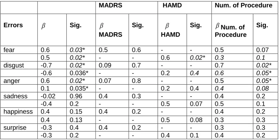
First a priori hypothesis: For each of the six emotions, a linear regression was calculated with the volume of overlap (between each patient’s lesions and Shackman’s negative affect binary mask) as the dependent variable (overlap negative affect, ONA), and number of emotion recognition errors as the first independent variable, depressive illness severity at the time of testing (MADRS or HAM-17) as the second independent variable and number of ACING procedures as the third independent variable. These covariates were included as both illness severity and number of operations correlated with total emotion recognition errors.
Analyses - Study 2 - Stroop Tasks
Between groups: For patients (SPG2), Stroop effect total errors for the CS and EFS tasks were tested using an ANOVA with the main effects group (SPG2, NSPG2 and control) and task type (CS vs EFS) and their interaction with covariates MADRS, HAM-17, HAD-A and number of ACING operations.
Second a priori hypothesis: A linear regression was done with the volume of overlap (between each patient’s lesions and Shackman’s cognitive control binary mask) as the dependent variable (overlap cognitive control, OCC), and the total CS and EFS Stroop effect error rate as the first independent variable, depressive illness severity (MADRS or HAM-17) as the second independent variable and number of ACING procedures as the third independent variable.
Specificity of Shackman masks
Third hypothesis: This was tested with linear regressions as above, but with total lesion volume as the dependent variable.
illness severity (MADRS or HAM-17) as the second independent variable and number of ACING procedures as the third independent variable. We also tested whether the total volume of overlap (between each patient’s ACING and the negative affect binary mask, ONA) was significantly related to the total number of Stroop errors, taking account of depressive illness severity (MADRS or HAM-17) and number of ACING procedures.
Correction for post hoc testing
Results Participants
Groups were matched on the bases of male/female ratio, age and NART. In Study 1, there were no significant differences in male/female ratio (Chisq=1.7, p=0.19), age (t=0.13, p=0.89) or NART (t=1.5, p=0.1). Similarly, in Study 2 there were no significant differences in male/female ratio (Chisq=2.1, p=0.3), age (F=1.6, p=0.2) or NART (F=0.3, p=0.7).
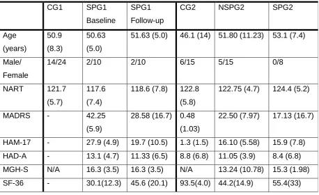
As shown in Table 1, MGH-S scores of 15.9 to 17.3 for patients receiving ACING indicated a very high level of treatment-resistance, similar to previous studies of patients attending the AIS (15.6) (Steele et al., 2008), in contrast to much less treatment-resistant secondary care 6.9 and primary care patients 0.54 (Hazari et al., 2013). SF-36 ratings (30±12 and 44±15) indicated marked quality of life impairments in patients with treatment-resistant depression before surgery (SPG1) (Table 1).
Patients satisfied ICD 10 criteria for recurrent unipolar depressive disorder unless in remission. With ACING ‘response’ defined as a reduction of 50% or more at one year compared to baseline HAM-17 and MADRS scores (Steele et al., 2008), ACING responders (n=6 Study 1, n=5 Study 2) had SF-36 scores of 60±13 and 88±10 respectively and non-responders (n=6 Study 1, n=3 Study 2) 31±14 and 35±20 respectively. Combined responder and non-responder scores are shown in Table 1. These scores can be compared with other severe chronic illnesses: 72, 55, 36 for congestive heart failure (New York Heart Association, NYHA, classes 1, 2, 3 respectively), 62 for hepatitis and in addition, 51 for non-treatment resistant depression with 89 for controls (Juenger et al., 2002).
Analyses - Study 1 - Emotional Facial Recognition Task
Pre-operative baseline and follow-up illness severity (MADRS, HAM-17, HAD-A) plus estimated IQ (NART) are shown in Table 1. This indicates that patients had a depressive illness in the ‘moderate-severe’ range before their operation and illness severity had on average decreased significantly at long term follow up (MADRS, t=3.04, p=0.014, HAM-17, t=2.5, p=0.02). There was no significant change in estimated IQ.
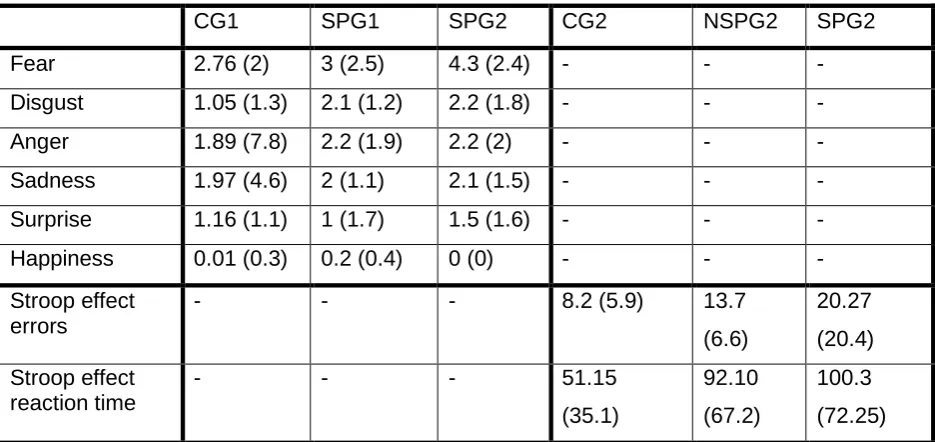
(p>0.05). Only post-operative impairment in fear recognition remained significant after Bonferroni correction (p=0.048).
Within patient group correlations: As depressive illness can be associated with cognitive impairment, we did exploratory tests for correlations between errors in recognising different emotional expressions and illness severity at baseline and follow-up. At pre-operative baseline, fear, anger and disgust recognition errors correlated positively with HAM-17 illness severity (r= 0.62, p=0.05; r=0.74, p=0.01; r=0.67, p=0.05 respectively). At follow-up, HAM-17 and MADRS also correlated positively with anger recognition errors (r=0.63, p= 0.05; r=0.58, p= 0.05). There were no significant correlations with anxiety (HAD-A) scores. There was a positive correlation between increased fear recognition errors and number of ACING procedures (r=0.78, p=0.02). There were no correlations with NART IQ score at either baseline or follow up.
First a priori hypothesis: When illness severity (quantified by either MADRS or HAM-17) and number of ACING procedures were ‘covaried out’, there was a significant relationship (Table 3) between ONA and impairment in identifying negative emotions of fear, disgust and anger, but not sadness, and no relationship between overlap and expressions of happiness or surprise. Bonferroni correction was not used as these were a priori planned tests. Fig. 2 shows partial regression plots for ONA versus total errors for fear, disgust and anger.
Analyses - Study 2 - Stroop Tasks
Clinical and demographic details for controls (CG2), patients with depression who had not received ACING (NSPG2), and patients who had received ACING as treatment (SPG2), are shown in Table 1.
Between groups’ analyses: There was a significant effect of group on Stroop effect errors (F=4.4, p=0.017) and post-hoc pair-wise testing with Bonferroni correction identified significant differences between SPG2, NSPG2 and CG2 (p=0.013). There was also a significant effect of Stroop effect on reaction time (F=3.7, p=0.032).
Within SPG2 group correlations: Linear regressions were used to test for a relationship between Stroop effect errors and symptom severity scores in the SPG2 group alone. Stroop effect errors correlated significantly with MADRS (t=2.7, p=0.018), HAM-17 (t=5.9, p=0.001) and HAD-A (t=2.9, p=0.023) scores. This indicates that more ill patients made more Stroop effect errors. There was no relationship with number of ACING procedures (t=1.4, p=0.2). Second a priori hypothesis: Similar to emotional facial recognition, we tested the hypothesis of a significant positive relationship between OCC and total Stroop errors, taking account of illness severity and number of operations. There was a significant positive relationship between cognitive control mask overlap and Stroop effect errors (
β
=1.2, p=0.03) as shown in Fig. 2.Specificity of masks
Third hypothesis: We tested whether lesion volume was significantly associated with emotional facial expression recognition accuracy or Stroop task accuracy, taking into account illness severity and number of ACING procedure. There were no significant correlations between total lesion volume and recognition of fear (r =-1.6, p=0.15), disgust (r =0.3, p=0.78), anger (r=-1.56, p=0.16), sadness (r=-0.198, p=0.85), surprise (r=-0.83, p=0.43) or happiness (r=0.75, p=0.48). Similarly, there were no significant correlations between total lesion volume and Stroop effect errors (r =2.2, p=0.09). This indicates that total lesion volume, which includes both grey and white matter, was not systematically related to impairments in emotional facial recognition or cognitive control.
Discussion
Negative affect, cognitive control and pain have been strongly linked to the aMCC (Peyron et al., 2000, Shackman et al., 2011, Vogt, 2014). Clinically, ACING lesions within the aMCC, have long been used as treatment for otherwise treatment-resistant mood and anxiety disorders (e.g. OCD) (Dougherty et al., 2002, Steele et al., 2008) and chronic pain syndromes (Pereira et al., 2014).
Consistent with our hypotheses, we found that increased volume of overlap between ACING lesions and the negative affect map (ONA) predicted impaired recognition of negative emotions of fear, disgust and anger and no impairment in recognising facial expressions of surprise or happiness. In addition, we found that increased volume of overlap between ACING lesions and the cognitive control map predicted increased Stroop effect errors. Only the overlap between ACING lesions and the negative affect map was associated with impairments in recognising facial expressions of negative affect and Stroop effect errors, not total ACING lesion volume or when using the cognitive control mask, supporting the specificity of Shackman’s grey matter-linked map functions.
(MNI y = 0-18) aMCC transient electrical stimulation was reported to induce ‘complex’, ‘stereotyped’ behaviours, ‘invariably involuntary, sometimes the subject was able to resist performing them, sometimes not’, but usually without clear effects on emotion (Talairach et al., 1973). Talairach’s description of induced behaviours is reminiscent of compulsions occurring in Obsessive Compulsive Disorder (OCD) and ACING is also used to treat otherwise intractable OCD. The anterior vs. posterior MCC stimulation effects are consistent with reports of the anterior but not the posterior MCC being associated with emotion (Vogt et al., 2003).
Theoretical accounts of the MCC have considered whether this structure is simply involved in detecting conflict or whether it resolves and controls conflict. Shackman and colleagues proposed that the aMCC processes both punishment-linked information and facilitates actions which avoid future punishment (Shackman et al., 2011). They formulated this as the Adaptive Control Hypothesis (ACH) noting that negative affect, anxiety and pain tend to engage the same processes described by theories of cognitive control to resolve similar challenges (Shackman et al., 2011). In particular, cognitive control tends to be engaged when habitual responses are insufficient to allow goal directed behaviour, particularly when there is uncertainty and conflict about the optimal course of action. Notably, studies on patients receiving ACING for treatment-resistant psychiatric disorders have reported individual neurones in the aMCC encoding response conflict information (Davis et al., 2005, Sheth et al., 2012) and individual neuronal firing that was predictive of motor actions (Williams et al., 2004). This and the results of the present study suggests that the MCC is involved in resolution and control of conflict supporting the ACH.
Behavioural errors are associated with an electrophysiological event-related potential, the Error Related Negativity (ERN). This signal is thought to reflect a discrepancy between representations of actual and correct actions and the ERN has been linked to MCC function (Riesel et al., 2013, Cavanagh and Shackman, 2015). The ERN was reported to be abnormally reduced in a patient with a rare naturally occurring lesion of the MCC (Swick and Turken, 2002). A recent review concluded that the ERN is abnormally increased in OCD, depression and generalised anxiety disorder, and an increased ERN is associated with high trait anxiety and high negative affect (Weinberg et al., 2012).
optimally adjusting behaviour to uncertainty, a characteristic of events that cause anxiety and require cognitive control (Cavanagh and Shackman, 2015). Recently a large international study has concluded that patients with depressive illness have abnormally increased frontal and rostral anterior cingulate theta compared to controls, which the authors suggest is a potential endophenotype of depressive illness (Arns et al., 2015).
Consequently, there is convergent evidence that the aMCC is an important part of a network linked to the experience of negative affect which engages cognitive control processes for optimising behaviour. In this context, our finding that increased lesion volume was linked to increased impairment in recognising facial expressions of negative affect, but not positive or neutral affect, and impairment in Stroop cognitive control, supports the assertion that the aMCC has a causal role in these processes. Importantly though, these abnormalities were only detectable on neuropsychological testing and not clinically. The therapeutic benefit of a reduction in negative affect may be associated with a reduction in cognitive control. However depressive illness has been reported to be associated with Stroop impairments independent of any lesion and we found correlations with illness severity in the present study. Therefore with ACING it is possible that any reduction in cognitive control is offset by an improvement in mood. Before receiving ACING, patients were ill long-term, highly treatment-resistant and had an SF-36 measured quality of life comparable to patients with very disabling diseases. AIS patients with treatment-resistant depression typically have an ACING ‘response’ rate of 62% (Steele et al., 2008) and for those who responded, this was associated with a substantially improved quality of life. Further work on understanding the normal function of the aMCC, and abnormal function in depressive illness, may allow the development of improved surgical techniques and outcomes.
A possible limitation is the small number of subjects. ACING is however a treatment of last resort, done in a limited number of centres world-wide, so the number of patients available for testing is limited. Furthermore, recruitment criteria and ACING procedures differ between centres complicating independent replication. To our knowledge we report data on more patients who have received ACING than previously. Medication exposure, quantified using the MGH-S score, has potential limitations. It’s not known if different medications given long term and in combination have different effects on facial affect recognition independent of illness severity in treatment-resistant illness.
Acknowledgements
We thank Christine Matthews for helping with accessing data and Mairi Stirling, Karen Walker and Craig Adam for their help during recruitment.
Funding
The project was supported by awards from an Anonymous Trust and by the Scottish Mental Health Research Network (SMHRN). The funding sources played no part in the design of the study, analysis and interpretation of the data, nor manuscript preparation.
Financial Disclosures
References
Arns M, Etkin A, Hegerl U, Williams LM, DeBattista C, Palmer DM, et al. Frontal and rostral anterior cingulate (rACC) theta EEG in depression: implications for treatment outcome? Eur Neuropsychopharmacol. 2015;25(8):1190-200.
Austin MP, Mitchell P, Goodwin GM. Cognitive deficits in depression: possible implications for functional neuropathology. Br J Psychiatry. 2001;178:200-6.
Brinley-Reed M, Mascagni F, McDonald AJ. Synaptology of prefrontal cortical projections to the basolateral amygdala: an electron microscopic study in the rat. Neurosci Lett. 1995;202(1-2):45-8.
Cavanagh JF, Shackman AJ. Frontal midline theta reflects anxiety and cognitive control: meta-analytic evidence. Journal of physiology, Paris. 2015;109(1-3):3-15.
Cohen RA, Kaplan RF, Moser DJ, Jenkins MA, Wilkinson H. Impairments of attention after cingulotomy. Neurology. 1999;53(4):819-24.
Cohen RA, Kaplan RF, Zuffante P, Moser DJ, Jenkins MA, Salloway S, et al. Alteration of intention and self-initiated action associated with bilateral anterior cingulotomy. J Neuropsychiatry Clin Neurosci. 1999;11(4):444-53.
Davis KD, Taylor KS, Hutchison WD, Dostrovsky JO, McAndrews MP, Richter EO, et al. Human anterior cingulate cortex neurons encode cognitive and emotional demands. J Neurosci. 2005;25(37):8402-6.
Dougherty DD, Baer L, Cosgrove GR, Cassem EH, Price BH, Nierenberg AA, et al. Prospective long-term follow-up of 44 patients who received cingulotomy for treatment-refractory obsessive-compulsive disorder. Am J Psychiatry. 2002;159(2):269-75.
Fava M. Diagnosis and definition of treatment-resistant depression. Biol Psychiatry. 2003;53(8):649-59.
Foltz EL, White LE, Jr. Pain "relief" by frontal cingulumotomy. J Neurosurg. 1962;19:89-100. Friston KJ, Ashburner JT, Kiebel SJ, Nichols TE, Penny WD. Statistical Parametric Mapping.
London: Academic Press; 2007.
Graeff FG. Serotonin, the periaqueductal gray and panic. Neurosci Biobehav Rev. 2004;28(3):239-59.
Hazari H, Christmas D, Matthews K. The clinical utility of different quantitative methods for measuring treatment resistance in major depression. J Affect Disord.
2013;150(2):231-6.
Juenger J, Schellberg D, Kraemer S, Haunstetter A, Zugck C, Herzog W, et al. Health related quality of life in patients with congestive heart failure: comparison with other chronic diseases and relation to functional variables. Heart. 2002;87(3):235-41. Milad MR, Quirk GJ, Pitman RK, Orr SP, Fischl B, Rauch SL. A role for the human dorsal
anterior cingulate cortex in fear expression. Biol Psychiatry. 2007;62(10):1191-4. Ochsner KN, Kosslyn SM, Cosgrove GR, Cassem EH, Price BH, Nierenberg AA, et al.
Deficits in visual cognition and attention following bilateral anterior cingulotomy. Neuropsychologia. 2001;39(3):219-30.
Parvizi J, Rangarajan V, Shirer WR, Desai N, Greicius MD. The will to persevere induced by electrical stimulation of the human cingulate gyrus. Neuron. 2013;80(6):1359-67. Pereira EA, Paranathala M, Hyam JA, Green AL, Aziz TZ. Anterior cingulotomy improves
malignant mesothelioma pain and dyspnoea. Br J Neurosurg. 2014;28(4):471-4. Peyron R, Laurent B, Garcia-Larrea L. Functional imaging of brain responses to pain. A
review and meta-analysis (2000). Neurophysiol Clin. 2000;30(5):263-88. Rauch SL, Kim H, Makris N, Cosgrove GR, Cassem EH, Savage CR, et al. Volume
reduction in the caudate nucleus following stereotactic placement of lesions in the anterior cingulate cortex in humans: a morphometric magnetic resonance imaging study. J Neurosurg. 2000;93(6):1019-25.
Ridout N, O'Carroll RE, Dritschel B, Christmas D, Eljamel M, Matthews K. Emotion recognition from dynamic emotional displays following anterior cingulotomy and anterior capsulotomy for chronic depression. Neuropsychologia. 2007;45(8):1735-43. Riesel A, Weinberg A, Moran T, Hajcak G. Time Course of Error-Potentiated Startle and its
Relationship to Error-Related Brain Activity. Journal of Psychophysiology. 2013;27(2):51-9.
Schiltenwolf M, Akbar M, Hug A, Pfuller U, Gantz S, Neubauer E, et al. Evidence of specific cognitive deficits in patients with chronic low back pain under long-term substitution treatment of opioids. Pain physician. 2014;17(1):9-20.
Shackman AJ, Salomons TV, Slagter HA, Fox AS, Winter JJ, Davidson RJ. The integration of negative affect, pain and cognitive control in the cingulate cortex. Nat Rev
Neurosci. 2011;12(3):154-67.
Sheth SA, Mian MK, Patel SR, Asaad WF, Williams ZM, Dougherty DD, et al. Human dorsal anterior cingulate cortex neurons mediate ongoing behavioural adaptation. Nature. 2012;488(7410):218-21.
Swick D, Turken AU. Dissociation between conflict detection and error monitoring in the human anterior cingulate cortex. Proc Natl Acad Sci U S A. 2002;99(25):16354-9. Talairach J, Bancaud J, Geier S, Bordas-Ferrer M, Bonis A, Szikla G, et al. The cingulate gyrus and human behaviour. Electroencephalogr Clin Neurophysiol. 1973;34(1):45-52.
Vogt BA, editor. Cingulate Neurobiology and Disease. New York: Oxford University Press; 2009.
Vogt BA. Submodalities of emotion in the context of cingulate subregions. Cortex. 2014;59:197-202.
Vogt BA, Berger GR, Derbyshire SW. Structural and functional dichotomy of human midcingulate cortex. Eur J Neurosci. 2003;18(11):3134-44.
Weinberg A, Riesel A, Hajcak G. Integrating multiple perspectives on error-related brain activity: The ERN as a neural indicator of trait defensive reactivity. Motivation and Emotion. 2012;36:84-100.
Williams ZM, Bush G, Rauch SL, Cosgrove GR, Eskandar EN. Human anterior cingulate neurons and the integration of monetary reward with motor responses. Nat Neurosci. 2004;7(12):1370-5.
Yang JC, Ginat DT, Dougherty DD, Makris N, Eskandar EN. Lesion analysis for cingulotomy and limbic leucotomy: comparison and correlation with clinical outcomes. J
Neurosurg. 2014;120(1):152-63.
Young AW, Perrett DL, Calder AJ, Sprengelmeyer R, Ekman P. Facial Expressions of Emotion: Stimuli and Tests (FEEST). Bury St. EDmonda, Suffolk: Thames Valley Test Company; 2002.
Table 1. Demographic and clinical characteristics
SPG1: Surgical Patient Group 1, CG1: Control Group1, SPG2: Surgical Patient Group 2, CG2: Control Group, NSP: Non-Surgical Patients 2. Values are mean (standard deviation); NART, National Adult Reading Test; MADRS, Montgomery-Asberg Depression Rating Scale; HAM-17, Hamilton Depression Rating Scale; HAD-A, Hospital anxiety and depression scale; Massachusetts General Hospital Staging (MGH-S), not significant (NS), not applicable (N/A), not available (-)
CG1 SPG1
Baseline
SPG1 Follow-up
CG2 NSPG2 SPG2
Age (years)
50.9 (8.3)
50.63 (5.0)
51.63 (5.0) 46.1 (14) 51.80 (11.23) 53.1 (7.4)
Male/ Female
14/24 2/10 2/10 6/15 5/15 0/8
NART 121.7
(5.7)
117.6 (7.4)
118.6 (7.8) 122.8 (5.8)
122.75 (4.7) 124.4 (5.2)
MADRS - 42.25
(5.9)
28.58 (16.7) 0.48 (1.03)
22.50 (7.97) 17.13 (16.7)
Table 2. Errors on Emotional Facial Recognition and Stroop tasks. Values are mean (standard deviation), not available (-)
CG1 SPG1 SPG2 CG2 NSPG2 SPG2
Fear 2.76 (2) 3 (2.5) 4.3 (2.4) - - -
Disgust 1.05 (1.3) 2.1 (1.2) 2.2 (1.8) - - -
Anger 1.89 (7.8) 2.2 (1.9) 2.2 (2) - - -
Sadness 1.97 (4.6) 2 (1.1) 2.1 (1.5) - - -
Surprise 1.16 (1.1) 1 (1.7) 1.5 (1.6) - - -
Happiness 0.01 (0.3) 0.2 (0.4) 0 (0) - - -
Stroop effect errors
- - - 8.2 (5.9) 13.7
(6.6)
20.27 (20.4) Stroop effect
reaction time
- - - 51.15
(35.1)
92.10 (67.2)
Table 3 Emotional facial recognition lesion overlap.
Regression parameters (
β
), significance (Sig.)MADRS HAMD Num. of Procedure
Errors
β
Sig.β
MADRS
Sig.
β
HAMDSig.
β
Num. of ProcedureSig.
fear 0.6 0.03* 0.5 0.6 - - 0.5 0.07
0.5 0.02* - - 0.6 0.02* 0.3 0.1
disgust -0.7 0.02* 0.09 0.7 - - 0.7 0.02*
-0.6 0.036* - - 0.2 0.4 0.6 0.05*
anger 0.6 0.02* 0.07 0.8 - - 0.5 0.05*
0.1 0.035* - - 0.2 0.4 0.4 0.08
sadness -0.02 0.96 0.4 0.3 - - 0.4 0.2
-0.4 0.2 - - 0.5 0.07 0.5 0.1
happiness 0.4 0.15 0.4 0.2 - - 0.4 0.2
0.4 0.13 - - 0.5 0.08 0.3 0.3
surprise -0.3 0.4 0.4 0.2 - - 0.3 0.3