www.wjpr.net Vol 7, Issue 10, 2018. 1
ANALYSIS OF THE MONOSODIUM GLUTAMATE ON
NEUROTRANSMITTERS USING QUANTUM METHOD
Manuel Aparicio-Razo2,3, Oscar Sánchez-Parada1, David César Malpica-Moreda2,
Manuel González-Pérez*3,4
1
Escuela de Medicina Universidad Popular Autónoma del Estado de Puebla..
2
Benemérita Universidad Autónoma de Puebla, Facultad de Ciencias de la Electrónica
3
Universidad Popular Autónoma del Estado de Puebla A.C. (UPAEP). Centro
Interdisciplinario De Posgrados (CIP). Posgrado en Ciencias de la Ingeniería Biomédica.
4
Sistema Nacional De Investigadores. Nivel 1.
ABSTRACT
Monosodium glutamate (GMS) is a sodium salt used as enhancer
additive flavor. This GMS is one of the chemicals responsible for
maintaining the flavor of long-lasting food. According to studies
carried out, consumption of this additive can cause various side effects.
Among the most common side, effects of GMS are headaches, nausea,
migraines and muscle spasms; in extreme cases may have epilepsy,
and heart irregularities. The objective of the study is to determine,
using the semi-empirical method of chemical quantum SE-ZINDO/1),
which neurotransmitter has a higher affinity with the GMS.
Hyperchem Professional software performed Molecular Modelling and
analysis of the GMS and nine of the major neurotransmitters.
(Hyperchem, Hypercube, Multi on for Windows, series
12-800-1501800080. Multi On, southern 1236-301 Tlacoquemecatl insurgent
Col. del Valle, Mexico, Benito Juárez, CP 03200). The result of the
simulations of the quantum poles reveals that this additive can oxidize Adrenaline, as well as
cross bands related to this neurotransmitter.
KEYWORDS: Monosodium Glutamate, Neurotransmitters, Quantum Method, Hyperchem,
SE-ZINDO/1.
Volume 7, Issue 10, 1-12. Research Article ISSN 2277–7105
Article Received on 21 March 2018,
Revised on 10 April 2018, Accepted on 01 May 2018
DOI: 10.20959/wjpr201810-12277
*Corresponding Author
Dr. Manuel
González-Pérez
Universidad Popular
Autónoma del Estado de
Puebla A.C. (UPAEP).
Centro
Interdisciplinario De
Posgrados (CIP). Posgrado
en Ciencias de la Ingeniería
www.wjpr.net Vol 7, Issue 10, 2018. 2 INTRODUCTION
Glutamate is a nonessential amino acid that is producing the degradation and metabolism of
glucose in the mitochondria inside. Today it is known to be a neuroexcitatory of the nervous
system and the central mediator of sensory, motor, cognitive, and emotional information. In
addition, it is present in 80-90% of the synapses of the brain.[1, 2]
In the industry, glutamate is used as salt in its form monosodium (monosodium glutamate
"GMS"). The GMS is as conservative in many of products, mainly added in food canned or
packaged soft drinks, medicines, sausages, sauces, among others.[3, 4] In the last five years has
been discovered that the GMS has related the development of obesity, since it could influence
the appetite regulation, favoring a high consumption of foods containing it.[5, 6]
Studies in monkeys in 1969, showed that GMS causes brain damage and acute neuronal death
when it supplies him subcutaneously.[7] Other studies in Wistar rats showed that prolonged
intake of GMS affects kidney functions.[8] As well as cause infertility by histological
alterations of the seminiferous tubules, interstitial tissues, and exfoliation of the
spermatocytes and the spermatids[9], as well as generate memory loss in rodents as many
males as females in different ways.[10]
Other articles suggest that the GMS is a substance genotoxic for lymphocytes from peripheral
blood human performed in vitro, causing damage to the DNA in different concentrations.[11]
The GMS can produce injury "excitotoxic " that much resemble the neurodegenerative
disorders that today suffer from many people around the world. Abnormally increased
glutamatergic neurotransmission can cause cellular damage excitotoxic and lead to neuronal
death associated with atrophy olivopontocerebellar, Huntington's disease, status epilepticus,
hypoxia/ischemia and the Hypoglycemia.[12]
The objective of this study is to determine, using the semi-empirical method of quantum
chemistry (SE-ZINDO/1), that neurotransmitter has a high affinity for the GMS. In this study
were analyzed nine neurotransmitters which they are: adrenaline, serotonin, dopamine,
GABA, glutamic acid, histamine, glycine, noradrenaline, and acetylcholine.
HyperChem is a program for molecular modeling graphic interface, which allows researchers
to carry out chemical simulations that facilitate multiple data entry. Through the program, it
www.wjpr.net Vol 7, Issue 10, 2018. 3
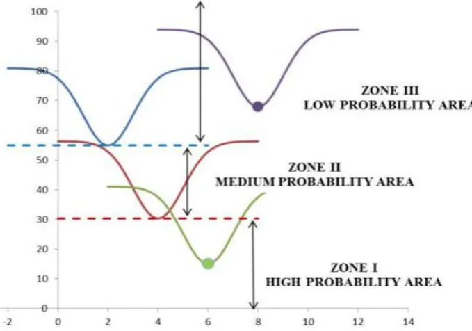
The theory of ETC is based on defining the band gap or Bandgap (BG), which is the energy
difference between the valence band and the conduction band. In quantum theory, it is known
as HOMO and LUMO, and in the old method known as E - and E +. On the other hand, the
quantum well is defined as the area in which can drop the value of, etc. These areas are
divided into 3 (figure. 1): 1) the area of high probability (zone I), 2) The area's average
[image:3.595.121.461.212.450.2]probability (ZONE II). 3) The area of low-probability (zone III).[13, 14]
Figure 1. Areas of probability in quantum well, according to the theory of ETC´s.
MATERIALS AND METHODS
The simulation of the GMS molecule was carried out using the Hyperchem Software
(Hypercube, Multi On for Windows, series 12-800-1501800080. Multi On, south 1236-301
Col. Insurgentes Tlacoquemecatl del Valle, Benito Juarez, Mexico City, Mexico CP 03200).
We utilized the Semiempirical method to perform the BG calculation, the Electrostatic
Potential (EP) and the ETC. When the complete molecule is drawn, the values of HOMO (-),
LUMO (+), E- and E + are obtained, in the amount at zero and with a density of 0.015. The
values that are recorded will be captured in an Excel sheet, and he will perform operations to
obtain BG, EP, ETC. We get the cross band of the compounds; it is done taking the HOMO
and E- the value of the first compound and the value of LUMO and E + the second
compound. The lower ETC of the transverse band will be the value that will determine which
www.wjpr.net Vol 7, Issue 10, 2018. 4
quantum well graphs To set the limits of the charts, the highest ETC will be put in the upper
limit and the ETC lower as, the lower limit of the compounds to be compared.[14, 15]
RESULTS AND DISCUSSION
Table 1 shows the interaction of the GMS against vital neurotransmitters with their respective
ETC´s, stressing that the adrenaline has one minor for electrons hop energy.
Table 1. GMS vs Neurotransmisores
No. Antioxidant Oxidant HOMO LUMO BG E- E+ EP ETC
1 GMS GMS -5.674 0.288 5.962 -0.156 0.190 0.346 17.232 9 ADRENALIN ADRENALIN -8.998 0.092 9.090 -0.117 0.198 0.315 28.858
Option 1 GMS ADRENALIN -5.674 0.092 5.766 -0.156 0.198 0.354 16.289
Option 2 ADRENALIN GMS -8.998 0.288 9.286 -0.117 0.190 0.307 30.248
In the case of the crossbands, we found that all the neurotransmitters that interact with
adrenalin, are oxidized by the GMS. I.e., have a 16.289 ETC, which means that the GMS has
a high probability of oxidizing all neurotransmitters analyzed here. In Table 2, we can see the
values of the ETC´s..
Table 2. Crossed bands vs GMS
No. Antioxidant Oxidant HOMO LUMO BG E- E+ EP ETC
1 GMS SEROTONIN--ADRENALIN -5.674 0.092 5.766 -0.156 0.198 0.354 16.289 2 GMS DOPAMINE--ADRENALIN -5.674 0.092 5.766 -0.156 0.198 0.354 16.289 3 GMS GABA--ADRENALIN -5.674 0.092 5.766 -0.156 0.198 0.354 16.289 4 GMS GLUTAMIC ACID--ADRENALIN -5.674 0.092 5.766 -0.156 0.198 0.354 16.289 5 GMS GLYCINE--ADRENALIN -5.674 0.092 5.766 -0.156 0.198 0.354 16.289 6 GMS HISTAMINE--ADRENALIN -5.674 0.092 5.766 -0.156 0.198 0.354 16.289 7 GMS NORADRENALINE--ADRENALIN -5.674 0.092 5.766 -0.156 0.198 0.354 16.289 8 GMS ACETYLCHOLINE--ADRENALIN -5.674 0.092 5.766 -0.156 0.198 0.354 16.289 9 GMS ADRENALIN--ADRENALIN -5.674 0.092 5.766 -0.156 0.198 0.354 16.289
Below we can see the interactions of neurotransmitters in the quantum wells. In these graphs
www.wjpr.net Vol 7, Issue 10, 2018. 5 Graph 1. Interaction of the GMS against Adrenalin-Adrenalin.
Graph 2. Interaction of the GMS against Serotonin-Adrenalin.
Graph 3. Interaction of the GMS against Dopamine-Adrenalin.
www.wjpr.net Vol 7, Issue 10, 2018. 6 Graph 5. Interaction of the GMS against Acid Glutamic-Adrenalin.
Graph 6. Interaction of the GMS against Glycine-Adrenalin.
Graph 7. Interaction of the GMS against Histamine-Adrenalin.
www.wjpr.net Vol 7, Issue 10, 2018. 7 Graph 9. Interaction of the GMS against Acetylcholine-Adrenalin
CONCLUSIONS
In the analysis of quantum wells and the interaction of the ETC´s, tells us there is an excellent
probability of oxidation the neurotransmitters by the presence of GMS. Both concepts may
suggest that this additive may be associated with neurological disorders that lurk in our
society. There is a growing amount of experimental evidence that oxidative stress is a causal
factor in the neuropathology of several neurodegenerative diseases in adults, as well as
stroke, trauma, and seizure disorders. Note that this type of quantum analysis-based
probabilistic systems.
The product of the oxidation of adrenaline is also called Adrenochrome and is responsible for
various mental disorders, including Alzheimer's disease[18, 19] and schizophrenia.[16,17] Also,
high doses of MSG can cause Neurodegeneration in the brain most vulnerable regions such as
the hypothalamic arcuate nucleus and generate acute cerebral disorders such as cardiac arrest
and hypoxia pre/perinatal.[20]
In the case of Serotonin, we can say it is a monoaminergic neurotransmitter participates in a
plethora of physiological processes and behavior, including emotionality, dream, locomotion,
perception, cognition, aggression, sexual behavior, and appetite. Most essential disorders of
the degradation of serotonin are associated with depression, addictions to substances,
attention deficit, irregular sexual cycles, inflammation and dysfunction of gastrointestinal
tract, disorders of sleep cycles, anxiety, Fibromyalgia, dizziness, nausea, obesity, chills,
tremors, confusion, delirium and tachycardia or fluctuations of blood pressure.[21, 22]
The oxidation of dopamine at high concentrations is correlated with the specific loss of
dopaminergic terminals. This oxidation may be related to Parkinson's disease disorder.[23, 24]
www.wjpr.net Vol 7, Issue 10, 2018. 8
glutamate function, which may have implications for neurodegenerative processes as
ischemia, induced toxicity by methamphetamine and disease of Parkinson.[25]
The neurotransmitter GABA deficiency is rare but can be very serious when it is experienced.
This neurotransmitter performs a variety of activities that are strictly related to the operation
of the parasympathetic nervous system. Some authors have reported that inhibition or
oxidation of the neurotransmitter GABA, may be linked to the regulation of overweight,
obesity, hypertension, and hyperglycemia.[26] GABA deficiency can make a person more
susceptible to several other diseases. Epilepsy, Huntington's disease, disease, Cushing,
meningitis, and psycho-organic syndrome are affected by levels of GABA.[27]
Glutamic acid oxidation also brings severe consequences for health. In 1987, the Academy of
Neurology American reported that patients who had amyotrophic lateral sclerosis showed a
low concentration of this neurotransmitter in the region of the brain and cervical cord. Brain
glutamate deficiency is not explained, but the production or insufficient release of this
neurotransmitter Exciter could have significant effects on motor neurons.[28]
Glycine is a neurotransmitter that performs several functions as a transmitter in the central
nervous system. As an inhibitory neurotransmitter participates in the processing of motor and
sensory information that allows movement, vision, and hearing. Also, the glycine modulates
excitatory neurotransmission to potentiate the action of glutamate in N-methyl-D-aspartate
receptors.[29] As we see this neurotransmitter, it is vital for the brain, and the oxidation of the
same could affect some related functions.
In the case of the histamine, studies have revealed that there are alterations in the
histaminergic system in neurological and psychiatric diseases. Brain histamine levels
decrease in patients with Alzheimer's disease, while abnormally high concentrations of
histamine are found in the brains of Parkinson's disease and schizophrenic patients. Low
histamine levels are associated with seizures.[30]
It has been shown that the low level of the neurotransmitter norepinephrine in the central and
peripheral, nervous system cause a defective hydroxylation of dopamine. This low level
suggests that it provoked a primary genetic defect in the enzyme dopamine-β-hydroxylase or
the presence of a highly specific inhibitor of this enzyme. Associated with this deficiency of
www.wjpr.net Vol 7, Issue 10, 2018. 9
system and an increase in the active absorption of tyrosine and other amino acids that share a
common mechanism of absorption with Tyrosine) through the blood-brain barrier.[31] Finally,
it is well known that this neurotransmitter deficiency is closely related to Parkinson's disease.
The significant deterioration of the neurotransmitter acetylcholine is associated with dementia
cases, usually caused by Parkinson's disease and Alzheimer's disease.[32] Also, this
neurotransmitter is responsible for regulates the vasodilation of the smooth muscles, which its
oxidation can be linked with various disorders of the structures related to the autonomic
nervous system.
In conclusion, we can say that the oxidation of these neurotransmitters may be the origin of
any pathology. The proper functioning of our neurotransmitters gives us a healthy balance in
all of our functions of the nervous system. Now, either by our genes or the environment,
production or erroneous processing of various neurotransmitters can lead to disorders or
physical and psychological disorders.
The study of the quantum grounds between substances and chemicals created by the human
body gives us the possibility to study and analyze the interactions among these. In the
particular case of the oxidation of neurotransmitters by the oxaloacetic monosodium
(Graphics 1 to 9), we can conclude that such alteration is correlated with the side effects of
such additive, giving rise to future research.
ACKNOWLEDGEMENTS
Appreciation to the Universidad Popular Autónoma del Estado de Puebla for allowing us to
use its postgraduate facilities to conduct our research.
REFERENCES
1. HAYASHI, T. (1954). Effects of sodium glutamate on the nervous system. The Keio
Journal of Medicine, 3(4): 183-192.
2. Pin, J. P., & Duvoisin, R. (1995). The metabotropic glutamate receptors: structure and
functions. Neuropharmacology, 34(1): 1-26.
3. El glutamato: de nutriente cerebral a neurotóxico, Carlos Beas Zárate, julio-septiembre
2005, revista de la academia Mexicana de ciencia,
www.wjpr.net Vol 7, Issue 10, 2018. 10
4. Bajaj, I., & Singhal, R. (2011). Poly (glutamic acid)–an emerging biopolymer of
commercial interest. Bioresource technology, 102(10): 5551-5561.
5. Carbonero-Carreño, R. (2013). Glutamato monosódico “la trampa de los alimentos
sabrosos”. Trastornos de la conducta alimentaria, 17: 1863-1876.
6. Savcheniuk, O. A., Virchenko, O. V., Falalyeyeva, T. M., Beregova, T. V., Babenko, L.
P., Lazarenko, L. M., & Spivak, M. Y. (2014). The efficacy of probiotics for monosodium
glutamate-induced obesity: dietology concerns and opportunities for prevention. EPMA
Journal, 5(1): 2.
7. Olney, J. W., & Sharpe, L. G. (1969). Brain lesions in an infant rhesus monkey treated
with monosodium glutamate. Science, 166(3903): 386-388.
8. Mahieu, S., Klug, M., Millen, N., Fabro, A., Benmelej, A., & del Carmen Contini, M.
(2016). Monosodium glutamate intake affect the function of the kidney through NMDA
receptor. Life sciences, 149: 114-119.
9. Alalwani, A. D. (2014). Monosodium glutamate induced testicular lesions in rats
(histological study). Middle East Fertility Society Journal, 19(4): 274-280.
10.Araujo, P. C. O., Quines, C. B., Jardim, N. S., Leite, M. R., & Nogueira, C. W. (2017).
Resistance exercise reduces memory impairment induced by monosodium glutamate in
male and female rats. Experimental physiology, 102(7): 845-853.
11.Ataseven, N., Yüzbaşıoğlu, D., Keskin, A. Ç., & Ünal, F. (2016). Genotoxicity of
monosodium glutamate. Food and Chemical Toxicology, 91, 8-18.
12.Greenamyre, J. T. (1986). The role of glutamate in neurotransmission and in neurologic
disease. Arch Neurol, 43(10): 1058-1063.
13.González-Pérez, M. chemical-quantum analysis of the aggressiveness of glucose and its
appeasement with atp inside the cell, and water as an excellent antioxidant, 2017.
14.Ibarra Medel, D., Meléndez Gámez, P., López Oglesby, J. M., & González Pérez, m.
molecular analysis of strychnine and the glycine receptor using quantum chemistry
methods, 2016
15.Perez, M. G., Barrera, F. A. G., Diaz, J. F. M., Torres, M. G., & Oglesby, J. M. L.
Theoretical calculation of electron transfer coefficient for predicting the flow of electrons
by PM3, using 20 amino acids and nicotine. European Scientific Journal, ESJ, 2014;
10(27).
16.El metabolismo de la adrenalina en algunos desórdenes mentales- Revisión del problema
con particular interés en la esquizofrenia, Manuel Almeida V. Pag: 35-59, Biblioteca
www.wjpr.net Vol 7, Issue 10, 2018. 11
Salud, Rev. Neuropsiquiatr, 22(1): p.35-59, mar. 1959,
http://www.bvsde.paho.org/documentosdigitales/bvsde/texcom/revneuropsiquiatr/1959/M
Almeida.pdf.
17.Saiz Ruiz, J., Sánchez, V., Diego, C., & Sánchez Páez, P. (2010). Bases neurobiológicas
de la Esquizofrenia. Clínica y Salud, 21(3): 235-254.
18.Kerchner, G. & Nicoll, R. (2008). Silent synapses and the emergence of a postsynaptic
mechanism for LTP. Nature Reviews Neuroscience, 9(11): 813-825.
http://dx.doi.org/10.1038/nrn2501
19.Papouin, T. & Oliet, S. (2014). Organization, control and function of extrasynaptic
NMDA receptors.Philosophical Transactions Of The Royal Society B: Biological
Sciences, 369(1654): 20130601-20130601. http://dx.doi.org/10.1098/rstb.2013.0601
20.Hardingham, G. & Bading, H. (2010). Synaptic versus extrasynaptic NMDA receptor
signalling: implications for neurodegenerative disorders. Nature Reviews Neuroscience,
11(10), 682-696. http://dx.doi.org/10.1038/nrn2911
21.Lin, M. T., Chern, Y. F., Chow, C. F., & Li, Y. P. (1979). Effects of brain serotonin
alterations on hypothermia produced by chlorpromazine in rats. Pharmacology, 18(3):
128-135.
22.Southwick, S. M., Paige, S., Bremner, J. D., Krystal, J. H., & Charney, D. S. (1999,
October). Neurotransmitter alterations in PTSD: catecholamines and serotonin.
In Seminars in clinical neuropsychiatry (Vol. 4, No. 4, pp. 242-248).
23.Hastings, T. G., Lewis, D. A., & Zigmond, M. J. (1996). Role of oxidation in the
neurotoxic effects of intrastriatal dopamine injections. Proceedings of the National
Academy of Sciences, 93(5): 1956-1961.
24.Hastings, T. G. (2009). The role of dopamine oxidation in mitochondrial dysfunction: implications for Parkinson’s disease. Journal of bioenergetics and biomembranes, 41(6):
469-472.
25.Berman, S. B., & Hastings, T. G. (1997). Inhibition of glutamate transport in
synaptosomes by dopamine oxidation and reactive oxygen species. Journal of
neurochemistry, 69(3): 1185-1195.
26.Sistema de inhibición GABAérgico implicando en la regulación de la ingesta alimentaria
y obesidad, Sandoval-Salazar, Ramírez-Emiliano, Solis-Silvia, Departamento de ciencias
médicas, división de ciencias de la salud del campo León, Univesidad de Guanajuato,
Agosto 2 del 2013, Rev Mex Neuroci, 14(5): 262-271,
www.wjpr.net Vol 7, Issue 10, 2018. 12
27.Ugalde, O. (2010). Guía clínica para el tratamiento de los trastornos
psicogeriátricos. México: Instituto Nacional de Psiquiatría Ramón de la Fuente.
28.Perry, T. L., Hansen, S., & Jones, K. (1987). Brain glutamate deficiency in amyotrophic
lateral sclerosis. Neurology, 37(12): 1845-1845.
29.Lopez-Corcuera, B., Geerlings, A., & Aragon, C. (2001). Glycine neurotransmitter
transporters: an update. Molecular membrane biology, 18(1): 13-20.
30.Nuutinen, S., & Panula, P. (2010). Histamine in neurotransmission and brain diseases.
In Histamine in Inflammation (pp. 95-107). Springer US.
31.Hunt, D. M., & Johnson, D. R. (1972). An inherited deficiency in noradrenaline
biosynthesis in the brindled mouse. Journal of neurochemistry, 19(12): 2811-2819.
32.Förstl, H., Gratz, S., Hahn, U., Schwarz, J., & Jarnig, M. (2008). Dementia with Lewy
bodies and reduced dopamine transporter binding indicates significant acetylcholine