Acta Cryst.(2001). E57, o1039±o1040 DOI: 10.1107/S1600536801016415 Bond and Davies C10H16
o1039
organic papers
Acta Crystallographica Section E
Structure Reports
Online ISSN 1600-5368
(1
S
)-(±)-
a
-Pinene
Andrew D. Bond* and John E. Davies
Department of Chemistry, University of Cambridge, Lensfield Road, Cambridge CB2 1EW, England
Correspondence e-mail: [email protected]
Key indicators
Single-crystal X-ray study T= 203 K
Mean(C±C) = 0.003 AÊ Rfactor = 0.046 wRfactor = 0.103
Data-to-parameter ratio = 14.5
For details of how these key indicators were automatically derived from the article, see http://journals.iucr.org/e.
#2001 International Union of Crystallography Printed in Great Britain ± all rights reserved
The crystal structure of (1S)-(ÿ)--pinene, C10H16, has been
determined at 203 (2) K by in situ crystal growth from the liquid.
Comment
-Pinene, (I), is very widely distributed in nature. It is present in the majority of essential oils derived from the Coniferae and it is the principal constituent of oil of turpentine. An account of its history and the determination of its structure using the techniques of classical organic chemistry is given by Simonsen & Owen (1947). This work forms part of a continuing study devoted to improving the techniques for determining the crystal structures of substances which are liquids at room temperature [see, for example, Davies & Bond (2001)].
Experimental
(1S)-(ÿ)--Pinene (99%) was obtained from the Aldrich Company and used without further puri®cation. The crystal was grown in a 0.4 mm glass capillary tube at 203 K (a temperature only slightly less than the melting point of the solid in the capillary tube). With the axis of the capillary parallel to the'axis and horizontal on the instru-ment, the crystal was obtained by moving a plug of solid material up and down the tube [the movement being controlled with the standard Z(height) adjustment of the goniometer head]. The data are 90.2% complete because the crystal melted during an attempt to move it into a different orientation for the ®nal set of frames. Previous attempts to reduce the temperature further for data collection resulted in the crystals being destroyed. Data were collected therefore at 203 (2) K. Crystal data
C10H16
Mr= 136.23
Orthorhombic,P212121
a= 7.1944 (6) AÊ
b= 7.5920 (3) AÊ
c= 15.9190 (15) AÊ
V= 869.49 (11) AÊ3
Z= 4
Dx= 1.041 Mg mÿ3
MoKradiation Cell parameters from 2466
re¯ections = 1.0±25.0
= 0.06 mmÿ1
T= 203 (2) K Cylinder, colourless 0.20 mm (radius)
Data collection
Nonius KappaCCD diffractometer Thin-slice!and'scans 3727 measured re¯ections 1379 independent re¯ections 1194 re¯ections withI> 2(I)
Rint= 0.050
max= 25.0
h=ÿ5!8
k=ÿ7!7
l=ÿ16!18 Re®nement
Re®nement onF2
R[F2> 2(F2)] = 0.046
wR(F2) = 0.103
S= 1.06 1379 re¯ections 95 parameters
H-atom parameters constrained
w= 1/[2(F
o2) + (0.0279P)2
+ 0.1574P]
whereP= (Fo2+ 2Fc2)/3
(/)max= 0.001
max= 0.13 e AÊÿ3
min=ÿ0.15 e AÊÿ3
Extinction correction:SHELXL97 Extinction coef®cient: 0.096 (12)
H atoms were placed geometrically and re®ned using a riding model with an isotropic displacement parameter ®xed at 1.2Ueqof the
carbon to which they are attached. The absolute con®guration could not be determined reliably and was assigned according to the known con®guration of the sample. Friedel pairs were merged, therefore, prior to merging of other equivalent re¯ections in P212121; the
reported value ofRintcorresponds to merging in this space group.
Data collection:COLLECT(Nonius, 1998); cell re®nement:HKL SCALEPACK(Otwinowski & Minor, 1997); data reduction: HKL DENZO (Otwinowski & Minor, 1997) and SCALEPACK; program(s) used to solve structure: SIR92 (Altomare et al., 1994); program(s) used to re®ne structure:SHELXL97 (Sheldrick, 1997); software used to prepare material for publication:SHELXL97.
We thank the EPSRC for ®nancial assistance towards the purchase of the Nonius CCD diffractometer.
References
Altomare, A., Cascarano, G., Giacovazzo, C., Guagliardi, A., Burla, M. C., Polidori, G. & Camalli, M. (1994).J. Appl. Cryst.27, 435.
Davies, J. E. & Bond, A. D. (2001).Acta Cryst.E57, o947±o949. Nonius (1998).COLLECT. Nonius BV, Delft, The Netherlands.
Otwinowski, Z. & Minor, W. (1997). HKL DENZO and SCALEPACK. University of Texas, Southwestern Medical Center at Dallas, USA. Sheldrick, G. M. (1993).XP. University of GoÈttingen, Germany. Sheldrick, G. M. (1997).SHELXL97. University of GoÈttingen, Germany. Simonsen, J. L. & Owen, L. N. (1947). The Terpenes, Vol. I, p. 105ff.
Cambridge: Cambridge University Press.
Watkin, D. J., Prout, C. K. & Pearce, L. J. (1996).CAMERON. Chemical Crystallography Laboratory, University of Oxford, England.
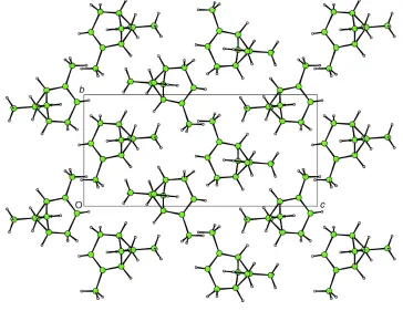
Figure 2
Projection onto (100) of the crystal structure of (I) (CAMERON; Watkin et al., 1996).
Figure 1
supporting information
sup-1
Acta Cryst. (2001). E57, o1039–o1040supporting information
Acta Cryst. (2001). E57, o1039–o1040 [doi:10.1107/S1600536801016415]
(1
S
)-(
–
)-
α
-Pinene
Andrew D. Bond and John E. Davies
S1. Comment
α-Pinene, (I), is very widely distributed in nature. It is present in the majority of essential oils derived from the Coniferae and it is the principal constituent of oil of turpentine. An account of its history and the determination of its structure using the techniques of classical organic chemistry is given by Simonsen & Owen (1947). This work forms part of a continuing study devoted to improving the techniques for determining the crystal structures of substances which are liquids at room temperature [see, for example, Davies & Bond (2001)].
S2. Experimental
(1S)-(-)-α-Pinene (99%) was obtained from the Aldrich Company and used without further purification. The crystal was grown in a 0.4 mm glass capillary tube at 203 K (a temperature only slightly less than the melting point of the solid in the capillary tube). With the axis of the capillary parallel to the φ axis and horizontal on the instrument, the crystal was obtained by moving a plug of solid material up and down the tube [the movement being controlled with the standard Z
(height) adjustment of the goniometer head]. The data are 90.2% complete because the crystal MELTED during an attempt to move it into a different orientation for the final set of frames. Previous attempts to reduce the temperature further for data collection resulted in the crystals being destroyed. Data were collected therefore at 203 (2) K.
S3. Refinement
H atoms were placed geometrically and refined using a riding model with an isotropic displacement parameter fixed at 1.2Ueq for the carbon to which they are attached. The absolute configuration could not be determined reliably and was
assigned according to the known configuration of the sample. Friedel pairs were merged therefore prior to merging in
Figure 1
The asymmetric unit in (I) showing displacement ellipsoids at the 50% probability level (XP; Sheldrick, 1993).
[image:4.610.124.488.380.661.2]supporting information
sup-3
Acta Cryst. (2001). E57, o1039–o1040(1S)-(-)-α-Pinene
Crystal data
C10H16 Mr = 136.23
Orthorhombic, P212121 a = 7.1944 (6) Å
b = 7.5920 (3) Å
c = 15.9190 (15) Å
V = 869.49 (11) Å3 Z = 4
F(000) = 304
Dx = 1.041 Mg m−3
Mo Kα radiation, λ = 0.7107 Å Cell parameters from 2466 reflections
θ = 1.0–25.0°
µ = 0.06 mm−1 T = 203 K
Cylinder, colourless 0.20 mm (radius)
Data collection
Nonius KappaCCD diffractometer
Radiation source: fine-focus sealed tube Thin–slice ω and φ scans
3727 measured reflections 1379 independent reflections
1194 reflections with I > 2σ(I)
Rint = 0.050
θmax = 25.0°, θmin = 3.7° h = −5→8
k = −7→7
l = −16→18
Refinement
Refinement on F2
Least-squares matrix: full
R[F2 > 2σ(F2)] = 0.046 wR(F2) = 0.103 S = 1.06 1379 reflections 95 parameters 0 restraints
Primary atom site location: structure-invariant direct methods
Secondary atom site location: difference Fourier map
Hydrogen site location: inferred from neighbouring sites
H-atom parameters constrained
w = 1/[σ2(F
o2) + (0.0279P)2 + 0.1574P]
where P = (Fo2 + 2Fc2)/3
(Δ/σ)max = 0.001
Δρmax = 0.13 e Å−3
Δρmin = −0.15 e Å−3
Extinction correction: SHELXL97, Fc*=kFc[1+0.001xFc2λ3/sin(2θ)]-1/4
Extinction coefficient: 0.096 (12)
Special details
Experimental. Crystal grown in a 0.4 mm Lindemann tube at 203 K. Friedel pairs were merged prior to merging in P212121; the value of Rint reported corresponds to merging of the data in this space group. The absolute configuration
was assigned from the known configuration of the sample. The data are only 90.2% complete because the crystal MELTED during an attempt to move it into a different orientation for the final set of frames!
Geometry. All e.s.d.'s (except the e.s.d. in the dihedral angle between two l.s. planes)
are estimated using the full covariance matrix. The cell e.s.d.'s are taken into account individually in the estimation of e.s.d.'s in distances, angles and torsion angles; correlations between e.s.d.'s in cell parameters are only used when they are defined by crystal symmetry. An approximate (isotropic) treatment of cell e.s.d.'s is used for estimating e.s.d.'s involving l.s. planes.
Refinement. Refinement of F2 against ALL reflections. The weighted R-factor wR and goodness of fit S are based on F2,
conventional R-factors R are based
on F, with F set to zero for negative F2. The threshold expression of
F2 > σ(F2) is used only for calculating R-factors(gt) etc. and is not relevant to the choice of reflections for refinement. R
-factors based on F2 are statistically about twice as large as those based on F, and R- factors based on ALL data will be
Fractional atomic coordinates and isotropic or equivalent isotropic displacement parameters (Å2)
x y z Uiso*/Ueq
C1 0.8184 (3) −0.0414 (3) 0.34005 (13) 0.0381 (5) H1 0.8467 −0.1469 0.3058 0.046* C2 0.7443 (3) −0.0758 (3) 0.42735 (12) 0.0389 (5) C3 0.7112 (3) 0.0642 (3) 0.47423 (13) 0.0432 (6)
H3 0.6684 0.0501 0.5296 0.052*
C4 0.7418 (3) 0.2465 (3) 0.43954 (13) 0.0434 (5) H4A 0.8282 0.3111 0.4758 0.052* H4B 0.6236 0.3107 0.4385 0.052* C5 0.8211 (3) 0.2352 (3) 0.35071 (13) 0.0397 (5)
H5 0.8524 0.3495 0.3243 0.048*
C6 0.7035 (3) 0.1064 (3) 0.29525 (12) 0.0363 (5) C7 0.9778 (3) 0.0946 (3) 0.34910 (14) 0.0449 (5) H7A 1.0603 0.1021 0.3002 0.054* H7B 1.0483 0.0850 0.4016 0.054* C8 0.4934 (3) 0.1089 (3) 0.30463 (14) 0.0439 (5) H8A 0.4397 0.0156 0.2709 0.066* H8B 0.4457 0.2217 0.2859 0.066* H8C 0.4608 0.0911 0.3631 0.066* C9 0.7516 (3) 0.1208 (3) 0.20215 (13) 0.0531 (6) H9A 0.6926 0.0254 0.1717 0.080* H9D 0.8853 0.1135 0.1951 0.080* H9B 0.7075 0.2327 0.1805 0.080* C10 0.7134 (4) −0.2629 (3) 0.45502 (15) 0.0563 (7) H10D 0.6757 −0.2644 0.5135 0.084* H10A 0.8278 −0.3292 0.4485 0.084* H10B 0.6168 −0.3159 0.4208 0.084*
Atomic displacement parameters (Å2)
U11 U22 U33 U12 U13 U23
C1 0.0322 (10) 0.0374 (12) 0.0447 (11) 0.0054 (8) −0.0009 (10) −0.0060 (9) C2 0.0323 (10) 0.0383 (13) 0.0461 (11) 0.0045 (10) −0.0050 (9) 0.0055 (9) C3 0.0411 (11) 0.0525 (15) 0.0361 (10) 0.0028 (11) 0.0007 (9) 0.0066 (9) C4 0.0455 (13) 0.0395 (13) 0.0452 (11) −0.0026 (10) −0.0005 (11) −0.0066 (8) C5 0.0380 (11) 0.0343 (11) 0.0468 (11) −0.0041 (9) 0.0015 (10) 0.0029 (9) C6 0.0347 (10) 0.0345 (11) 0.0396 (10) 0.0019 (9) −0.0009 (9) 0.0015 (9) C7 0.0293 (9) 0.0570 (13) 0.0485 (12) −0.0006 (11) 0.0012 (9) 0.0006 (12) C8 0.0339 (10) 0.0456 (13) 0.0520 (12) 0.0018 (10) −0.0041 (9) 0.0022 (11) C9 0.0535 (13) 0.0614 (15) 0.0445 (12) 0.0019 (13) 0.0015 (11) −0.0002 (11) C10 0.0542 (15) 0.0461 (15) 0.0685 (16) −0.0007 (12) −0.0062 (13) 0.0144 (11)
supporting information
sup-5
Acta Cryst. (2001). E57, o1039–o1040C1—C6 1.566 (3) C7—H7A 0.9800
C1—H1 0.9900 C7—H7B 0.9800
C2—C3 1.320 (3) C8—H8A 0.9700
C2—C10 1.504 (3) C8—H8B 0.9700
C3—C4 1.506 (3) C8—H8C 0.9700
C3—H3 0.9400 C9—H9A 0.9700
C4—C5 1.527 (3) C9—H9D 0.9700
C4—H4A 0.9800 C9—H9B 0.9700
C4—H4B 0.9800 C10—H10D 0.9700
C5—C7 1.553 (3) C10—H10A 0.9700
C5—C6 1.566 (3) C10—H10B 0.9700
C5—H5 0.9900
C2—C1—C7 106.91 (16) C8—C6—C5 118.33 (17) C2—C1—C6 110.86 (15) C9—C6—C5 112.36 (16) C7—C1—C6 87.46 (14) C1—C6—C5 84.57 (14)
C2—C1—H1 116.0 C1—C7—C5 85.55 (13)
C7—C1—H1 116.0 C1—C7—H7A 114.4
C6—C1—H1 116.0 C5—C7—H7A 114.4
C3—C2—C10 124.64 (19) C1—C7—H7B 114.4 C3—C2—C1 116.38 (17) C5—C7—H7B 114.4 C10—C2—C1 118.99 (18) H7A—C7—H7B 111.5 C2—C3—C4 120.40 (18) C6—C8—H8A 109.5
C2—C3—H3 119.8 C6—C8—H8B 109.5
C4—C3—H3 119.8 H8A—C8—H8B 109.5
C3—C4—C5 110.04 (16) C6—C8—H8C 109.5
C3—C4—H4A 109.7 H8A—C8—H8C 109.5
C5—C4—H4A 109.7 H8B—C8—H8C 109.5
C3—C4—H4B 109.7 C6—C9—H9A 109.5
C5—C4—H4B 109.7 C6—C9—H9D 109.5
H4A—C4—H4B 108.2 H9A—C9—H9D 109.5
C4—C5—C7 108.97 (17) C6—C9—H9B 109.5 C4—C5—C6 110.80 (16) H9A—C9—H9B 109.5 C7—C5—C6 87.34 (15) H9D—C9—H9B 109.5
C4—C5—H5 115.5 C2—C10—H10D 109.5
C7—C5—H5 115.5 C2—C10—H10A 109.5
C6—C5—H5 115.5 H10D—C10—H10A 109.5 C8—C6—C9 108.69 (17) C2—C10—H10B 109.5 C8—C6—C1 119.33 (17) H10D—C10—H10B 109.5 C9—C6—C1 111.94 (16) H10A—C10—H10B 109.5